
Transfection system

Inactive Publication Date: 2004-03-04
UNIVERSITATSKLINIKUM FREIBURG
View PDF4 Cites 7 Cited by
  • Summary
  • Abstract
  • Description
  • Claims
  • Application Information

AI Technical Summary

Benefits of technology

[0058] The advantage of the present invention resides in the rapid availability of transfected, autologous / allogenic cells which have the potency of repairing cell defects and simultaneously provide the required substances by autosynthesis in optimal form. Moreover, by the homogenous distribution of the transfected cells it is possible to rapidly attain a therapeutic effect. This is particularly true of wound healing of the skin.
[0059] By aid of this system, the immediate availability of transfected cells is increased without requiring prior complex steps of laboratory technology and time-consuming culturing and selection methods which, moreover, harbour the risk of contamination and mean a great loss of time.
[0060] Furthermore, the present invention comprises a kit containing the components of the transfection system, i.e. a plasmid which comprises a gene / genes coding for a substance, in particular for a protein / peptide having a positive effect on the regeneration of the tissues target cells which are transfected, as well as the self-hardening polymer or a component thereof, respectively.
[0061] The preparation obtained by the method according to the invention may be used for the treatment of damaged or defective tissue, in particular full thickness wounds, e.g. of the skin. It also allows for the treatment of humans and animals suffering from damaged or defective tissue and full thickness wounds, in particular wounds of the skin. Examples of diseases or disorders preferably treated include burn wounds, bone, muscle, nerve or cartilage defects, chronic wounds or tissue augmentations, especially preferred is a method for wound healing in the skin.
[0062] The composition obtained according to the invention allows for the rapid therapeutic treatment of injured or defective tissue. Particularly due to the homogenous distribution of the transfected cells it is possible to quickly and homogenously treat full thickness wounds. In particular, also the inaesthetic scar formation with full thickness wounds can be reduced or avoided.
[0063] According to a possible alternative, the target cells which are transfected by the transfection system according to the invention are already present in the matrix in transfected form. These cells are homogenously distributed in the matrix so that a homogenous healing may occur. Thus, according to the invention, a matrix is applied to the damaged tissue which contains the ex vivo transfected target cells which in turn contain genes coding for substances that have a positive effect on the regeneration of the defective tissue. According to a further alternative, however, transfection of the cells may also occur after application in the matrix. By the homogenous distribution of the plasmid and of the cells, a homogenous transfection of the cells is possible which then will lead to a homogenous expression of the factor that has a positive effect on the progress of regeneration.

Problems solved by technology

In medicine, tissue defects and their treatment represent a great problem.
On the other hand, these factors may also inhibit processes which counteract a rapid regeneration.
However, this has the disadvantage that high doses had to be supplied.
Moreover, most of the factors have only a short half-life in vivo so that multiple administrations had been necessary.
When administering factors externally, there is also a risk that they might contain contaminants.
Both, when purifying material from natural sources and in the recombinant production of the factors there is a risk of the preparations containing contaminants which may negatively affect the progression of regeneration.
Therefore, the external administration of mediators, such as growth factors, proved to be an inefficient system, mainly also from the point of view that some mediators cannot at all be administered externally.
Moreover, a specific transfection is not possible with this method.
Yet in doing so, also the carrier gets into the cell, which may have negative consequences.
In the transfection of eukaryotic cells, the greatest problem is the effectiveness of the transfection.
Especially with viral vectors, their tropism, i.e. their affinity relative to certain cell types, still is a major problem.
Viruses and viral vectors may have a high rate of polymorphism, and this may lead to inactive vectors or genes.
As has already been mentioned, the tropism of the viral vectors is a further problem.
Adenoviruses, although capable of transfecting non-proliferating cells, also have the afore-mentioned disadvantages of viral vectors, i.e. they integrate randomly into the genome of the target cell.
A disadvantage of these liposomes is, e.g., a remaining cytotoxicity of the liposomal transfection agent concerning the target cells.
This means that the cells to be transfected (target cells) are damaged by the transfection agent itself, causing them to die.
Although this method partially allows for the transfection of cells without a carrier, it is, however, a random transfection of the cell without allowing to determine the amount of material transfected.
The transfection by means of electroporation has the disadvantage that the transfection is performed in vitro in a special apparatus.
This requires various method steps, and the risk of a contamination of the cell suspension is high.
Yet with this approach, the transfection efficiency is too low.
A permanent transfection of the cells, however, is not always desirable, instead a transient transfection of the cells is preferred so that expression of the factor encoded by the plasmid will occur only for a certain period of time.
When wound healing has been finished, continued expression of this factor is not desirable.
On the contrary, this could, e.g., promote scar formation and an abnormal formation of the tissue structure which is unwanted.

Method used


Image

  • Transfection system
  • Transfection system
  • Transfection system

Examples


example 2

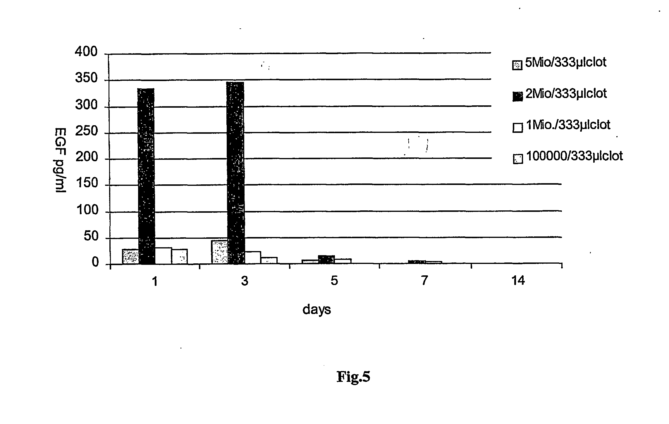
[0110] Determination of the optimal keratinocyte concentration for a given buffer. EGF-plasmid, Tissucol.RTM. and cell preparations were used as described in Example 1. Only the amount of keratinocytes was varied between 100,000, 1 million, 2 millions and 5 millions. The highest expression rates of EGF were obtained in using a cell number of 2 millions / 333 .mu.l of fibrin clot, which amounts to about 6 millions cells / ml of clot (see FIG. 5).

example 3

[0111] Optimization of a gene activated matrix for the treatment of full thickness wounds of nude mice.

[0112] Full thickness wounds of nude mice (12 mice per group) were treated with different combinations (groups 1-4). The amount of fibrin in each case was 333 .mu.l, the amount of plasmid 200 .mu.g. In each case a pre-incubation of appropriate cells with the plasmid was carried out in 5.7 .mu.l PBS for 3 hours. The amount of keratinocytes used was 2 millions. Biopsies were taken on days 1, 3, 5, 7, 9 and 12 and histologies starting with day 5.

1 Group 1: Fibrin and EGF-plasmid Group 2: Fibrin and keratinocytes Group 3: Fibrin, EGF-plasmid and endothelial cells (=support cells) Group 4: Fibrin, EGF-plasmid and keratinocytes (=repair cells)

[0113] The results of histologies clearly show that only group 4 leads to a full re-epithelialization of a full thickness wounds in nude mice, having a completely regenerated epithelium consisting of 9-11 layers of cells (see FIGS. 9 and 10).

[0114] ...

example 4

[0115] Transfection of Various Cell Types 200,000 cells, namely muscle cells, Schwann cells, endothelial cells, preadipocytes and fibroblasts, where transfected with 10 .mu.g of EGF-plasmid each, the amount of fibrin used was 333 .mu.l. Expression of EGF was measured after day 1, 2, 3, 4 and 5 and is shown in FIG. 11.

[0116] Examples for Isolation of Cells

[0117] Schwann Cells

[0118] Cells were prepared with modification according to the method of Shahar et al., (1989) in which Schwann cells were harvested from the sciatic nerve of neonatal rats. In brief, the Schwann cells were harvested from 7 mm segments of the sciatic nerve. Nerves were collected in HBSS, stripped of their epineurium and chopped into 1 mm.sup.2 pieces. The nerve pieces were dissociated by incubating the chunks for 30 minutes at 37.degree. C. with 0.3% trypsin and 0.1% collagenase. The cells were then triturated, washed and cultured with DMDM containing 10% FCS and penicillin / streptomycin on poly-D-lysine coated fla...


PUM

PropertyMeasurementUnit
Compositionaaaaaaaaaa
Ratioaaaaaaaaaa

Abstract

The present invention relates to a method of preparing a composition for wound healing, and for repairing and regenerating human and animal tissue, said method comprising the following steps: a. providing a plasmid DNA in substantially pure form, which encodes a gene that has a positive effect on the progression of the regeneration of the tissue, b. providing a component / components of a self-hardening bio-polymer, and c. providing a cell suspension with cells which promote regeneration, characterized in that components (a), (b) and (c) are incubated with each other simultaneously or successively so that the plasmid and the cell suspension are obtained homogenously distributed in one of the biopolymer components. Furthermore, transfection systems containing a plasmid DNA, a component of a self-hardening biopolymer and a cell suspension with cells promoting regeneration are disclosed. This transfection system does not contain any further transfection-promoting or transfection-mediating substances. Moreover, therapeutical kits, pharmaceutical compositions and their use for the treatment of tissue defects, in particular burn wounds, and for wound healing in the skin are described. In particular, the present invention relates to a transfection system containing a plasmid DNA, a component of the fibrin adhesive and a cell suspension.

Description

[0001] The present invention relates to matrix-mediated transfection systems, wherein the matrix consists of a self-hardening biopolymer. Moreover, the invention relates to a method of producing a preparation containing plasmid DNA, a component of the self-hardening material, and a cell suspension of the cells to be transfected, the preparation itself and its use for treating tissue injuries and changes.[0002] In particular, the present invention relates to a fibrin-mediated transfection system for cells for improved wound healing, tissue regeneration and tissue repair.[0003] In medicine, tissue defects and their treatment represent a great problem. Both in surgical interventions and also as a consequence of wear, of external influences such as injuries caused by burns, a stroke, etc., corresponding traumas of the tissue occur the healing and regeneration of which is decisive. Also in many illnesses tissue is damaged, psoriasis and arthritis being mentioned by way of example.[0004] ...

Claims


Application Information

Patent Timeline
IPC IPC(8): A61K9/10A61K35/12C12N15/09A61K35/32A61K35/36A61K47/30A61K47/36A61K47/42A61K48/00A61L26/00A61L27/00A61P17/02A61P19/00A61P41/00A61P43/00
CPCA61L26/0057A61K48/00A61P17/00A61P17/02A61P19/00A61P19/02A61P19/08A61P21/00A61P25/00A61P41/00A61P43/00
Inventor ANDREE, CHRISTOPHVOIGHT, MATTHIASSTARK, BJOERN G.
Owner UNIVERSITATSKLINIKUM FREIBURG
Learn More