

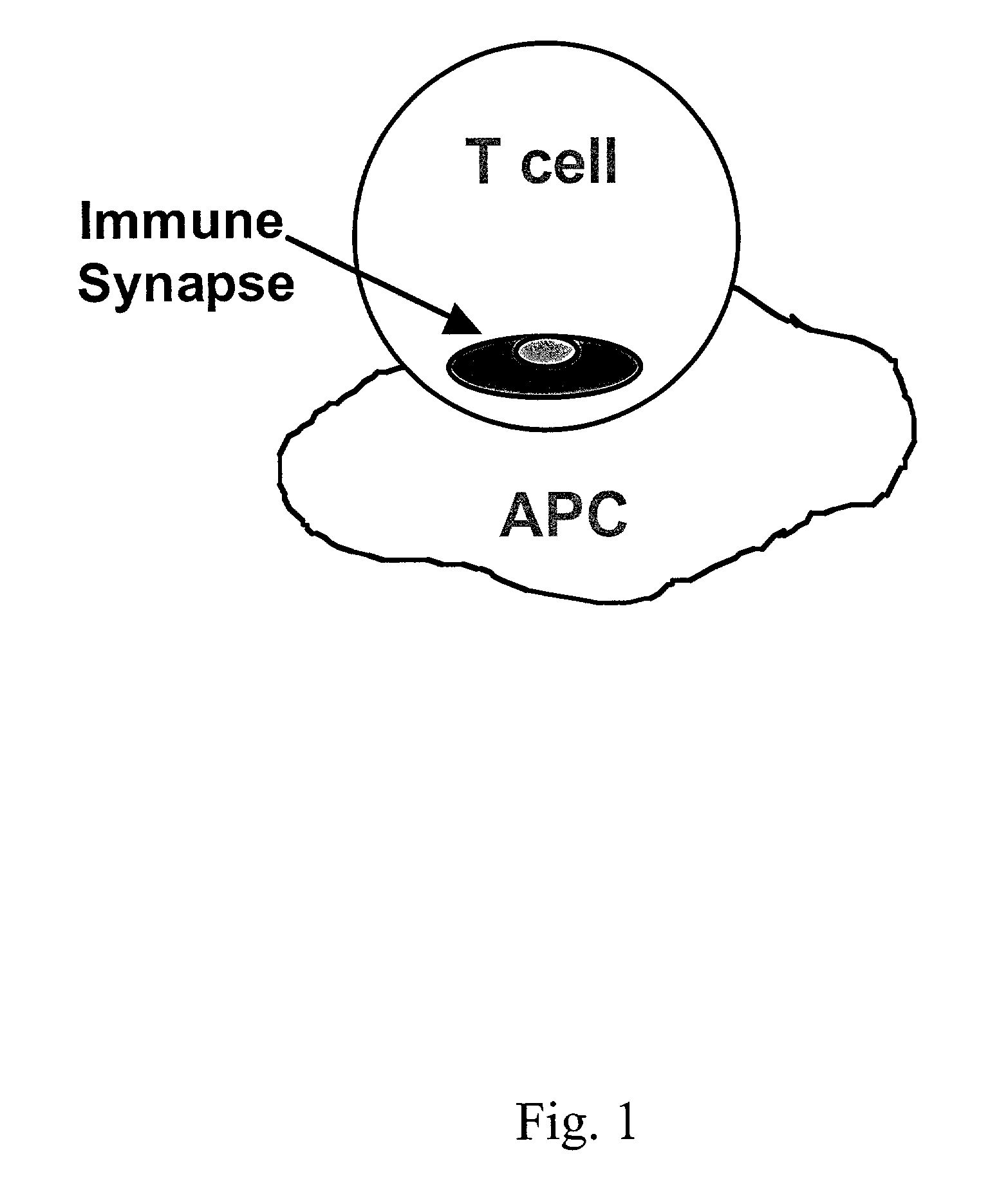
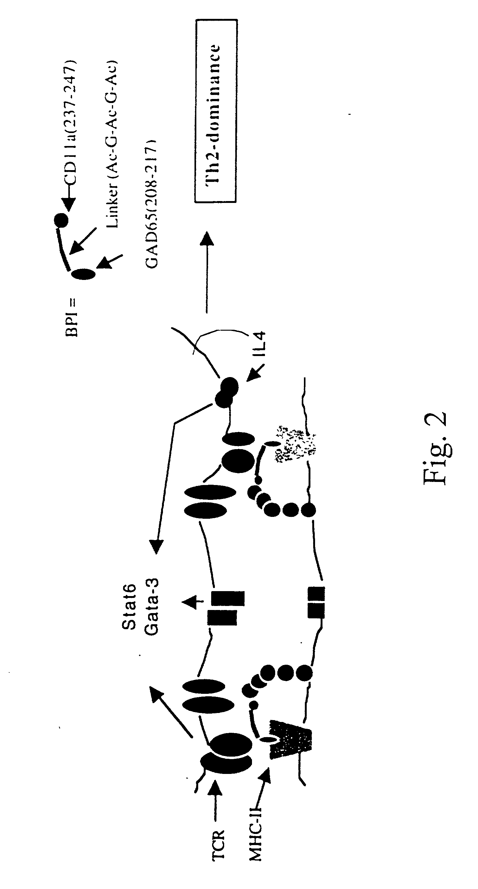
Signal-1/signal-2 bifunctional peptide inhibitors
a technology of peptide inhibitors and peptides, which is applied in the direction of immunological disorders, lyases, antibody medical ingredients, etc., can solve the problems of end-stage immune synapse alteration, and achieve the effect of preventing the translocation step of immune synapse formation and reducing lfa-1:icam-1 signaling
- Summary
- Abstract
- Description
- Claims
- Application Information
AI Technical Summary
Benefits of technology
Problems solved by technology
Method used
Image

Examples
example 1
[0080] This example describes the methods used to generate the BPI.
Materials and Methods:
[0081] Synthesis of peptides was via Fmoc on chlorotrity resins. Protected amino acids were double coupled at 8-fold excess for one hour. Resins were dimethylforamide (DMF) and methanol (MeOH) washed and cleaved in Reagent R: trifluorolacetic acid (TFA), ethylene diamine tetraacetic acid (EDTA), Thioanisole, Anisole. The TFA mixture containing the peptide in solution is precipitated in ether and washed extensively. Preparative HPLC of peptides was accomplished by a gradient of 0-80% acetonitrile in 0.1% TFA. Lyophilization of the various fractions and verification by mass spectroscopy yielded the synthetic peptides as a TFA salt. Modeling, crystallography and binding studies are as described above.
Results:
[0082] The peptides produced in this example are provided in Table 1 and are also listed as SEQ ID Nos. 1-46. These peptides include the Signal-1 moiety, the Signal-2 moiety and the non-s...
example 2
[0090] This example uses biotinylated BPI to test for competitive inhibition of BPI binding by unlabeled peptides or monoclonal antibodies to MHC-II and ICAM-1 on live APC, and to verify antigenic peptide binding to live APC. Additionally, it was shown that monoclonal antibodies to MHC-II or ICAM-1 effectively block binding of the diabetes BPI (GAD65(208-217)—[Ac-G-Ac-G-Ac]-CD11a (237-247)) (hereinafter referred to as EGAD-BPI) to NOD spleenocytes.
Materials and Methods:
[0091] To obtain biotinylated BPI, the synthesized EGAD BPI was biotinylated with NHS-Biotin as described in Murray et al., 24 Eur. J. Immunol. 2337-2344 (1994). Spleen cell density-gradient fractions from normal (unimmunized) NOD, BALB / c and other MHC congenic strains were incubated in round bottom 96-well plates with increasing concentrations of individual biotinylated peptides at 37° C.,5% CO2 for 16 hours. Following binding of the BPI to the APC, Avidin-FITC was incubated with the cells on ice for 30 minutes, f...
example 3
[0097] This example utilizes co-capping experiments to demonstrate simultaneous binding of the BPI to MHC-II and ICAM-1 molecules.
Materials and Methods:
[0098] Further support for simultaneous binding of the BPI to MHC-II and ICAM-1 molecules has been observed in co-capping experiments using biotinylated mAb 10-3.62 and streptavidin to cap MHC-II in the presence or absence of the BPI. To test the ability of the BPI peptide to link MHC-II and ICAM-1 molecules on the APC surface, we used a modification of a co-capping experiment originally described for monoclonal antibodies. Briefly, biotinylated monoclonal antibody to MHC-II (10-3.62) is incubated with freshly-isolated APC from NOD mice previously treated by intravenous (i.v.) injection of a given BPI variant or saline. Antibody-bound cells are then incubated with streptavidin (37 C×15 min.) to cap the MHC-II molecules on the APC surface. The cells were transferred to ice and labeled with a fluorescent (PE) monoclonal antibody to ...
PUM
| Property | Measurement | Unit |
|---|---|---|
| Fraction | aaaaa | aaaaa |
| Fraction | aaaaa | aaaaa |
| Fraction | aaaaa | aaaaa |
Abstract
Description
Claims
Application Information