
Bead bound combinatorial oligonucleoside phosphorothioate and phosphorodithioate aptamer libraries

a technology of phosphorothioate and aptamer libraries, which is applied in the field of aptamer libraries, can solve the problems of odns possessing high fractions of phosphorothioate or phosphorodithioate linkages losing some of their specificity, and limited to substrates

Inactive Publication Date: 2005-06-09
BOARD OF RGT THE UNIV OF TEXAS SYST
View PDF52 Cites 24 Cited by
  • Summary
  • Abstract
  • Description
  • Claims
  • Application Information

AI Technical Summary

Benefits of technology

[0043] The present invention uses a two dimensional (2D) robotic spot picker to identify a target, e.g., a protein, on an individual bead of a combinatorial bead library that had been incubated with a target to for an aptamer-bead-target complex. The subject invention may use, e.g., a combinatorial bead library / protein incubation step of the existing methodology. One advantage of the present invention is that it does not require flow cytometry to isolate those beads that have bound target protein, often a bottleneck for high-throughput analysis. Instead, the invention scans and picks target bead spots, e.g., a pre-incubated target and aptamer bead library in a polyacrylamide gel, which is then cast onto a 2D plate whose bead “spots” are then imaged, analyzed and manipulated using a robotic scanning spot picker. The target and the aptamer-beads may be pre-incubated, or the target and / or the aptamer-beads may be incubated in situ. The aptamer-beads and the target are generally distributed uniformly throughout the two-dimensional matrix or agent, e.g., a gel, a gel plate, a glass plate into, e.g., a single layer. In one example, the aptamer-beads are 20-80 microns in diameter, but may be larger or smaller depending on factors such as the necessary signal-to-noise ration, amount of sample, number of beads in an aptamer-bead library, etc.
[0044] To capture, scan, image, identify and locate the spots to be picked, software available with robotic scanners and pickers, known in the art, is used to determine the XY coordinates and intensity of each bead in the 2D gel matrix, and directs and controls the hardware for manipulation of aptamer-beads and their target. For example, a high speed optical scanner based on a positional scanning excitation source (e.g. laser, diode laser) and an emission sensor (e.g. CCD camera) may be used to create a 2D image / map of candidate beads. Examples of instruments for use with the present invention include commercially available robotic spot pickers, e.g., a Bruker Daltonics Proteineer spII spot-picker (Bruker Daltonics website). Such spot pickers may be operated in conjunction with a 2D matrix, e.g., an polyacrylamide electrophoretic gel, performing 2D gel imaging, detection of protein spots of the gel, software selection, directing the robotic head to cut out (“core”) a relevant spot (either automatically or based on manual review of the image map) and transfer of the spot(s) to a sample array for MS analysis. An example of a 2D imaging system is available commercially from Oncosis (Oncosis website), which uses “Photosis” technology (WO 98 / 42356, 1998; WO 01 / 40454, 2001). The Oncosis system is capable of high speed laser-based optical scanning to image a 2D matrix of cells embedded in a polymeric matrix, however, the system then re-addresses and destroys cells that are not of interest via high intensity laser photolysis.
[0045] The following are general requirements of a coring mechanism for use with the robotic head of the spot picker for use with the present invention. The coring mechanism of the robotic head of the spot picker, will generally transfer a candidate bead from the cast gel matrix to, e.g., a mass spectrometer inlet. To do so it will generally capture only one bead at a time. As such, the coring mechanism will generally be designed to have a diameter less than that of two beads. To introduce a bead into the interior of a cylinder, e.g., a needle, the inner diameter of the cylinder has to be greater than that of one bead. However, if a bead or plug is not to be fully captured within the cylinder, then the item may be held outside the tip of the cylinder using, e.g., application of a vacuum to the other end of the cylinder to cause a vacuum within the cylinder. Thus, there is a suitable range between one bead diameter and less than two bead diameters.
[0046] For example, if the beads are 60 micron diameter, the coring mechanism must have a diameter between 80-100 microns. If this requirement is not met, the possibility exists that the coring mechanism will transfer more than one bead in a single “core.” The “bystander” bead(s) transferred may also be candidate beads (also bind target protein and thus have appropriate fluorescent signature) or may be non-candidates (do not bind protein, do not have fluorescent signature). Where the single “core” contains “bystander” candidate beads, the mass spectrum of the multiple proteins (or peptide digests if on-gel digestion is used) will be difficult to interpret. Where the single “core” contains “bystander” non-candidate beads, the identification of the binding thioaptamer of the candidate bead will be difficult.

Problems solved by technology

Current DNA array technology is problematic in that it is focused on the identification and quantification of a single mRNA species, and does not provide information on the more relevant level of functional protein expression and in particular protein-protein interactions such as between heterodimers and homodimers.
Unfortunately, ODNs possessing high fractions of phosphorothioate or phosphorodithioate linkages may lose some of their specificity and are “stickier” towards proteins in general than normal phosphate esters, an effect often attributed to non-specific interactions.
The identification of specific S—ODN and S2—ODN aptamers (“thioaptamers”) that bind proteins based upon in vitro combinatorial selection methods, however, is limited to substrates only accepted by polymerases required for reamplification of selected libraries by the polymerase chain reaction (PCR).
One disadvantage of using the polymerization of substituted nucleoside 5′-triphosphates into ODN aptamers are the restrictions on the choice of P-chirality by the enzymatic stereospecificity.
Therefore, using current methods it is not possible to select [Sp]-phosphorothioate stereoisomers along with achiral S2—ODN analogous since both [Rp]-diastereoisomers of dNTP(αS) and nucleoside dNTP(αS2) are not substrates of polymerases.
Additionally, these in vitro combinatorial selection methods require many iterative cycles of selection and reamplification of the bound remaining members of the library by the PCR, which are quite time consuming.

Method used


Image

  • Bead bound combinatorial oligonucleoside phosphorothioate and phosphorodithioate aptamer libraries
  • Bead bound combinatorial oligonucleoside phosphorothioate and phosphorodithioate aptamer libraries
  • Bead bound combinatorial oligonucleoside phosphorothioate and phosphorodithioate aptamer libraries

Examples


example 1

S—ODN, S2—ODN and Monothio-RNA Split and Pool Synthesis

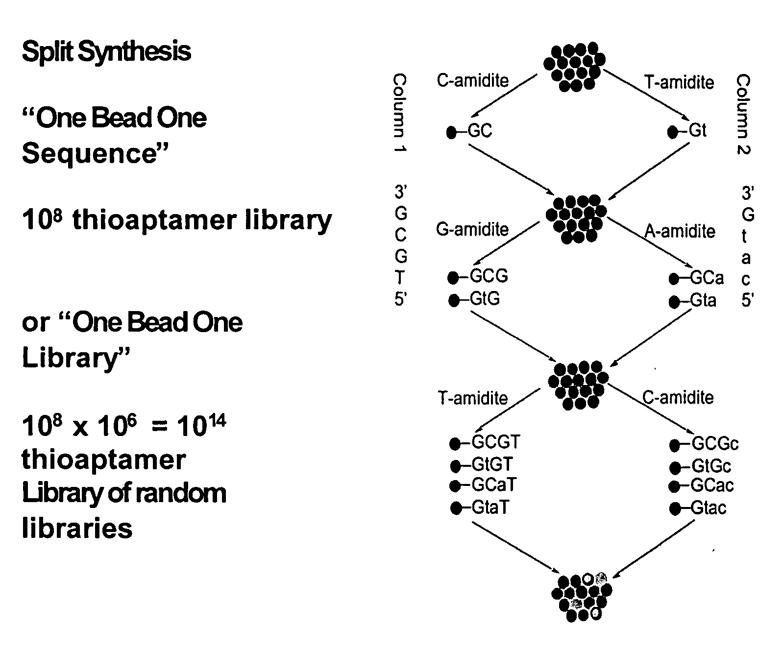
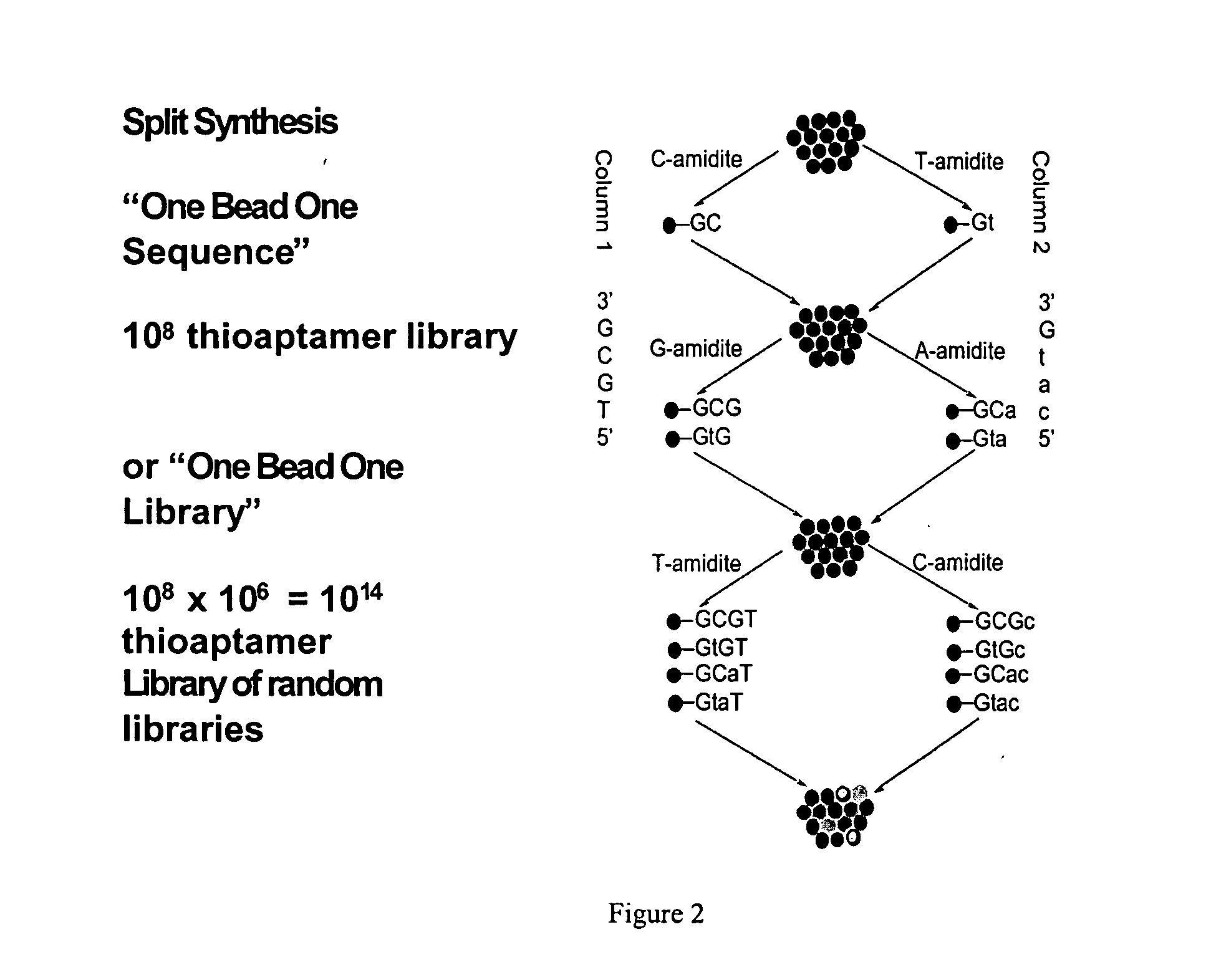
[0071] A split and pool synthesis combinatorial chemistry method was developed for creating combinatorial S—ODN, S2—ODN and monothio-RNA libraries (and readily extended to unmodified ODNs-whether single strand or duplex). In this procedure each unique member of the combinatorial library was attached to a separate support bead. Targets that bind tightly to only a few of the potentially millions of different support beads can be selected by binding the targets to the beads and then identifying which beads have bound target by staining and imaging techniques. The methodology of the present invention allowed the rapid screening and identification of aptamers that bind to proteins such as NF-κB using a novel PCR-based identification tag of the selected bead.

[0072] The dA, dG, dC and dT phosphoramidites were purchased from Applied Biosystems (Palo Alto, Calif.) or Glen Research (Sterling, Va.). The Beaucage reagent (3H-1,2-Benzodit...

example ii

ELISA Based Thioaptamer Selection-Indirect ELDIA

[0106] Although the beads were screened against a target protein labeled with a fluorescent dye, the beads can also been screened directly against the transcription factor. The binding of the NF-κB to a specific sequence can be detected using a primary anti-NF-κB antibody (Rabbit IgG antibody, Santa Cruz Biotechnology, Inc.) followed by a secondary antibody conjugated with Alexa Fluor 488 (goat anti-rabbit IgG from Molecular Probes). Next, several beads were selected for sequencing. The sequencing result were as follows:

E008 Selected sequences5′-CGCCAGCCGaAGGTGCTGTCAG-3′(SEQ ID NO:31)5′-ATGTAGCCAaAGGTGgaACCCC-3′(SEQ ID NO:32)5′-CGCCcAGTgaAGGTGCTGTCAG-3′(SEQ ID NO:33)5′-CGCCcAGTAGCTAGTCTGTCAG-3′(SEQ ID NO:34)

[0107] It was observed that the phosphorodithioate linkage (s) in the selected above sequences were different from those of the screening against the fluorescently labeled NF-κB p50. This result suggests that some of the binding...

example iii

Labeling of the ODN with Fluorescent Dyes

[0108] When synthesizing combinatorial libraries or specific thioaptamer sequences on beads, one may also identify beads by attaching 2 or more fluorescent dyes to the ODN either at the 5′ or 3′ ends or internally by using phosphoramidites with specific fluorophors attached. By using 1-3 (or more) fluorophors at 2-3 or more different levels (individual nucleosides), it is possible to identify dozens or more of the sequences or libraries by multicolor flow cytometry. (Each bead can thus be identified by dye A, B and / or C at levels high, medium, low in various combinations: thus bead with A(hi), B(medium) and C(low) would be one of dozens of different possible combinations.)

[0109] Thus it is possible to multiplex using flow cytometry or by randomly placing beads onto, e.g., the Texas tongue with hundreds or thousands or more of different microwell holders, random assortment of thioaptamer beads specific for binding different analytes. Altern...


PUM

PropertyMeasurementUnit
Thixotropic indexaaaaaaaaaa
Colorimetric evaluationaaaaaaaaaa

Abstract

The present invention includes composition and methods for making and using a combinatorial library having two or more beads, wherein attached to each bead is a unique nucleic acid aptamer that have disposed thereon a unique sequence. The library aptamers may be attached covalently to the one or more beads, which may be polystyrene beads. The aptamers may include phosphorothioate, phosphorodithioate and / or methylphosphonate linkages and may be single or double stranded DNA, RNA or even PNAs.

Description

TECHNICAL FIELD OF THE INVENTION [0001] The present invention relates in general to the field of aptamer libraries, and more particularly, to enhancing availability and use of aptamers for screening, including high-throughput screening, of primary or secondary target molecules by using thioated aptamers bound to a substrate. BACKGROUND OF THE INVENTION [0002] This application is a continuation in part application based on U.S. patent application Ser. No. 10 / 272,509, filed Oct. 16, 2002, and a continuation in part of U.S. Provisional Patent Application No. 60 / 334,887, filed on Nov. 15, 2001. This work was supported by the following United States Government grants DARPA (9624-107 FP), NIH (AI27744) and NIEHS (ES06676). Without limiting the scope of the invention, its background is described in connection with oligonucleotide agents and with methods for the isolation of sequences that are bound by nucleic acid binding molecules and the like. [0003] Virtually all organisms have nuclease...

Claims


Application Information

Patent Timeline
IPC IPC(8): C07H19/00C07H21/00C12NC12N1/00C12Q1/68C40B40/06C40B60/14G01N33/53G01N33/543G01N33/68
CPCB01J2219/00378G01N33/6851B01J2219/00497B01J2219/005B01J2219/00529B01J2219/00585B01J2219/0059B01J2219/00592B01J2219/00596B01J2219/00608B01J2219/0061B01J2219/00612B01J2219/00626B01J2219/00637B01J2219/00641B01J2219/00648B01J2219/00659B01J2219/00689B01J2219/00707B01J2219/00722C07B2200/11C07H21/00C12N15/1048C40B20/04C40B40/06C40B40/08C40B60/14G01N33/6803B01J2219/00387
Inventor GORENSTEIN, DAVIDYANG, XIANBINLUXON, BRUCEHERZOG, NORBERT
Owner BOARD OF RGT THE UNIV OF TEXAS SYST
Learn More