Protein micro-arrays and multi-layered affinity interaction detection

a protein microarray and affinity interaction technology, applied in the field of proteomics, can solve the problems of many technical and practical problems that remain unsolved, the 2d-page/ms-id approach is either very poorly addressed, or not at all addressed by the 2d-page/ms-id approach, and achieves high throughput, high throughput and quantitative analysis.

Inactive Publication Date: 2005-07-14
GEMBITSKY DMITRY S +1
View PDF2 Cites 17 Cited by
  • Summary
  • Abstract
  • Description
  • Claims
  • Application Information

AI Technical Summary

Benefits of technology

[0014] In one embodiment of the present invention there is provided a high throughput and quantitative method of analyzing post-translational protein modifications in a sample comprising the steps of preparing at least one of N identical arrays of immobilized protein capture agents, each of the capture agents binding specifically to a protein in the sample; and performing in any order the steps of applying the proteins of the sample to at least one of the N arrays of immobilized protein capture agents; and binding the proteins of the sample to at least one of X detectable affinity reagents to label the proteins, where X is an integer from 1 to N and where each of the X detectable affinity reagents specifically recognizes one of N post-translational protein modifications and measuring a signal associated with the detectable affinity reagents, wherein quantitation of the signal of the X detectable affinity reagent(s) provides a high throughput and quantitative analysis of post-translational protein modifications in the sample.

Problems solved by technology

Completion of the human genome project has brought with it a new round of challenges to characterize the components and understand the behavior of a cell.
However, compared to micro-arrays, the throughput and sensitivity of 2D-PAGE / MS analysis are orders of magnitude lower, and many technical and practical problems remain unsolved.
Moreover, post-translational modification analysis is either very poorly, or not at all, addressed by the 2D-PAGE / MS-ID approach.
Identifying dynamic covalent protein modifications in their proper biological context is clearly a biochemical problem. for example, reversible phosphorylation, one of dozens or hundreds of different estimated modifications, is critical for transmission of signals in all living cells.
Similar issues and questions can be raised for analysis of protein interactions.
However, it will take great effort and expense to produce 35,000 unique antibodies that are absolutely specific in recognition of their cognate human protein targets.
Two potential problems are evident.
First, antibody-antigen interactions are likely to be quite variable, making general ‘capturing’ conditions for 35,000 different proteins difficult to establish.
Because a number of cellular proteins will be fluorescently labeled, this could lead to over-representation of the amount of any particular antigen, creating a false positive-like situation.
Both concept and execution are straightforward, albeit very laborious and time consuming to get the necessary tools (2).
However, even though the above techniques are perfectly optimized, neither approach will provide any direct information on dynamic protein modifications, or protein-protein, protein-nucleic acid and protein-small molecule interactions in the cell.
The prior art is deficient in proteomic techniques that allow sensitive and high throughput analysis of protein modifications and interactions.

Method used


Image

  • Protein micro-arrays and multi-layered affinity interaction detection
  • Protein micro-arrays and multi-layered affinity interaction detection
  • Protein micro-arrays and multi-layered affinity interaction detection

Examples


example 1

Fabrication of Microarrays

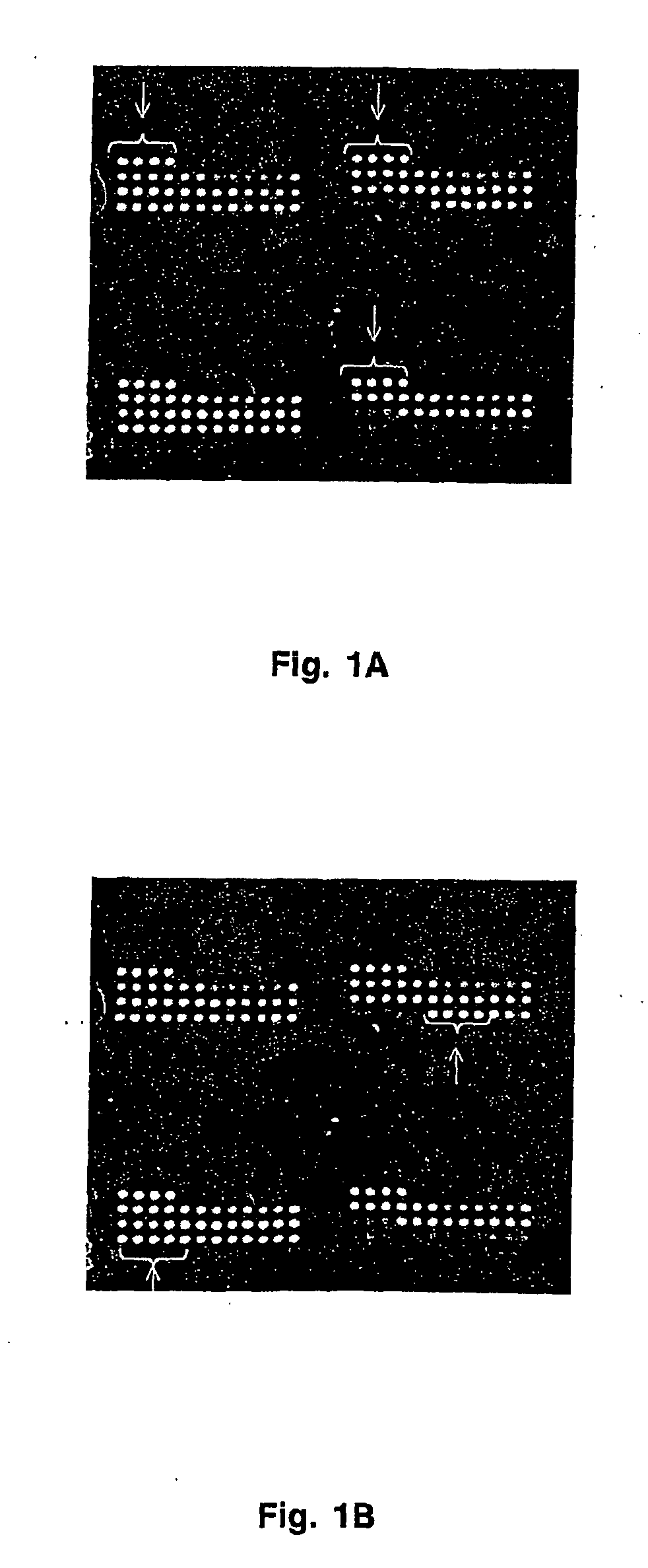
[0054] Antibodies were printed on HydroGel slides (Perkin Elmer Life Sciences) using MicroSpot 2500 pins and a MicroGrid II arrayer (BioRobotocs). Printing ink was PBS containing 0.2% gelatin and 0.1% sodium azide. Printing concentration of each antibody was 200 μg / mL. Each antibody was spotted onto the array at least 5 times. The spacing between spots was 300 microns. Quality control of antibody deposition was performed using Cy5-labeled non-specific antibody. Quality control of retention of antibodies was performed using deposition of mouse IgG which was detected at the completion of experiments with Cy5-labeled goat anti-mouse antibody.

[0055] After completion of a printing cycle, arrays were incubated in the dark at room temperature and 65% relative humidity for at least 48 hrs. They were washed with PBST (PBS supplemented with 0.01 to 0.1% tween-20) for 30 min 3 times on an orbital shaker. Finally they were dipped in PBS, centrifuged at 1,000 rpm for...

example 2

Preparation of Protein Extract

[0056] Human leukemia cells, R10+ (glucophorin A positive), were grown in IMDM medium supplemented with 20% (v / v) heat inactivated fetal bovine serum and a penicillin-streptomycin mixture at 37° C. in 5% CO2. Cells were collected and washed 4 times with ice-cold PBS without calcium and magnesium. The extraction buffer typically was carbonate buffer (pH 6.0 to 9.6) supplemented with EDTA (1 μM to 10 mM), IGEPAL (0.1 to 5%), NaF (1 μM to 10 mM), and Na3VO4 (1 μM to 10 mM). Ice-cold extraction buffer was added to cells. Proteins were extracted for 15 min on a rocking platform at 4° C. Cell debris was removed by centrifugation at 15,000 g for 30 min at 4° C. Protein content in the extract was determined using micro BCA reagent kit (Pierce).

example 3

Labeling of Cellular Proteins with Fluorescent Tag

[0057] In a typical experiment, 2.6 mL of protein extract (protein concentration 0.1 to 0.5 mg / mL) was labeled with Cy5 fluorescent dye. NHS-ester activated Cy-dyes were from Amersham Biosciences. The dye (200 nmoles) was dissolved in a total volume of protein extract to be labeled and incubated in the dark at room temperature and gentle rocking for 30 minutes. Separation of non-incorporated dye was performed by gel-filtration on a Sephadex G-25 column (Amersham Biosciences) that was previously equilibrated with PBST. An equal volume of non-labeled protein extract was also applied to a G-25 column to exchange the buffer for incubation with arrays.


PUM

PropertyMeasurementUnit
concentrationaaaaaaaaaa
v/vaaaaaaaaaa
pHaaaaaaaaaa

Abstract

The present invention provides proteomic techniques that extend sensitive and quantitative analysis of proteins to post-translational modifications. Protein micro-arrays and/or multiplex coded-microbeads are used in combination with multilayered affinity interaction detection (MAID) methods that permit high throughput analysis of cellular protein modifications and functional protein interactions.

Description

CROSS-REFERENCE TO RELATED APPLICATION [0001] This application claims benefit of priority of provisional application U.S. Ser. No. 60 / 335,645, filed Oct. 23, 2001, now abandoned.BACKGROUND OF THE INVENTION [0002] 1. Field of the Invention [0003] The present invention relates generally to the field of proteomics. More specifically, the present invention relates to protein micro-arrays and multi-layered affinity interaction detection procedures that allow high throughput and quantitative cellular protein profiling. [0004] 2. Description of the Related Art [0005] Completion of the human genome project has brought with it a new round of challenges to characterize the components and understand the behavior of a cell. Protein science will play a major role in this endeavor because proteins carry out most of the work in a cell, including control of growth and development. While genomics and functional genomics will continue to provide significant insights, it is likely to overlook many oth...

Claims


Application Information

Patent Timeline
Patent Type & Authority Applications(United States)
IPC IPC(8): C12Q1/68G01N33/48G01N33/50G01N33/53G01N33/68G06F19/00
CPCG01N33/6803
Inventor GEMBITSKY, DMITRY S.TEMPST, PAUL
Owner GEMBITSKY DMITRY S
Why Eureka
Try Eureka