Three dimensional cell protector/pore architecture formation for bone and tissue constructs

a cell protector and pore structure technology, applied in the field of three-dimensional cell protector/pore architecture formation of bone and tissue constructs, can solve the problems of no cells being mixed into the cpc paste, no prior art design of cell protector/pore architecture, etc., and achieve the effect of protecting from mechanical and chemical damag

Inactive Publication Date: 2006-07-20
ADA FOUND
View PDF1 Cites 8 Cited by
  • Summary
  • Abstract
  • Description
  • Claims
  • Application Information

AI Technical Summary

Benefits of technology

[0019] 1. That is capable of containing and maintaining living cells prior to the implant formation and placement. This is advantageous because the cells are distributed throughout the entire self-hardening implant and are protected from mechanical and chemical damage during paste mixing, manipulation and setting of the implant matrix material, such as a moldable and self-hardening calcium phosphate bone cement. Other methods that seed cells only after the implant is formed or hardened only have cells on the surface or in a shallow surface layer. Methods that directly mix cells into the implant materials risk cell damage or death due to mechanical, thermal and chemical damages.
[0020] 2. That has engineered three-dimensional lattice-like structures, zigzag or woven or curved structures, or three-dimensional web-like structures that can build three-dimensional pore architectures, thus allowing critically-important cell migration, in

Problems solved by technology

However, in these studies, no cells were mixed into the CPC paste.
Furthermore, there has been no mention in prior art of the design of cell protectors/pore architecture

Method used


Image

  • Three dimensional cell protector/pore architecture formation for bone and tissue constructs
  • Three dimensional cell protector/pore architecture formation for bone and tissue constructs
  • Three dimensional cell protector/pore architecture formation for bone and tissue constructs

Examples


example 1

[0046] An example of manufacture of this invention would be for medical, veterinary, dental, craniofacial and orthopedic bone repair. The CPC powder and liquid are mixed to form a paste that is filled into a stainless steel mold of 6 mm diameter and 3 mm depth. Distilled water is used as the cement liquid. Each specimen in the mold is sandwiched between two glass slides, and the assembly is incubated in a humidor with 100% relative humidity at 37° C. The hardening time is measured using a needle method with a load of 453.5 g and a flat tip diameter of 1.06 mm. A cement specimen is considered set when the needle loaded onto the specimen surface failed to leave a perceptible indentation. The time measured from the paste being mixed to this point is used as the setting time. Depending on composition and powder-to-water ratio, the setting time ranges from (14.5±1.3) min (82.8±4.4) min.

[0047] When the liquid contains water with sodium phosphate and hydroxypropyl methylcellulose, the cem...

example 2

[0048] An example of manufacture of this invention would be to incorporate living cells into a calcium phosphate cement for tissue repair. As an example, MC3T3-E1 osteoblast-like cells (Riken, Hirosaka, Japan) are cultured following established protocols (Simon et al., Preliminary report on the biocompatibility of a moldable, resorbable, composite bone graft consisting of calcium phosphate cement and poly(lactide-co-glycolide) microspheres. J Orthop Res 20:473-482, 2002; Xu et al., Self-hardening calcium phosphate composite scaffold for bone tissue engineering. Journal of Orthopaedic Research 22:535-543, 2004). Cells are cultured in flasks at 37° C. and 100% humidity with 5% CO2 (volume fraction) in a. modified Eagle's minimum essential medium (Biowhittaker, Walkersville, Md.). The medium is supplemented with 10% volume fraction of fetal bovine serum (Gibco, Rockville, Md.) and kanamycin sulfate (Sigma, St. Louis, Mo.), and changed twice weekly. Fifty thousand cells diluted into 2 m...

example 3

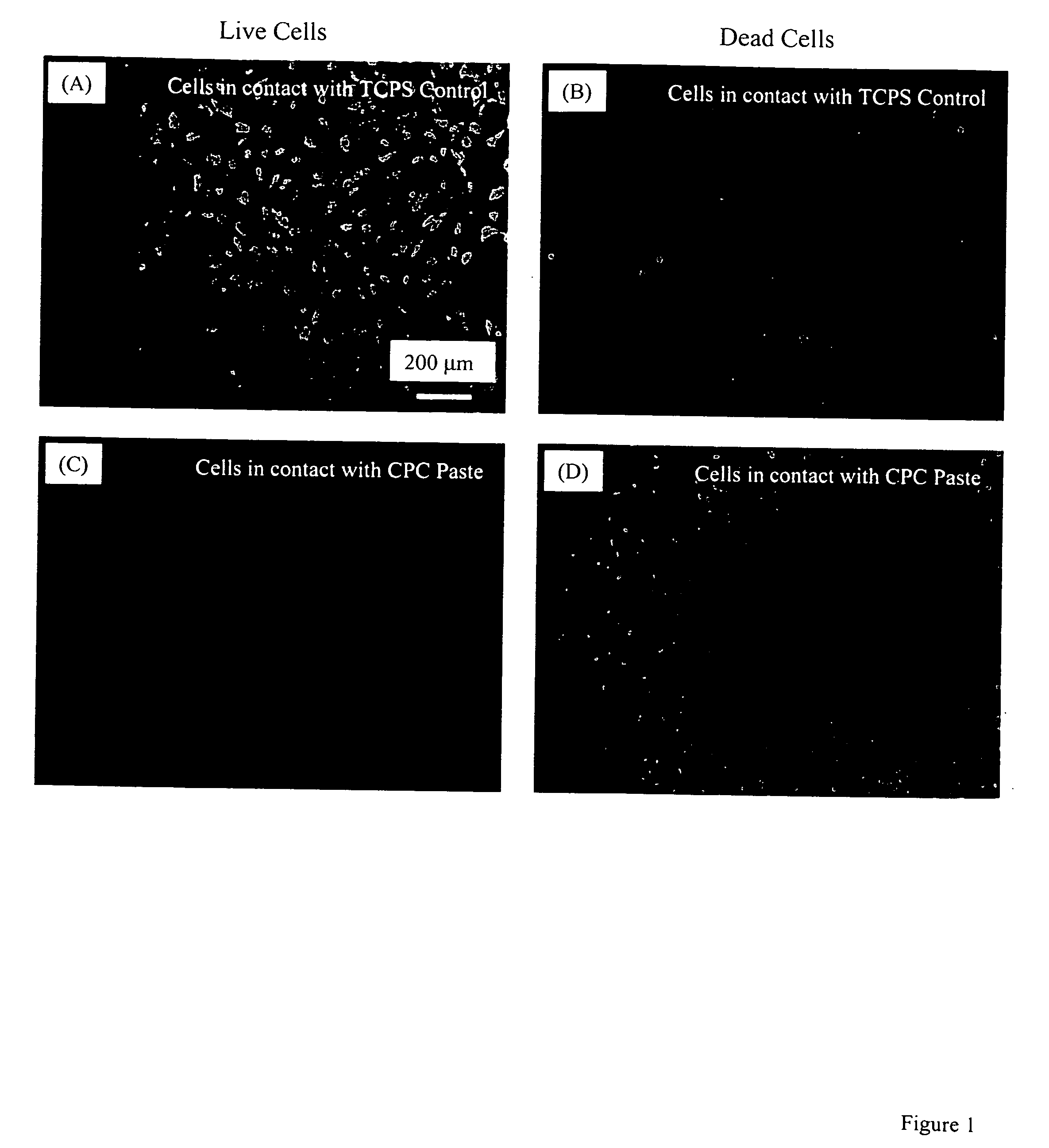
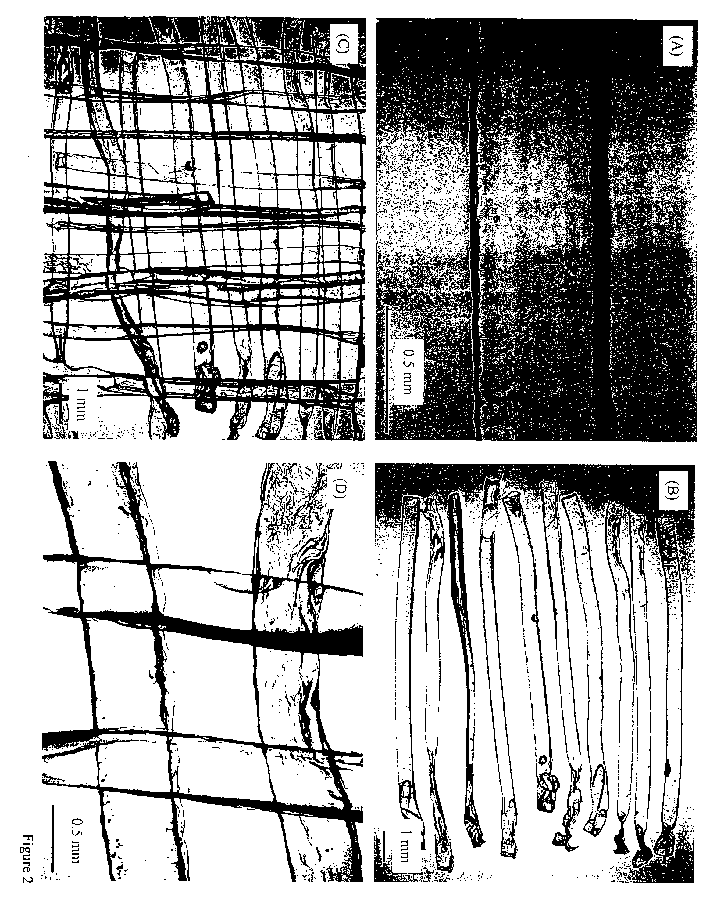
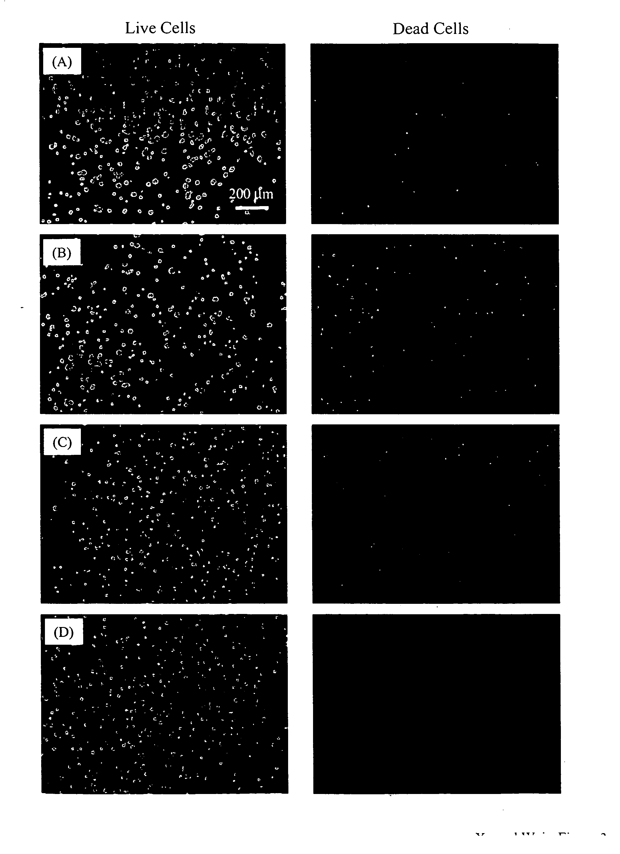
[0049] Another example of manufacture would be to produce in-situ setting CPC bone graft materials with living cells inside cell protectors that are mixed into the CPC paste. Biocompatible and degradable polymers, ceramics and composites can be used to construct the cell protectors. For example, a degradable polymer, poly(ethylene glycol)-anhydride dimethacrylate, is used as a rapidly degradable polymer for fabricating cell protectors. Structures such as those shown in FIG. 2 are fabricated by mixing osteoblast cells into the monomer liquid and then polymerizing the liquid into designed shapes inside molds. In the example in FIG. 2A, there are about 580 osteoblast cells inside each rod. These cell protector structures with living cells inside are mixed into the CPC paste, which is then mixed with an appropriate liquid and allowed to set to form an implant. The construct is immersed in water or in a physiological solution for the CPC to mature and for the cell protector to dissolve. ...


PUM

PropertyMeasurementUnit
Fractionaaaaaaaaaa
Fractionaaaaaaaaaa
Fractionaaaaaaaaaa

Abstract

Living cellular material is encapsulated or placed in a protective material (cell protector) which is biocompatible, biodegradable and has a three-dimensional form. The three dimensional form is incorporated into a matrix that maybe implanted in vivo, ultimately degrade and thereby by replaced by living cell generated material.

Description

CROSS-REFERENCE TO RELATED APPLICATION [0001] This is a utility application derived from and based upon Ser. No. 60 / 644,421 filed Jan. 14, 2005 entitled Three Dimensional Cell Protector / Pore Architecture Formation for Bone and Tissue Constructs incorporated herewith by reference and for which priority is claimed. STATEMENT REGARDING FEDERALLY SPONSORED RESEARCH OR DEVELOPMENT [0002] The subject matter of this invention was supported, at least in part, by grants from the National Institutes of Health, NIDCR grant R01 DE 14190 thereby resulting in potential use and / or license rights in the U.S. government and / or other entities pursuant to grant terms. BACKGROUND OF THE INVENTION [0003] In a principal aspect, the present invention relates to a construct for protecting biologics (such as cellular materials, particularly living cellular materials) by use of three-dimensional protective structures for such biologics using generally non-reactive, three dimensional containment materials so ...

Claims


Application Information

Patent Timeline
IPC IPC(8): A61K35/32
CPCA61K38/1825A61K38/1841A61K38/1858A61K38/1875A61L27/20A61L27/38A61L27/425A61L27/46A61L27/58A61L2400/06A61L2430/02C08L5/04C08L5/08C08L67/04C08L89/06A61P19/04A61P19/08
Inventor XU, HUAKUNWEIR, MICHAEL D.
Owner ADA FOUND
Why Eureka
Try Eureka