Eureka AIR delivers breakthrough ideas for toughest innovation challenges, trusted by R&D personnel around the world.

Clinically intelligent diagnostic devices and methods

a diagnostic device and intelligent technology, applied in the field of medical diagnosis, can solve the problems of difficult diagnosis based on only symptoms, time-consuming, cumbersome, slow, etc., and achieve the effects of reducing test costs, reducing test costs, and extending the spectrum of information

Inactive Publication Date: 2006-10-26
INTELLIGENT MEDICAL DEVICES
View PDF51 Cites 15 Cited by
  • Summary
  • Abstract
  • Description
  • Claims
  • Application Information

AI Technical Summary

Benefits of technology

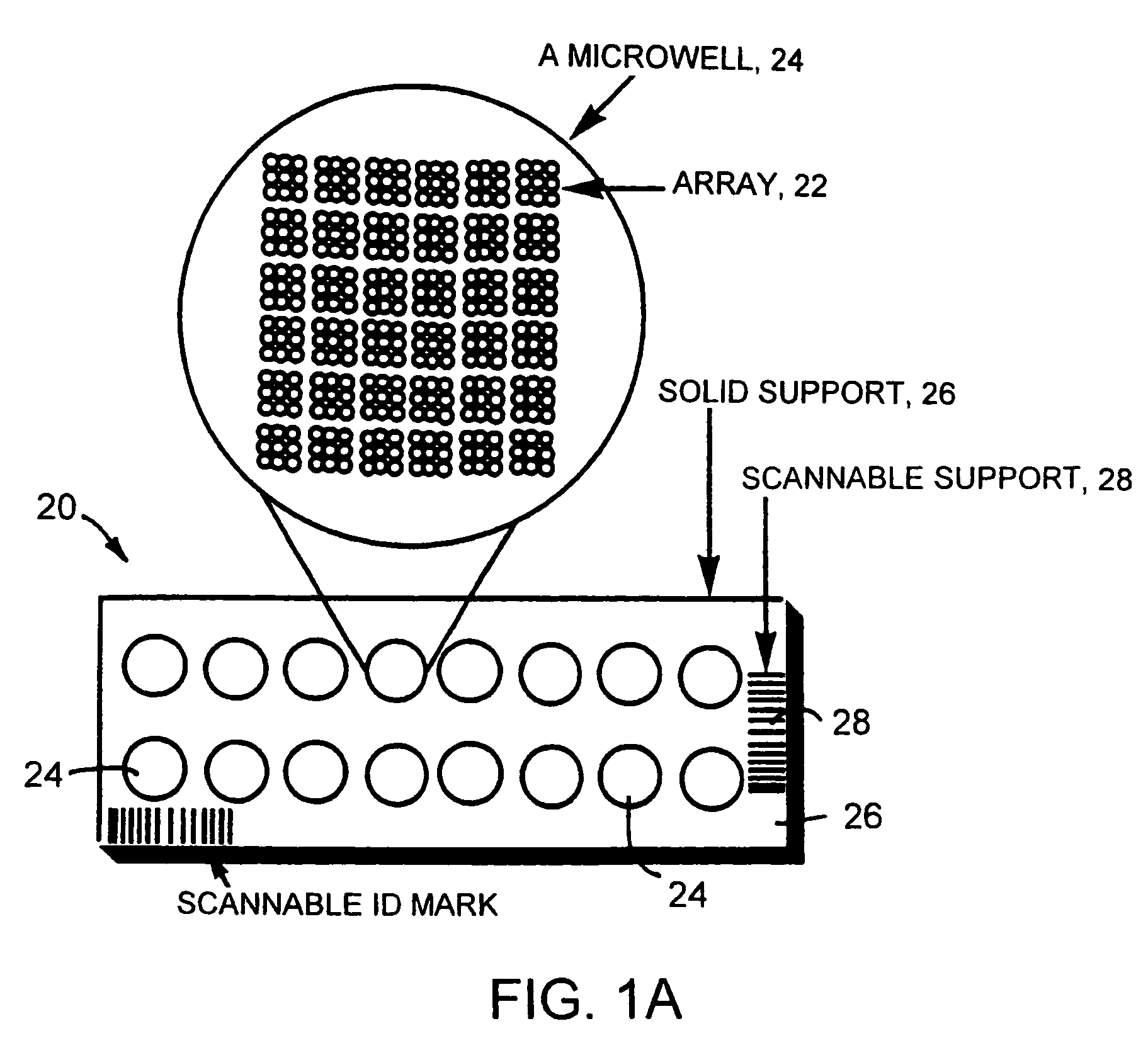
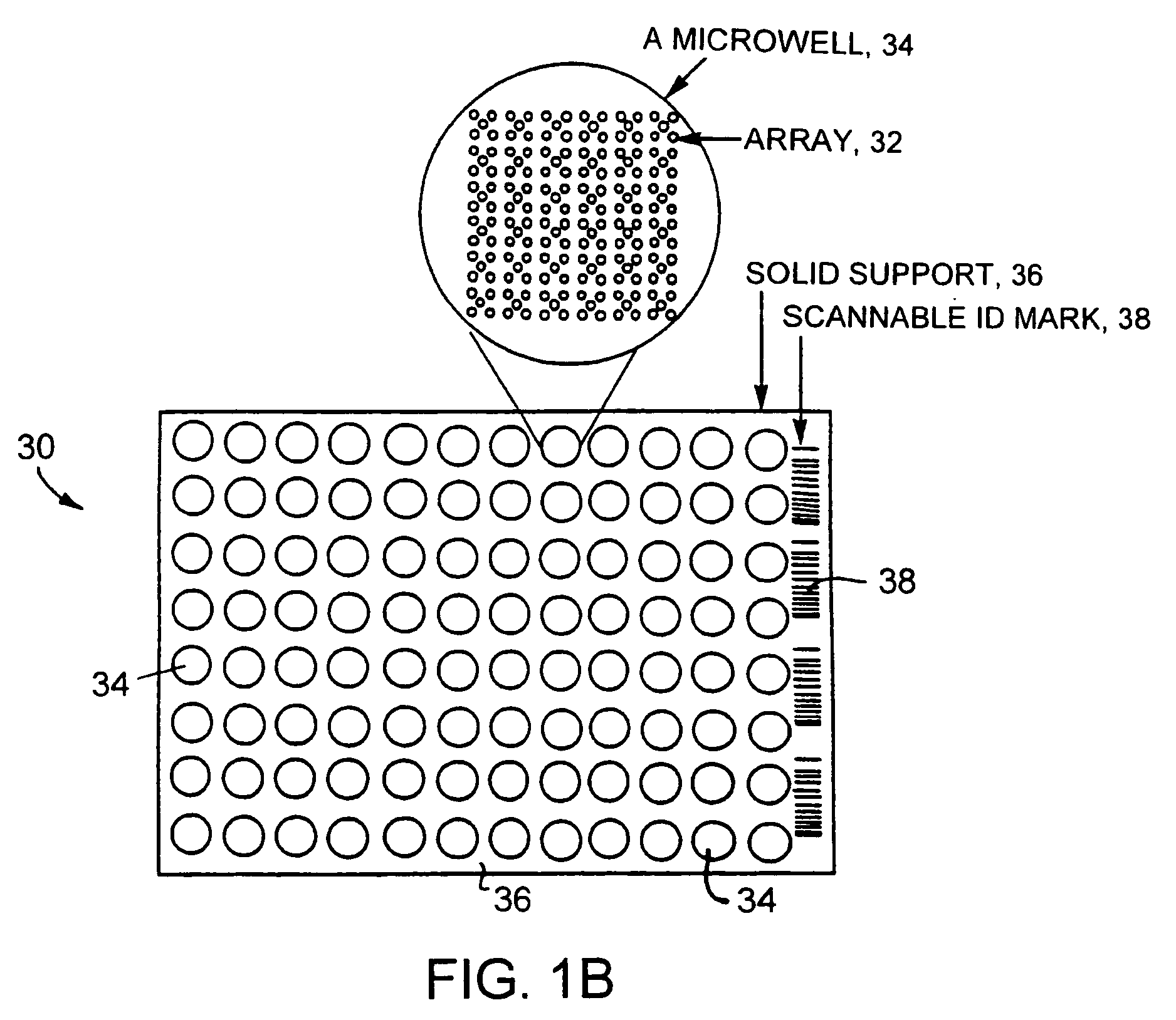
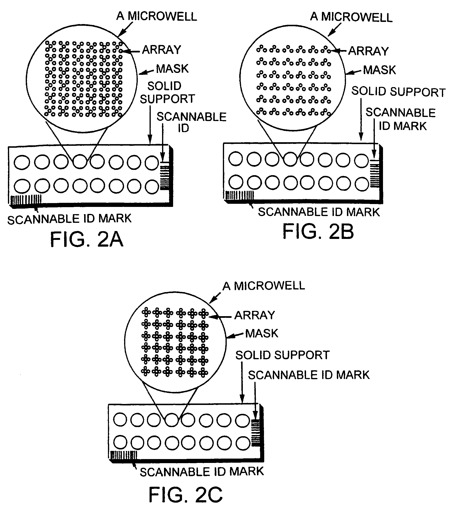
[0019] In other embodiments, the invention features methods for intelligently clustering probes in kit design; reagents and kits for use in the new devices / systems, methods for interpreting the data and results as well as making a recommendation for diagnosis; methods for immobilizing biomolecules in such a manner that their activity is largely preserved (e.g., by the use of cross-linked streptavidin and other protein layers for attaching capture probes, or spotting solutions with reagents that help in stabilizing / preserving biological activity of the probes); methods of dispensing probes in one or more replicates and in geometric patterns that provide a read-out that can be easily converted to a result by simple visual inspection (e.g., in “X” patterns); methods for image storage and processing, and manipulating stored signals to form a new image; new systems / devices that will combine the sample collection modules with the diagnostic devices into a single combination device / system (that circumvent the need to transfer biological samples from collection tubes to diagnostic devices); new systems / devices in which the sample collection module is separate from the diagnostic device module and is easily attached together at any stage, without having to take the sample manually out of the collection module; methods of preserving the developed slides (e.g., by keeping them sealed in aqueous buffer, e.g., containing BSA, milk proteins, glycerol, trehalose or other such reagents that preserve the activity of attached probes; methods for performing automated processing of slides (including screening, scanning and delivery of results); methods of selecting and using either single unique probes for each analyte or multiple unique probes for each analyte; and methods for placing optical positioning markers for automated image processing and read-out (e.g., that can include a row of dilution series for dynamic range determination and internal calibration on the biochip / micro array).
[0033] Another advantage is that the microarray-based diagnostic methods can be easily automated and the new devices can be used with the robots and technologies currently used in most clinical testing laboratories. This will cut down on the costs for incorporating this new technology into an existing laboratory. In addition, the microarray-based diagnostic methods can be carried out with portable devices.

Problems solved by technology

However, patients seldom present physicians with a diagnosis already made; instead, they present one or more symptoms.
However, patients typically present physicians with clinical symptoms that are confounding, which make diagnosis based on only the symptoms very difficult.
Currently, the diagnostic testing process is very tedious, time-consuming, cumbersome, and slow.
Moreover, because laboratories are constantly updating and adding new tests that facilitate medical diagnosis, physicians regularly confront dilemmas when ordering and interpreting these tests.
These advances in modern clinical laboratory medicine, though enormously helpful, also create new problems.
These tests are often not user friendly, and increase costs in an already heavily burdened health care system.
In common practice, physicians also complain about the delay in the processing of tests at the laboratories thus delaying accurate patient diagnosis.
Additionally, many tests are not available at all, are available at only one or two testing sites / laboratories, or are known only to specialists.
Sometimes the initially chosen set of tests present results that are not clear, which precludes an accurate diagnosis.
The nature and relatively non-specific symptoms of the disease can make a proper diagnosis challenging.
Thus, testing slows down the entire process of patient care and treatment, which is costly, and is detrimental to the patient's health and treatment plans.

Method used


Image

  • Clinically intelligent diagnostic devices and methods
  • Clinically intelligent diagnostic devices and methods
  • Clinically intelligent diagnostic devices and methods

Examples


example 1

Preparation of Clustered Probe Arrays on Various Solid Supports

[0195] A blank 3D-Link™ slide (Surmodics), which contains NHS-ester groups at the surface of the slides, is used in this example. The slide is placed in a chamber for arraying of probes onto the slide surface. Probe molecules (ZFPs and other polypeptides) are dissolved in a bicarbonate buffer at pH 8.3, and are spotted onto the glass slides using an arrayer. All the probe molecules contain an amine group for reaction with the slide surface. The spotting solution can also contain chemicals that have low vapor pressure (high boiling point) and that preserve the activity of probe molecules (such as glycerol, trehalose, and polyethylene glycol). The spotting is done under controlled conditions (such as humidity, in one instance around 70% relative humidity, temperature, for example around 4° C., pressure, and air-flow).

[0196] After the probes are spotted, the slides are put in the development chamber for 1-12 hours. The ch...

example 2

Jaundice (Liver Failure) Kit

[0208] This kit allows comprehensive, cost-effective, rapid diagnosis of numerous diseases / conditions based on a patient's clinical presentation of jaundice / liver failure. Diagnosis of genetic, autoimmune, and infectious diseases is based on the precise detection of specific gene mutations or other markers (such as microbial-specific sequences, or autoreactive antibodies) using DNA, RNA, and protein (spotted antigenic proteins and specific mAbs) chips. Jaundice kits are focused on the etiologic considerations of jaundice. In addition, therapeutic markers may be included to test for different potential therapeutic options. Briefly, one or more of three groups of etiological conditions are evaluated: A) autoimmune hepatitis, B) viral-induced hepatitis, and C) genetic diseases causing jaundice and / or liver enlargement. See, e.g., Feldman: Sleisenger & Fordtran's Gastrointestinal and Liver Disease, 6th ed. (W.B. Saunders Company 1998); McFarlane, “IG: The Re...

example 3

Fever / Skin Rash / Weight Loss (Autoimmune) Kit

[0229] This kit allows comprehensive, cost-effective, rapid diagnosis of numerous diseases / conditions based on a patient's clinical presentation of autoimmunity (systemic autoimmune diseases have frequently overlapping clinical pictures consisting of fever, skin rash, skin discoloration, and weight loss). Diagnosis of autoimmune diseases is based on the precise detection of autoreactive antibodies, specific gene mutations or other markers (such as autoimmune prone HLAs) using DNA, RNA, live cells, and protein (spotted antigenic proteins and specific mAbs) chips. Briefly, one or more of three groups of etiological conditions are evaluated: A) Systemic and organ-specific autoimmune diseases, B) HLAs that are associated with specific autoimmune diseases, C) Detection of gene mutations that result in the autoimmune syndrome, D) Deficiencies of early and late complement components associated with autoimmune diseases, and E) Therapeutic markers...


PUM

PropertyMeasurementUnit
pHaaaaaaaaaa
temperatureaaaaaaaaaa
concentrationaaaaaaaaaa

Abstract

The invention relates to the clinically intelligent design of diagnostic devices (such as microarrays) and methods of making and using such devices in differential diagnoses of specific clinical symptoms or sets of symptoms. In one aspect, the devices include various probes used to perform parallel screening of a number of analytes. The probes are clustered on the devices based on known clinical presentations of symptoms associated with specific diseases and disorders.

Description

CROSS-REFERENCE TO RELATED APPLICATIONS [0001] This application is a continuation of U.S. patent application Ser. No. 11 / 102,498, filed on Apr. 8, 2005, which is a continuation of U.S. patent application Ser. No. 09 / 996,056, filed on Nov. 27, 2001, now U.S. Pat. No. 6,905,816, which claims priority to U.S. Provisional Patent Application Ser. Nos. 60 / 253,284, filed on Nov. 27, 2000, 60 / 287,994, filed on May 1, 2001, and 60 / 308,870, filed on Jul. 30, 2001, which are all incorporated by reference herein in their entirety.FIELD OF THE INVENTION [0002] This invention relates to medical diagnosis. BACKGROUND OF THE INVENTION [0003] It is common for patients to seek the advice of a physician when experiencing discomfort. However, patients seldom present physicians with a diagnosis already made; instead, they present one or more symptoms. Selecting the most probable diagnosis from a list of alternatives (hypotheses) is a process called differential diagnosis. Certain signs or symptoms can s...

Claims


Application Information

Patent Timeline
Patent Type & Authority Applications(United States)
IPC IPC(8): C12Q1/68G01N33/53C12M1/34B01F13/00B01F13/08B01J19/00B01L3/00B81B1/00C40B40/06C40B40/10C40B40/12C40B40/18C40B60/14C40B70/00G01N33/543G01N33/68
CPCB01F13/0809B01F13/08B01J2219/00315B01J2219/00317B01J2219/00481B01J2219/00497B01J2219/00509B01J2219/00511B01J2219/00527B01J2219/00547B01J2219/00585B01J2219/00596B01J2219/00605B01J2219/0061B01J2219/00612B01J2219/00619B01J2219/00621B01J2219/00626B01J2219/0063B01J2219/00637B01J2219/00644B01J2219/00659B01J2219/00662B01J2219/00664B01J2219/00677B01J2219/00689B01J2219/00702B01J2219/0072B01J2219/00722B01J2219/00725B01J2219/00731B01J2219/0074B01J2219/00745B01L3/5027B01L3/5085B01L3/50857B01L2300/021B01L2300/0636B01L2300/069B01L2300/0819B01L2300/0864B82Y30/00C12Q1/6837C40B40/06C40B40/10C40B40/12C40B40/18C40B60/14C40B70/00G01N33/53G01N33/543G01N33/54366G01N33/6803G01N33/6845B01F13/0088B01J19/0046B01F33/3036B01F33/451B01F33/45
Inventor JACOBS, ALICE A.GUPTA, VINEETNIKOLIC, BORIS
Owner INTELLIGENT MEDICAL DEVICES
Why Eureka
Eureka Blog
Learn More