Pharmaceuticals That Promote Functional Regeneration of Damaged Tissues

a technology of functional regeneration and pharmaceuticals, applied in the field of pharmaceuticals that promote functional regeneration of damaged tissues, can solve the problems of difficult total removal of neuroglioma arising from cerebral parenchymal cells, bone marrow-derived stem cells, and the like, and achieve the effect of promoting functional tissue regeneration

Inactive Publication Date: 2009-08-13
OSAKA UNIV
View PDF17 Cites 20 Cited by
  • Summary
  • Abstract
  • Description
  • Claims
  • Application Information

AI Technical Summary

Benefits of technology

[0008]Once factors that mobilize cells differentiating into damaged tissues are revealed, the administration of such factors to lesion sites allows the mobilization of a large number of cells (which exist in peripheral blood or local tissues) that differentiate into damaged tissues. Thus, the development of novel regenerative medicines which promote functional tissue regeneration is made possible.

Problems solved by technology

1) HMGB1 regulates gene expression by intracellularly binding with DNA to control chromatin structure (Non-patent Document 1);
2) HMGB1 is secreted from monocytes or macrophages existing in inflammatory tissues by the action of inflammatory cytokines TNF-α, IL-1, and LPS, and extracellularly binds to RAGE (Receptor for Advanced Glycation End products) (Non-patent Document 2) to induce strong inflammatory reactions (Non-patent Document 3);
3) HMGB1 is released from hypoperfusion-induced necrosed cells to the surrounding tissues (Non-patent Document 4);
4) HMGB1 is associated with inflammation progress in patients with septicemia, a severe infectious disease (Non-patent Document 5);
5) HMGB1 administration to infracted areas of myocardial infarction models promotes the division / proliferation of stem cells existing in the myocardium so as to promote the regeneration / functional recovery of the myocardium (Patent Document 1);
6) HMGB1 administration to model animals with hypoperfusive liver failure prior to the induction of hypoperfusive conditions alleviates the degree of hepatic impairment (Non-patent Document 6);
7) HMGB1 administration to lesion sites of muscle injury models directs simultaneously-administered vascular progenitor cells to lesion sites so as to promote muscular tissue regeneration (Non-patent Document 7); and
8) HMGB1 induces neurite formation in nerve cells (Non-patent Document 8). However, no previous reports showed that bone marrow-derived stem cells, in particular those mesenchymal stem cells that can differentiate into osteoblasts, chondrocytes, adipocytes, and the like, were mobilized to damaged tissues.
Other causes can be injury resulting from brain surgeries such as brain tumor removal.
In particular, the total removal of neuroglioma arising from cerebral parenchymal cells is difficult, and there is no choice but to stop at partial removal to avoid damage to motor and language functions.
Moreover, malignant neuroglioma has a worse prognosis, and none of the treatments from chemotherapy and radiotherapy to immunotherapy / gene therapy that are actively researched these days, have achieved satisfactory effects.

Method used


Image

  • Pharmaceuticals That Promote Functional Regeneration of Damaged Tissues
  • Pharmaceuticals That Promote Functional Regeneration of Damaged Tissues
  • Pharmaceuticals That Promote Functional Regeneration of Damaged Tissues

Examples


example 1

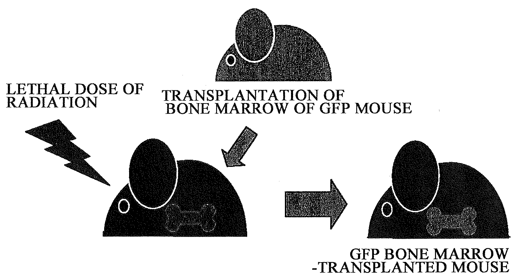
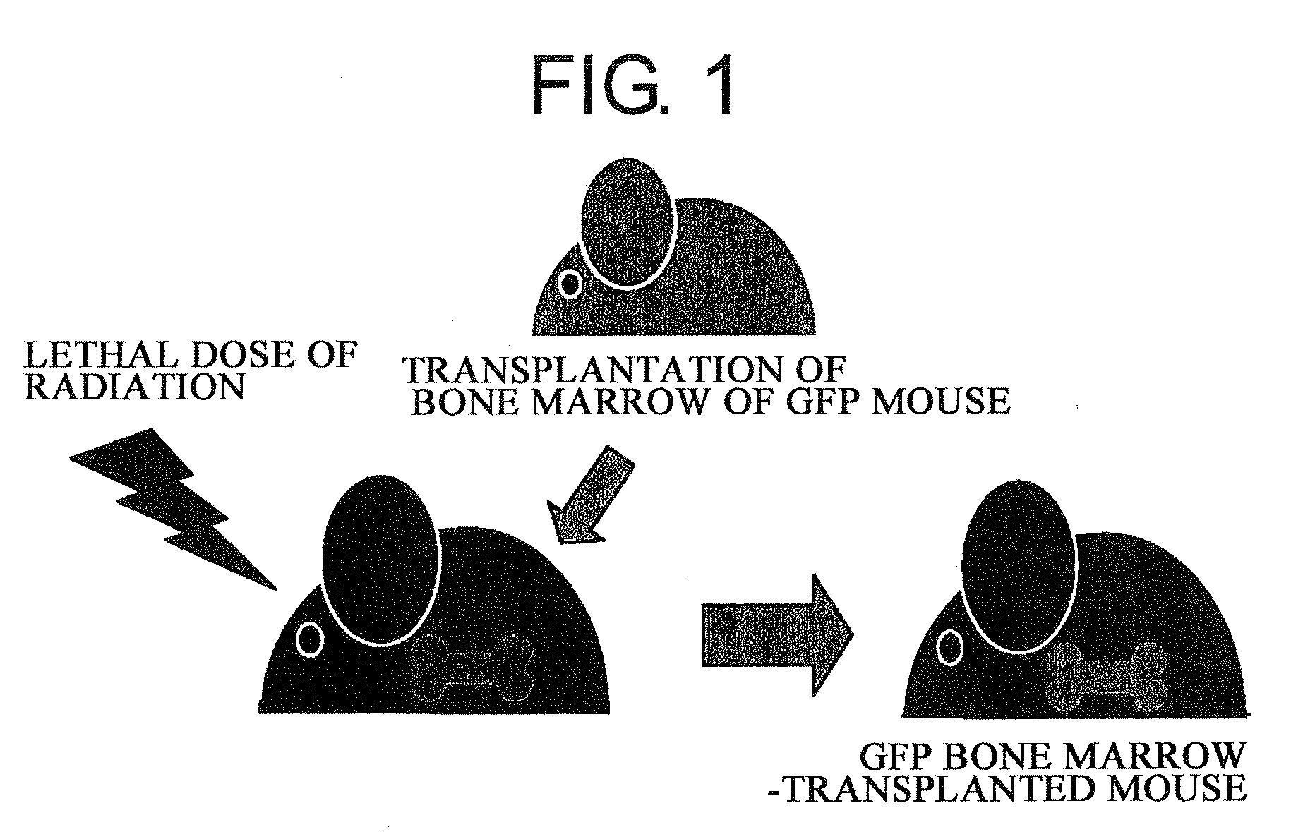
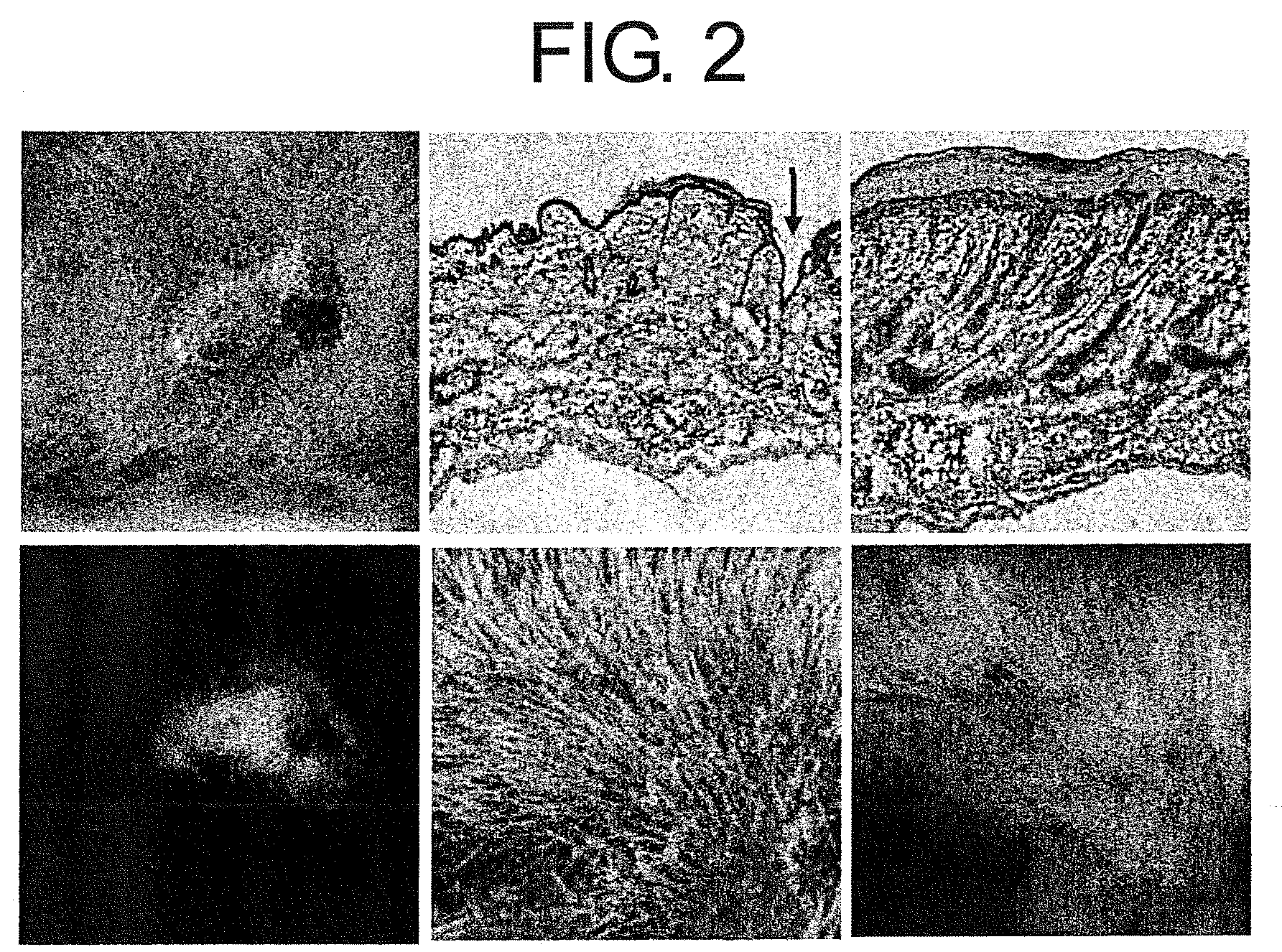
[0115]Objective: Assessment of the contribution of bone marrow-derived cells towards functional regeneration of skin tissue transplanted to a living body.

[0116]Method: In view of the above objective, studies were carried out by the following methods.

1) Utilizing the live skin transplant system of GFP bone marrow-transplanted mice, the degree of contribution of bone marrow-derived cells towards functional regeneration of grafted skin was examined. Specifically, 6 to 8-week-old male C57BL / 6 mice were irradiated with a lethal dose of radiation (10 Gy), and immediately after that, GFP (green fluorescent protein) transgenic mouse-derived bone marrow cells (5×10⁶ cells / 0.1 ml of physiological phosphate buffer solution at pH 7.4) were transplanted through the caudal vein (FIG. 1)

2) The transplanted bone marrow cells were allowed to engraft (for 6 weeks), and as a result, a GFP bone marrow-transplanted mice was obtained. Then, skin of a neonatal mouse (female) was transplanted to the dorsal...

example 2

[0120]Objective: Identification of a bone marrow-derived mesenchymal stem cell-inducing factor in a skin tissue extract

[0121]Method: With the objective of identifying a bone marrow-derived mesenchymal stem cell-mobilizing factor which is expected to be released from excised skin under hypoperfusive conditions, studies were carried out by the following methods.

1) To obtain mouse bone marrow-derived mesenchymal stem cells, bone marrow cells were collected from the femur or crus bone of C57BL / 6 mice, and then were spread on a cell culture plate having a 10% fetal bovine serum-containing D-MEM (Nacalai) as a cell culture medium, and then were cultured under the condition of 5% CO2 at 37° C. When the cells proliferated to the point of occupying 70 to 100% of the bottom area of the culture plate, the cells were peeled off from the culture plate using 0.25% trypsin 1 m MEDTA (Nacalai), and were then cultured under the above conditions. This passing and culturing procedure was repeated at l...

example 3

[0125]Objective: Identification of the HMGB1 family in the skin extract and examination of bone marrow mesenchymal stem cell-inducing activity

[0126]Method: Whether or not the neonatal mouse skin extract contained the HMGB protein family was confirmed using the Western blot method. Ten μl of the skin extract obtained in [Example 2] was used as a sample and subjected to SDS-PAGE electrophoresis. The proteins separated within the gel were transferred onto a PVDF membrane using a blotting device (ATTO). The membrane was incubated with PBS containing 3% skim milk and 0.1% Tween 20 (S-T-PBS) at room temperature for 1 hour, and then was allowed to react with each of rabbit anti-mouse HMGB1 antibody, rabbit anti-mouse HMGB2 antibody, or rabbit anti-mouse HMGB3 antibody which were diluted 1000-fold with S-T-PBS, at 4° C. for 16 hours. After the reaction, the PVDF membrane was washed with S-T-PBS five times for 5 minutes. Then, the PVDF membrane was incubated with 2000-fold diluted (diluted w...


PUM

PropertyMeasurementUnit
immersion timeaaaaaaaaaa

Abstract

The present inventors revealed the following for the first time in the world:
  • 1) a large amount of bone marrow-derived cells are mobilized to grafted skin;
  • 2) the mobilized bone marrow-derived cells are differentiated into any of dermal fibroblasts, adipocytes, muscle cells, vascular endothelial cells, and epidermal keratinocytes in the grafted skin, and the mobilized bone marrow derived cells include bone marrow-derived mesenchymal stem cells;
  • 3) the factors which mobilize bone marrow-derived mesenchymal stem cells from peripheral blood to the grafted skin are HMGB1, HMGB2, and HMGB3 released from the necrosed tissue of recipient skin;
  • 4) purified HMGB1, HMGB2, and HMGB3 promote the migration of mesenchymal stem cells isolated and cultured from bone marrow;
  • 5) activators containing HMGB1 which allows the migration of bone marrow mesenchymal stem cells can be conveniently purified from several organ extracts including skin, brain, and heart;
  • 6) activators which allow the migration of bone marrow mesenchymal stem cells can be conveniently extracted from cultured cells; and
  • 7) a heparin-column purified fraction of skin extract mobilizes a large amount of bone marrow-derived cells in case of brain injury.

Description

TECHNICAL FIELD[0001]The present invention relates to pharmaceuticals that promote functional regeneration of damaged tissues.BACKGROUND ART[0002]Recent years have revealed that various stem cells contribute towards the repairing processes of damaged tissues, and novel regenerative medicines that induce functional tissue regeneration by mobilizing a large number of stem cells to lesion sites are being progressively developed. To realize these novel regenerative medicines, it is necessary that: (i) stem cells that are mobilizable to lesion sites are abundantly present in vivo; and (ii) factors that mobilize stem cells to lesion sites have been isolated / identified.[0003]Examples of stem cells that are mobilizable to lesion sites include tissue stem cells existing in lesion areas or nearby tissues, and bone marrow-derived stem cells existing in peripheral blood. In recent years, contribution of bone marrow-derived cells to many types of damaged tissue regenerations has been reported, b...

Claims


Application Information

Patent Timeline
Patent Type & Authority Applications(United States)
IPC IPC(8): A61K35/12C12N5/00C07H1/00C12P19/44C12Q1/02A61K35/28A61K35/36A61K35/76A61P17/00A61P43/00C07K14/46C12N5/071C12N5/077C12N5/0775C12N15/09
CPCA61K31/711A61K38/00G01N2500/10G01N33/5073G01N33/6863C07K14/52A61P1/04A61P11/00A61P17/00A61P17/02A61P17/14A61P19/08A61P29/00A61P37/06A61P43/00A61P9/10A61K38/17A61K48/00
Inventor TAMAI, KATSUTOYAMAZAKI, TAKEHIKOKANEDA, YASUFUMI
Owner OSAKA UNIV
Why Eureka
Try Eureka