Single-Molecule-Format Probe And Utilization Thereof

a single-molecule, probe technology, applied in the direction of instruments, transferases, enzyme stabilisation, etc., can solve the problems of affecting the detection efficiency of fluorescent proteins, the difficulty of circular permutation, and the inability to perform circular permutation, so as to suppress the background enzyme activity and high sensorial efficiency

Inactive Publication Date: 2009-10-29
NAT INST OF ADVANCED IND SCI & TECH
View PDF1 Cites 24 Cited by
  • Summary
  • Abstract
  • Description
  • Claims
  • Application Information

AI Technical Summary

Benefits of technology

[0059]The present inventors intensively investigated the solution of the foregoing problems. Consequently, they invented a probe which can determine a ligand with high sensorial efficiency by dissecting an enzyme into two fragments and fabricating circular permutation. Particularly, they found that circular permutation of luciferase fragments inside a single-chain probe greatly suppresses the background enzyme activity.

Problems solved by technology

The circular permutation of GFP required great skills because of their monolithic cylindrical symmetry and complexity of their protein strands.
Thus, it was generally difficult to perform circular permutation.
Conventionally, the fluorescent proteins are suffered from an intrinsic problem that autofluorescence-caused elevation of background intensity.
Autofluorescence inevitably causes an elevation of background intensity and poor signal-to-noise contrast in case of Yellow Camelleons (Non Patent Literatures 2 through 4) for example.
In addition, the obtained results from fluorescent proteins are qualitative rather than quantitative because of the limited number of analyzable cell population at once (Non Patent Literature 5).

Method used


Image

  • Single-Molecule-Format Probe And Utilization Thereof
  • Single-Molecule-Format Probe And Utilization Thereof
  • Single-Molecule-Format Probe And Utilization Thereof

Examples


example 1

Plasmid Construction

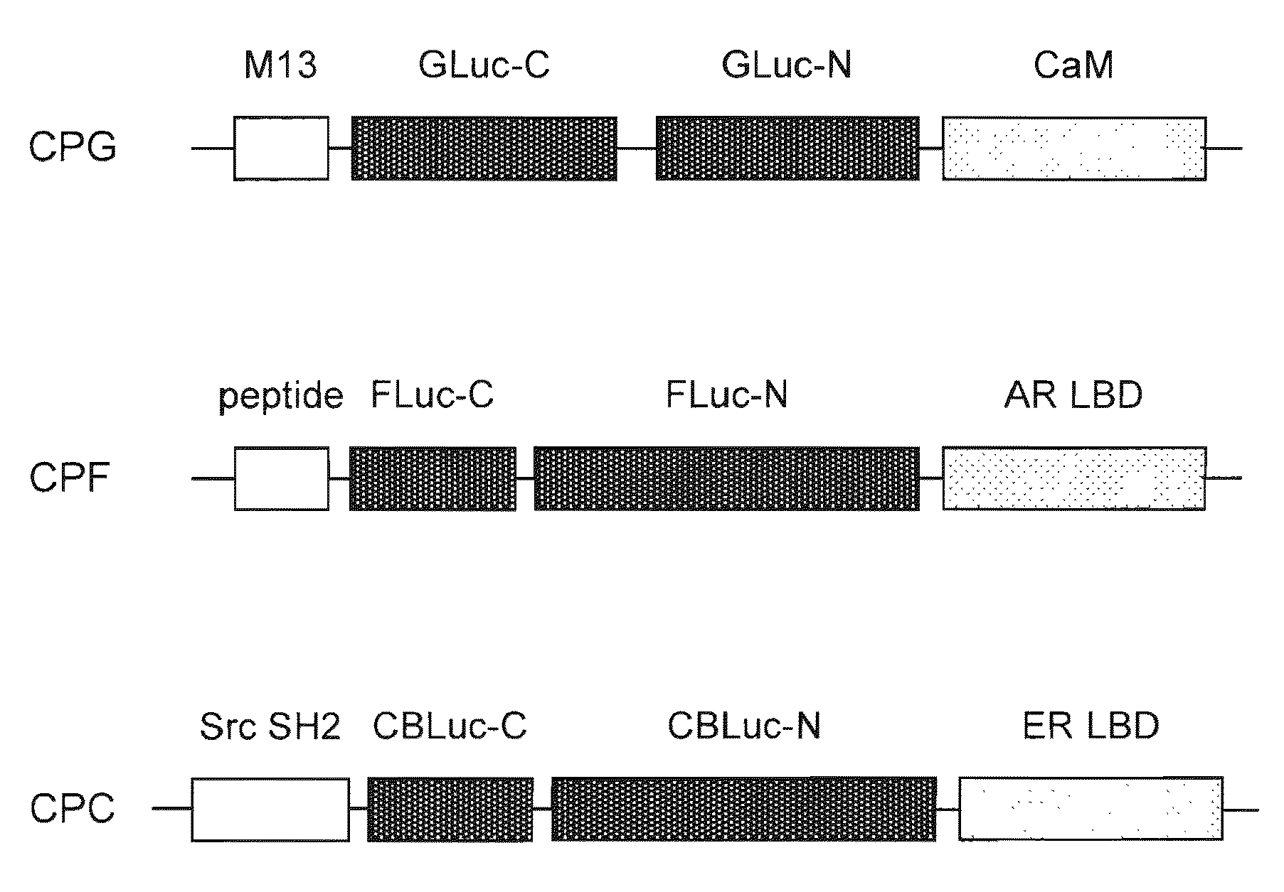
[0208]The respective cDNA fragments consisting of circularly permutated probes were generated by the polymerase chain reaction (PCR) to introduce each unique restriction site at each end of the fragments using adequate primers and templates. For example, a plasmid carrying Yellow Cameleon-3.1 (YC3.1) donated by Dr. Miyawaki was utilized for amplifying Xenopus laevis calmodulin (CaM) in PCR as a template (see Non Patent Literature 4). The specific restriction enzyme recognition sites were summarized in FIG. 5.

[0209]The restricted cDNA fragments were then ligated as shown in FIG. 5 and subcloned into the pcDNA 3.1(+) (Invitrogen). The plasmids were respectively named pCPC (cpCBLuc probe), pCPF (cpFLuc probe), and pCPG (cpGLuc probe), according to the kind of the luciferases circularly permutated in the probes. The probes expressed in the plasmids may be called CPC, CPF, CPG, respectively.

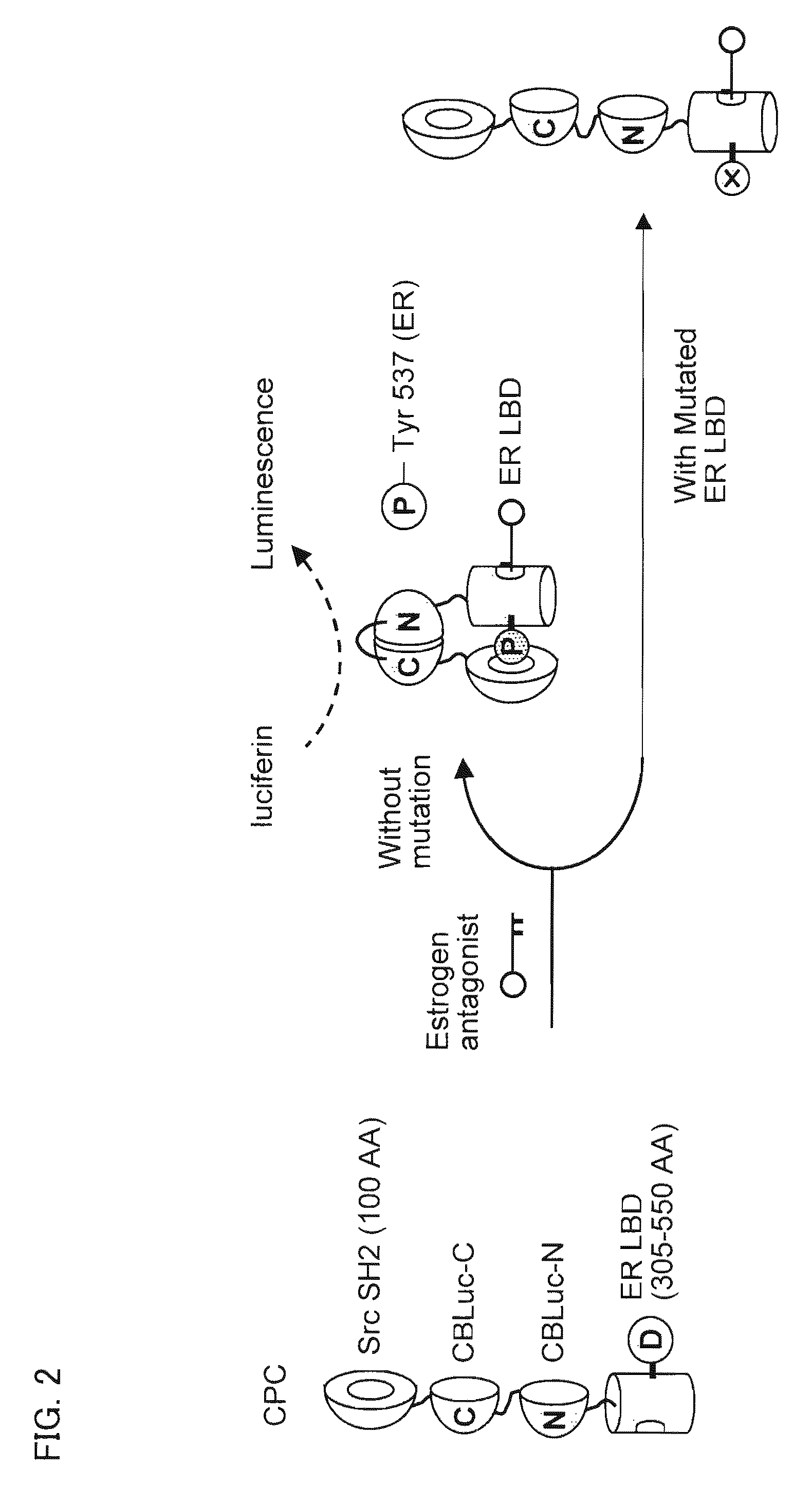
[0210]In addition, the cDNA of ligand binding domain of estrogen receptor (ER ...

example 2

Cell Culture and Transfection

[0214]COS-7 cells derived from African green monkey kidney were raised on a 24-well plate with Dulbecco's modified eagle's medium (DMEM; Sigma) supplemented with 10% steroid-free fetal bovine serum (FBS) and 1% penicillin-streptmycin (P / S) at 37° C. in a cell incubator maintaining 5% CO2 (Sanyo). The COS-7 cells were transiently transfected with pCFG, pCPF, or pCPC (0.2 μg per each well) using a plasmid transfection reagent, TransIT-LT1 (Mirus). The cells were extensively incubated in the 5% CO2 incubator for 16 hours.

example 3

Comparison of Relative Luminescence Intensities by CPC, CPF, or CPG

[0215]The relative luminescence intensities by CPC, CPF, or CPG were compared in the presence or absence of the ligand. The results are shown in FIG. 7.

[0216]First, the COS-7 cells carrying pCPC or pCPF were stimulated with 10−6 M of 4-hydroxy tamoxifen (OHT) or 5α-dihydroxytestosterone (DHT) (final concentration). The luminescence intensities were developed with a Bright-Glo substrate solution (Promega), and integrated for 15 seconds with a luminometer (Minilumat LB9506; Berthold). The brief procedure for the use of the Bright-Glo substrate solution is as follows.

[0217]The mammalian cells were washed once with PBS 20 minutes after stimulation with ligand. A 40 μL of the substrate (D-leuciferin) solution was added to each well of the plates. Three minutes after substrate addition, the plate was tapped gently, and the subsequent cell lysates were transferred to a test tube for determining the luminescence intensities....


PUM

PropertyMeasurementUnit
diameteraaaaaaaaaa
enzyme activityaaaaaaaaaa
luminescence intensityaaaaaaaaaa

Abstract

A single-chain probe of the present invention for detecting a ligand, comprises: a ligand binding protein for binding the ligand; a recognition protein for recognizing that the ligand is bound by the ligand binding protein; and C— and N-terminal fragments, generated by dissecting an enzyme, between the ligand binding protein and the recognition protein, wherein a carboxy terminal end of the C-terminal fragment is located upstream of an amino terminal end of the N-terminal fragment, and the C— and N-terminal fragments vary the enzyme activity via complementation in case where the recognition protein recognizes that the ligand is bound by the ligand binding protein. This makes it possible to achieve detection of a target protein-specific ligand using the single chain with a high efficiency.

Description

[0001]This Nonprovisional application claims priority under 35 U.S.C. §119(a) on Patent Application No. 2008-116098 filed in Japan on Apr. 25, 2008, the entire contents of which are hereby incorporated by reference.TECHNICAL FIELD [0002]The present invention relates to a fusion protein for detecting a ligand in a subject sample and to utilization thereof. More specifically, the present invention relates to a fusion protein used as a single-molecule-format probe for detecting a target protein-specific ligand and to utilization thereof.BACKGROUND ART [0003]Revolutionary advances in molecular imaging technologies have allowed researchers to carry out quantitative examination of molecular dynamics and cell signaling in living cells (Non Patent Literature 1). As described in Non Patent literature 2, one of the imaging technologies with lighting protein is circular permutation (CP) of fluorescent proteins such as green fluorescent protein (GFP) for construction of probes. CP of GFP is a m...

Claims


Application Information

Patent Timeline
Patent Type & Authority Applications(United States)
IPC IPC(8): G01N33/53C12N9/96C12N15/11C12N15/00C12N5/06C12N15/87
CPCC07K14/721C07K2319/61G01N33/542C12N15/62C12N9/12
Inventor KIM, SUNG-BAETAO, HIROAKISATO, MORITOSHI
Owner NAT INST OF ADVANCED IND SCI & TECH
Why Eureka
Try Eureka