

Use of heat shock activators for tissue regeneration
a technology of tissue regeneration and activator, which is applied in the direction of extracellular fluid disorder, metabolism disorder, therapy, etc., can solve the problem of limited supply of donor organs, and achieve the effect of reducing the probability of developing a disorder
- Summary
- Abstract
- Description
- Claims
- Application Information
AI Technical Summary
Benefits of technology
Problems solved by technology
Method used
Image

Examples
example 1
Recruitment of Stem Cells by Laser
[0145]Chimeric mice were constructed with GFP+ stems cells that were transplanted into mice that had undergone near lethal irradiation. Chimeric mice were made with gfp-expressing hematopoietic stem cells from gfp homozygous transgenic donors. These cells were transplanted into recipients that had undergone near lethal irradiation. These gfp chimeric mice (C57B16.gfp) were used in all subsequent laser studies. Using the 810 nm diode laser with a spot diameter of 75 μm, various levels of energy were delivered to the retina including energy of 5 mJ (50 mW, 0.1 msec) that did not produce visible laser tissue reaction in the retina (laser irradiance 1130 W / cm2). This was considered sub-visible threshold. Three weeks post-laser the animals were euthanized and the eyes were harvested.
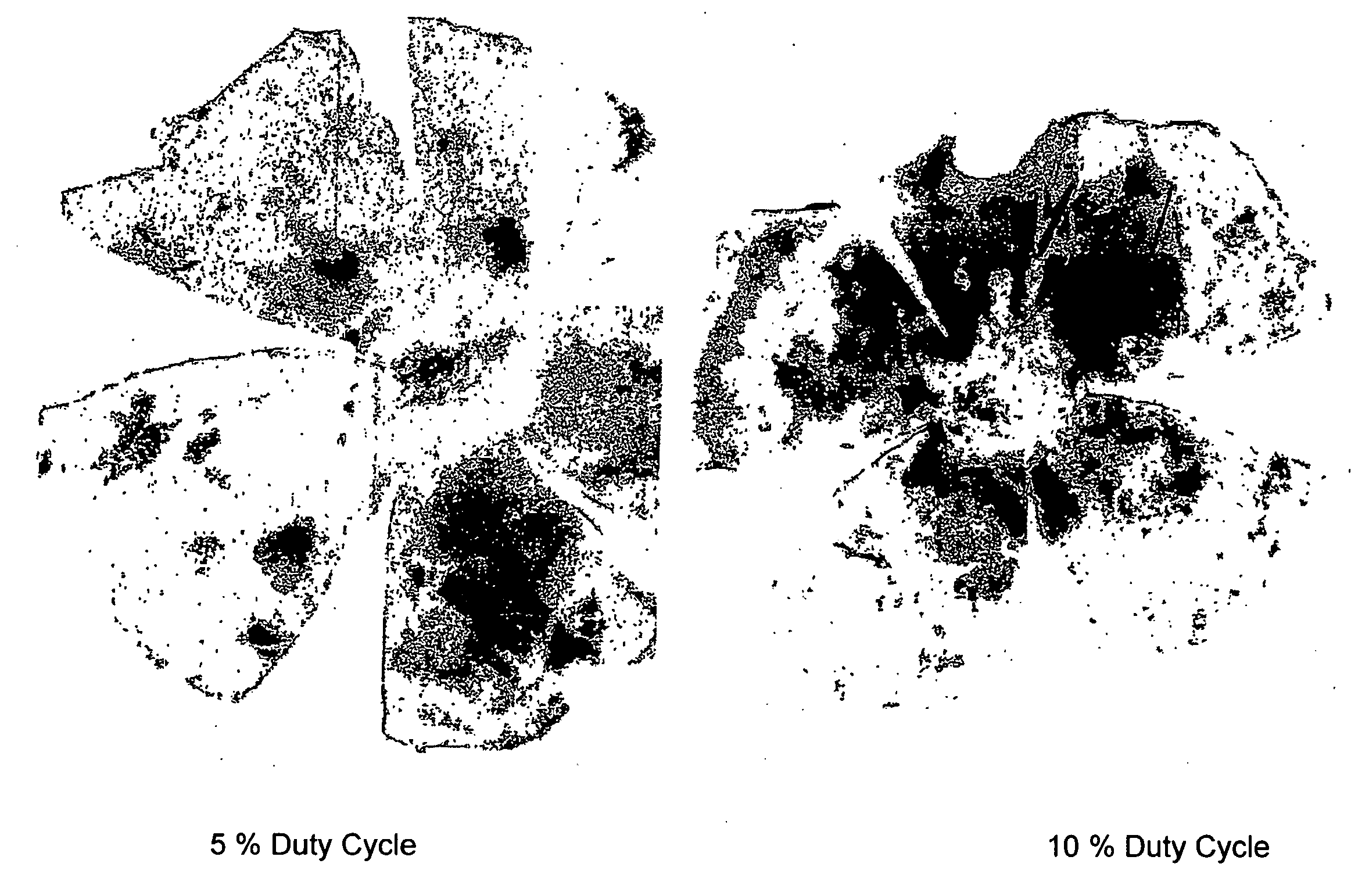
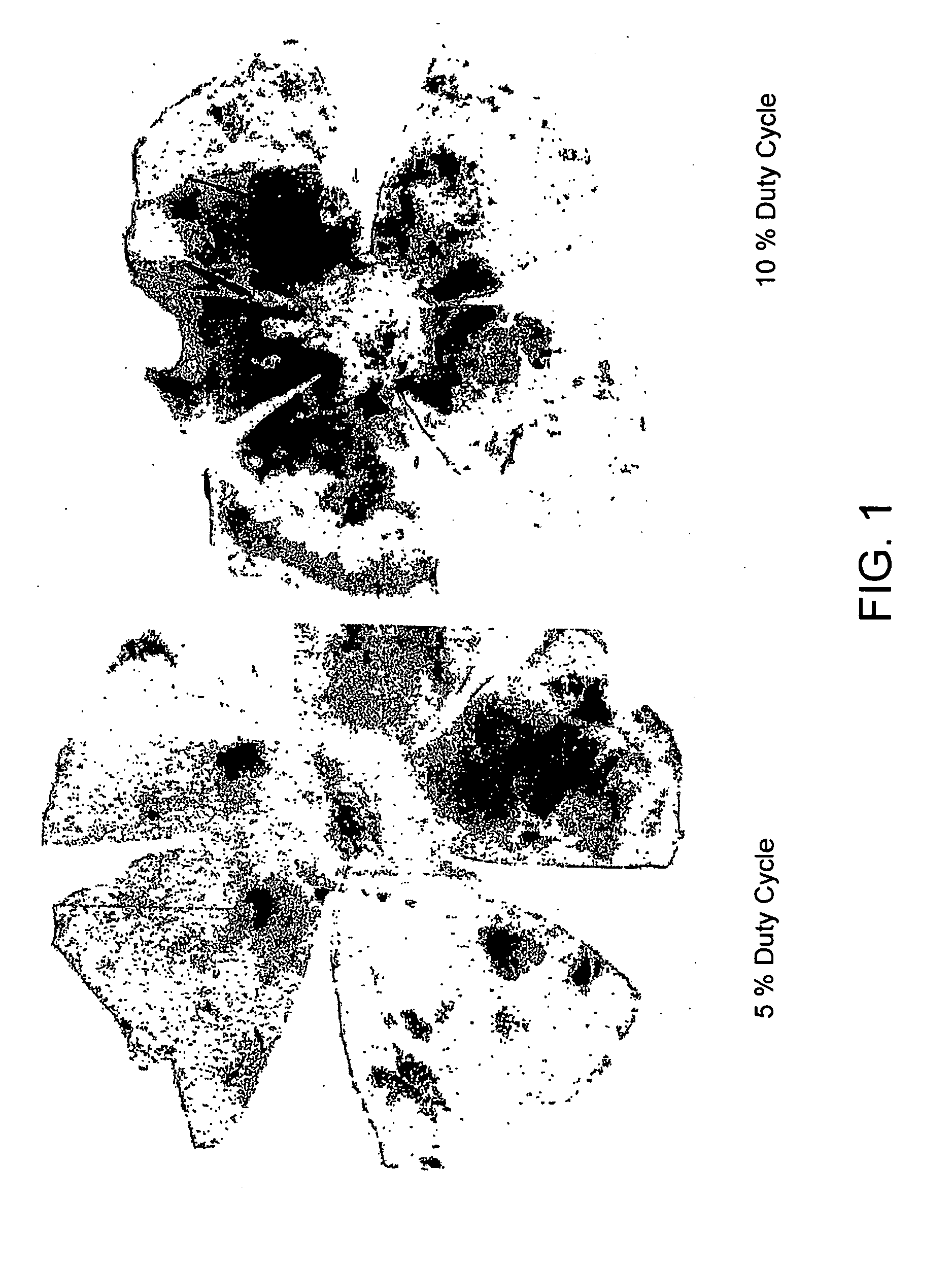
[0146]Eye cups were prepared and the neurosensory retina was removed. Eyes receiving subthreshold laser energy demonstrated robust recruitment of hematopoietic stem cells (HS...
example 2
HSC's Recruited to the Retina Express a Retinal Specific Marker
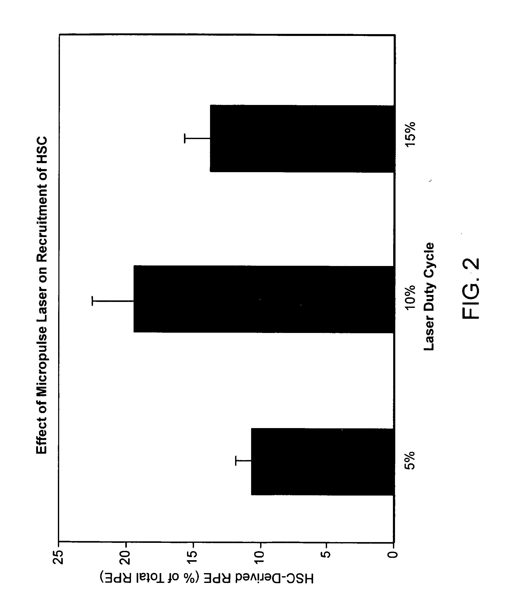
[0147]By confocal immunofluorescence microscopy, it was also shown that GFP positive cells co-localized with RPE65—a protein specific for the RPE, suggesting that the recruited hematopoietic stem cells have acquired RPE characteristics. These eyes also demonstrated diffuse endothelial cell recruitment as well. Eyes receiving high energy laser with noticeable retinal opacification (150 mW, 0.1 sec) showed focal recruitment of GFP+ HSC to the scar region along with endothelial cells. Sub-threshold laser induced HSC to migrate to and incorporate into the RPE. The degree of incorporation correlates with laser duty cycle. 15% duty cycle resulted in the greatest degree of HSC incorporation.
example 3
Subvisible Threshold Laser Increased Hsp70, Hsp90, and Induction of the Heat Shock Response Resulted in the Release of SDF-1 and VEGF
[0148]Ophthalmic lasers are an important tool for the treatment of various retinal disorders. In most instances, the effect has been attributed to visible changes in the retina (i.e., laser-induced photochemical burns). The diode 810 nanometer laser is believed to cause less damage to the neurosensory retina because the laser energy is absorbed by the RPE. FIG. 2 demonstrated the striking duty-cycle dependent localization of GFP+ cells at the level of the RPE. There is a maximal response at 10% duty-cycle. This was separately confirmed using adoptive transfer methods, a technique that closely resembles cellular therapy. Adoptive transfer involves the systemic administration of HSCs and results in the rapid homing of these cells to areas producing chemoattractants.
[0149]Micropulse lasering has been developed clinically to minimize photodestructive damag...
PUM
| Property | Measurement | Unit |
|---|---|---|
| Heat | aaaaa | aaaaa |
Abstract
Description
Claims
Application Information