

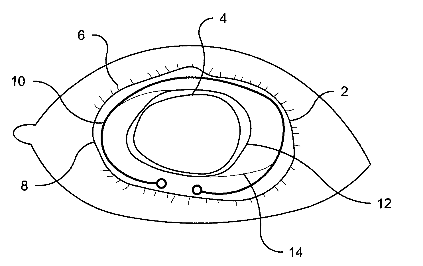
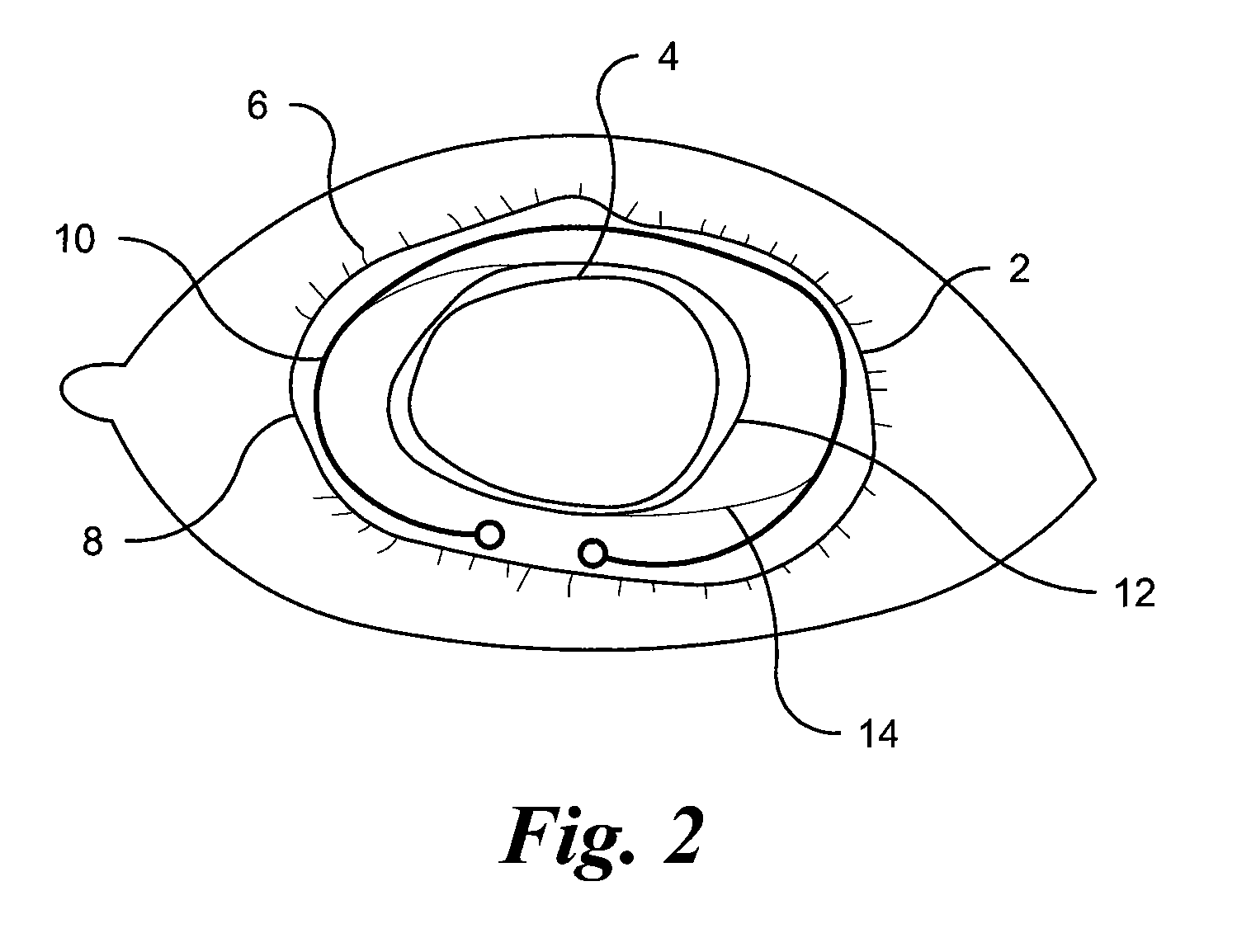
Methods and Devices for Preventing or Delaying Posterior Capsule Opacification
a technology of posterior capsule and opacification, which is applied in the direction of medical preparations, medical science, pharmaceutical delivery mechanisms, etc., can solve the problems of gradual vision loss, the form of cataract surgery does not lend itself as readily to intraocular lens implantation, and so as to prevent, minimize, or delay the formation of posterior capsule opacification
- Summary
- Abstract
- Description
- Claims
- Application Information
AI Technical Summary
Benefits of technology
Problems solved by technology
Method used
Image

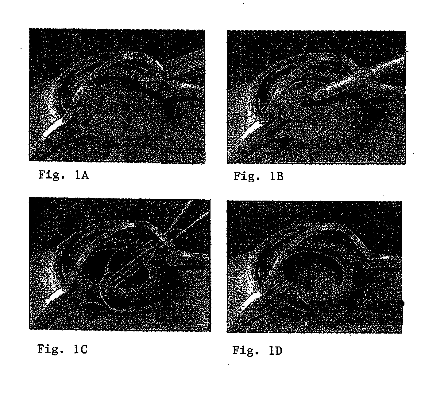
Examples
example 1
Modification of Acrylic IOL Surface
[0088]An acrylic IOL material is washed with an aqueous sodium dodecyl sulfate solution in an ultrasonic water bath to remove any contaminants, such as dirt and dust, which might be present on the IOL surface. The surface chemistry of the acrylic IOL is then determined using Attenuated Total Reflection Fourier Transform Infra-Red Spectroscopy (ATR-FTIR) in order to establish a chemical baseline signal that will allow measurement of the changes in the surface chemical functionality which is introduced by the subsequent argon plasma treatment. The IOL is also examined microscopically using an environment scanning electron microscope to establish the initial conditions of the surface morphology prior to the argon plasma treatment.
[0089]The IOL is then placed in an RF-plasma reactor and treated first with argon plasma and then grafted with an appropriate chemical moiety. Specifically, grafting is accomplished by the introduction of a selected organic v...
example 2
Covalent Attachment of Inhibitor Compound to the IOL
[0091]The acrylic IOL with carboxyl groups immobilized on its surface is covalently attached to the desired inhibitor compound. The protocol for covalent attachment depends on the functional groups present on the selected compound. For example, if the selected compound contains amino groups (i.e., an RGD mimetic or RGD peptide), the acrylic acid-grafted IOL is treated with a water-soluble carbodiimide to activate the carboxyl groups on the IOL and to facilitate its reaction with the amino group. A wash step using distilled water at room temperature is typically performed. The IOL is incubated in a pH-adjusted solution of the selected compound to allow the amino groups of the compound to react with the activated carboxyl groups on the surface to form amide bonds. Immobilization of compounds on PMMA has previously been demonstrated to be pH dependent (Kang; Biomaterial; 14:787-792 (1993)). Thus, the inhibitor compound is coupled onto...
example 3
Evaluation of Modified IOL
[0094]Following modification, changes in chemical stability and surface roughness of the modified IOL are determined as follows. The modified IOL is incubated in de-ionized water at 45° C., a temperature capable of breaking secondary bonds. When compared to the spectra before incubation, FTIR analysis is used to verify the presence of the introduced functional groups which remain after incubation. Specifically, the presence of carbonyl groups indicates that the flavonoid, RGD mimetic, or RGD peptide is covalently attached to the surface of the acrylic IOL and that the bond formation is stable. Similarly, ESCA analysis may also be used to confirm immobilization of these compounds onto the IOL. Peaks corresponding to the carbonyl and phenol groups from the ESCA survey scan spectrum will indicate that a flavonoid, RGD mimetic, or RGD peptide is covalently attached to the IOL. Changes in the ESCA spectrum will also identify if any chemical moieties, important f...
PUM
 Abstract
Description
Claims
Application Information