Method of treating glaucoma and intraocular hypertension

a technology of intraocular hypertension and glaucoma, which is applied in the field of methods, can solve the problems of long-term development of glaucoma, irreparable permanent blindness all over the world, and similar risks, and achieve safe and effective treatment of glaucoma, enhance the effect of insulin activity, and enhance the effect of insulin

Inactive Publication Date: 2011-12-01
SHANTHA TOTADA R +2
View PDF1 Cites 21 Cited by
  • Summary
  • Abstract
  • Description
  • Claims
  • Application Information

AI Technical Summary

Problems solved by technology

Ocular hypertension with the absence of visual field defects, may lead to development of glaucoma over a long time, also has a similar risk.
It is the leading cause of irreparable permanent blindness all over the world.
Many people who have the disease do not even know that that they have this serious blinding affliction of the eye.
Patients with congenital (developmental) glaucoma are born with growth deficiency of the iridocomeal angle, resulting in obstruction to the aqueous outflow resulting in this type of glaucoma.
When there is an obstruction to the drainage, the fluid builds up in the eye (called intraocular pressure or IOP) and presses against the optic nerve, resulting in glaucoma and vision changes.
If the intraocular pressure remains too high for long periods of time, the extra pressure damages parts of the optic nerve affecting the vision due to apoptosis of the ganglion cells of the retina.
However, the functional importance of each of these two pathways is still tentative.
Migration is likely to be a difficult event to study in vivo because cell loss has been calculated to be at most only in the region of 20 cells per day.
In addition, any chemo attractants found in aqueous humor are likely to be there in very small quantities.
In actuality, glaucoma is characterized by damage in the optic nerve that results in visual field loss regardless of the IOP level.
As the elevated IOP goes untreated, the damage to the optic nerve results in loss of peripheral and then central vision.
However, not all patients with elevated IOP develop glaucoma-related retinal eye damage.
By the time the vision is impaired, the visual loss is irreversible because once the nerve cells are dead; nothing can restore them.
This causes fluid pressure to build up behind the iris, further narrowing the TM angle.
If the pressure becomes sufficiently high, the iris is forced against the trabecular meshwork, blocking drainage, resulting in ACG.
This sudden rise in pressure can occur within a matter of hours and become very painful and can be associated with nausea and vomiting.
If untreated immediately, eyesight can be permanently destroyed with scarring of the trabecular meshwork and Cataracts development with optic nerve damage resulting in impaired vision.
Many of these sudden attacks occur in darkened rooms, such as movie theaters due to papillary dilatation further narrowing angle and trigger an attack.
Many drugs such as anti-depressants, cold medications, antihistamines, and some medications to treat nausea can also cause dilation of the pupil and can lead to an AG attack.
This causes the pigment layer of the eye to rub on the lens.
Both pigment and exfoliation material clogs the trabecular meshwork, leading to both open-angle glaucoma and angle-closure glaucoma.
The elevated IOP can cause the eyeball itself to enlarge and injury to the cornea, poor vision, light sensitivity, tearing, and blinking.
Herpes zoster ophthalmicus can be painful and result in complications centered on the destruction of the ocular structures that can lead to glaucoma and permanent loss of sight.
These agents interfere with DNA synthesis and inhibit viral replication.
Even today there is no effective therapy for the local effects of this condition.
Therapeutic agents administered to the eye can pass rapidly through the nasolacrimal duct into the nose and then through the highly vascular nasal mucosa to enter the systemic circulation bypassing the liver; may result in adverse effects, even death in rare instances.
1. Drugs to increase uveoscleral outflow of aqueous humor: Prostaglandin analogs like latanoprost (Xalatan), bimatoprost (Lumigan) and travoprost (Travatan) and Rescula. Bimatoprost in addition also increases trabecular meshwork outflow
2. Pharmacological therapeutic agents to decrease aqueous humor production by the ciliary body and ciliary process: Topical beta-adrenergic receptor antagonists such as: Timoptic, Timoptic (XE / GFS, or Ocudose), Betoptic, Optipranolol, Ocupress) timolol, levobunolol (Betagan), and betaxolol. They are either non selective like Timopitc or selective blockers such as Betopitc.
3. Therapeutic agents which have dual mechanism of decreasing aqueous production and increasing trabecular outflow: Alpha2-adrenergic agonists such as brimonidine (Alphagan, Iopidine, Propine).
4. Drugs which increase the outflow of aqueous humor through trabecular meshwork and possibly through uveoscleral outflow pathway, probably by a beta2-agonist action: Less-selective sympathomimetics like epinephrine and dipivefrin (Propine)
5. Medications which cause Contraction of the ciliary muscle (miotic), thus tightening the trabecular meshwork and allowing increased outflow of the aqueous humour. Miotic agents (parasympathomimetics) like pilocarpine and Ecothiopate used in chronic glaucoma.
6. Therapeutic agents that lower secretion of aqueous humor by inhibiting carbonic anhydrase in the ciliary body and process: Carbonic anhydrase inhibitors like dorzolamide (Trusopt), brinzolamide (Azopt), acetazolamide (Diamox). Oral carbonic anhydrase inhibitors are also available (Diamox, Neptazane).
7. Ophthalmic preparations that cause constriction of pupils to widen the iridocorneal angle: Physostigmine is used to treat glaucoma (and delayed gastric emptying). Instillation of 0.25% physostigmine sulphate eye drops caused a sustained miosis, with some deleterious action on the visual system.
8. Combination anti glaucoma therapeutic agent: Currently combinations of Timoptic and Trusopt (Cosopt) are available and are effective in twice a day dosing. Combinations of Xalatan and Timoptic as well as Alphagan and Timoptic are being studied and will be marketed soon. Combination eye drops may improve compliance if more than one drug is needed to control the IOP as it is more convenient to deal with just one bottle.
9. Combining our invention with the above described therapeutic agents will be more efficacious in controlling the glaucoma especially when it is resistant to single therapeutic agents and adverse systemic effects are anticipated.
Effect of Exercise on intra ocular Pressure: Physical exercise can reduce the IOP by 2 mm Hg and may not be effective in everyone.
Xalatan has the potential of causing premature labor, therefore may not be a first choice agent (Stamper.
Concomitant use with aspirin increases the risk of salicylate toxicity.
The color change is stable and may not be reversible with discontinuation of the drug.
Although most medications are applied topically to the eye, they can cause severe systemic side effects and adversely affect the quality of the patient's life.
The drug therapies for glaucoma are sometimes associated with significant side effects, such as headache, blurred vision, allergic re actions, potential interactions with other drugs and death from cardiopulmonary complications.
Argon Laser Trabeculoplasty (ALT): ALT is also an in-office laser therapy similar to that of SLT, but uses longer pulses that non-selectively target cells; more likely to result in unnecessary damage and scarring to surrounding internal eye structures, limiting treatment repeatability options for maintaining eye pressure control and vision preservation.
All surgical procedures have risks; such as infection, bleeding, swelling in the retina, development of fluid under the retina (choroidal detachment), retinal detachments etc.
The scarring over the conjunctival dissipation segment of the shunt may become too thick for the aqueous humor to filter through.
In spite of all the treatment modalities evolved over decades as described above, there is still no a cure for glaucoma and elevated IOP.
There are problems with the aforementioned approaches to treating glaucoma in that the treatments can be accompanied by side-effects.
For example, the instillation of a cholinergic agent, such as pilocarpine, into the eye of a subject can cause nausea, diarrhea, muscular spasms, sweating, lacrimation, salivation, etc.
The treatment with a sympathomimetics agent such as dipivalylepinephrine is known frequently to produce sensations of burning or irritation in a subject.
Another side-effect of these agents is the appearance of cardiac disturbances, e.g., palpitations, tachycardia, arrhythmia, etc.
The anti-inflammatory drugs acetylsalicylate and rednisolone are known to regulate the microglia responsible for the onset of photoreceptor apoptosis and retinal degeneration thereafter; both drugs are proved to be ineffective.
They are not known to be useful in treating or controlling glaucoma.

Method used


Image

  • Method of treating glaucoma and intraocular hypertension
  • Method of treating glaucoma and intraocular hypertension
  • Method of treating glaucoma and intraocular hypertension

Examples


example 1

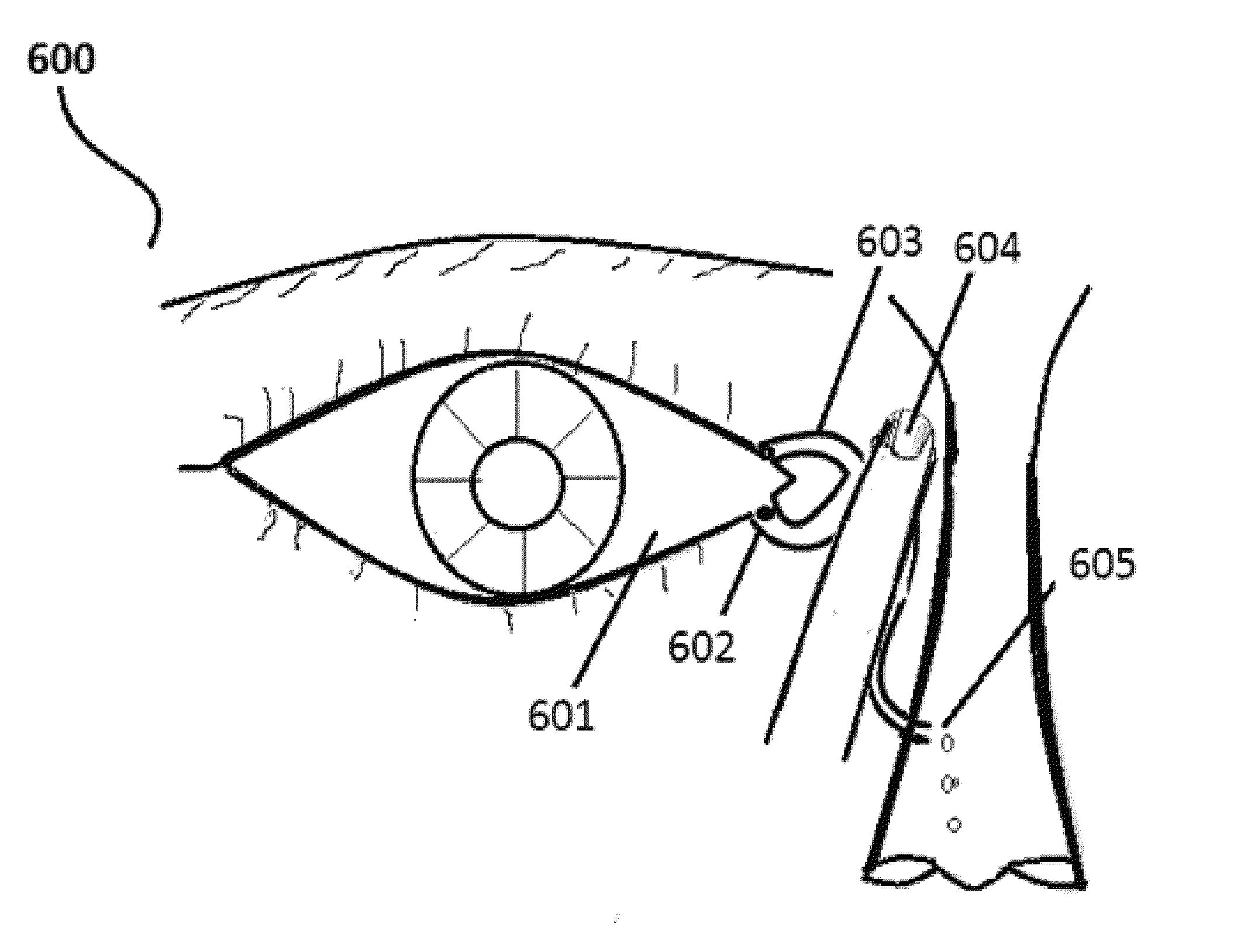
[0210]Select the patient and establish the type of glaucoma (definite diagnosis) the person is suffering. Record the degree of retinal damage by the thorough examination. Position the patient in supine posture with head extended on a support. Using a dropper, instill one drop of insulin preparation in each eye lower lid fornix and / or everted upper eyelid. Apply slight pressure at the nasal angle of eye pressing on the nasolacrimal canaliculi-sac-duct to prevent leaking of the therapeutic agents to the nose to avoid systemic absorption and its adverse effects (see FIG. 6). Stay still for 3-5 minutes and resume the desired posture.

example 2

[0211]Select the patient and establish the type of glaucoma the person is suffering (definite diagnosis) from. Record the degree of retinal damage by the examination. Position the patient in supine posture with head extended on a support. Using a dropper, instill one drop of IGF-1 preparation in each eye lower lid fornix and / or everted upper eyelid. Apply slight pressure at the nasal angle of eye pressing on the nasolacrimal canaliculi-sac-duct to prevent leaking of the therapeutic agents to the nose. Stay still for 3-5 minutes and resume the natural desired posture.

example 3

[0212]Select the patient and establish the type of glaucoma the person is suffering (definite diagnosis) from. Record the degree of retinal damage by the examination. the patient in supine posture with head extended on a support. Using a dropper, instill one drop of insulin preparation in each eye lower lid fornix and / or everted upper eyelid. Then apply slight pressure at the nasal angle of eye pressing on the nasolacrimal canaliculi-sac-duct to prevent leaking of the therapeutic agents to the nose. Following this, instill a drop in each afflicted eye Prostaglandin analogs like latanoprost (Xalatan), bimatoprost (Lumigan) and travoprost (Travatan) and Rescula which increase uveoscleral outflow of aqueous humor: Bimatoprost in addition also increases trabecular mesh work outflow. Stay still for 3-5 minutes and resume the natural desired posture. The dose of these medications can be reduced because the insulin has augmentation—amplification effects on these therapeutic agents anti gla...


PUM

PropertyMeasurementUnit
thicknessaaaaaaaaaa
pressureaaaaaaaaaa
pressuresaaaaaaaaaa

Abstract

A safe and effective treatment for glaucoma for mammalian species comprises the steps of applying insulin and / or insulin like growth factors (IGF-1) to an eye. In addition to the insulin, another therapeutic agent may be applied to enhance the activity of the insulin. The therapeutic agent may be a pharmaceutical agent or a biochemical pharmaceutical agent. The therapeutic agents include prostaglandin analogs, topical beta-adrenergic receptor antagonists-β blockers, Alpha2-adrenergic agonists hair growth therapeutic agents, beta2-agonist action agents, parasympathomimetic miotic agents, carbonic anhydrase inhibitors, and Physostigmine. In another embodiment, a combination of at least two agents are applied to the eye. To enhance the effect of the insulin, uptake facilitators may be used. Additionally, an antibacterial agent may be applied to control bacterial infection.

Description

FIELD OF THE INVENTION[0001]The invention relates to method for treating ocular hypertension, glaucoma with retinal damage and loss of vision in human and mammals.BACKGROUND OF THE INVENTION[0002]Glaucoma is a disease characterize by the increase of intraocular pressure (IOP) due to varied pathogenesis with damage to the retina and atrophy of the optic nerve resulting in the abnormal visual field, with reduced visual acuity. The optic nerve does not recover once atrophy occurs, even if the raised IOP is corrected. Elevated intraocular pressure (IOP) is a very significant risk factor for the development of the majority of common forms of glaucoma (Sommer A, et al., “Relationship Between Intraocular Pressure and Primary Open Angle Glaucoma Among White and Black Americans,” Arch. Ophthalmol, 109:1090-1095 (1991)). Ocular hypertension with the absence of visual field defects, may lead to development of glaucoma over a long time, also has a similar risk.[0003]Some of the Risk Factors for...

Claims


Application Information

Patent Timeline
Patent Type & Authority Applications(United States)
IPC IPC(8): A61K38/28A61P27/06
CPCA61K38/28A61K38/30A61K2300/00A61P27/06
Inventor SHANTHA, TOTADA R.SHANTHA, ERICA MAYASHANTHA, JESSICA GOWRAMMA
Owner SHANTHA TOTADA R
Why Eureka
Try Eureka