

Medical device for placement into a lumen and manufacturing method thereof
a medical device and lumen technology, applied in the field can solve the problems of difficult preparation of medical devices for placement into lumens, difficult to attach a sufficient amount of drugs to carriers, and low adhesion of particles to negative-charged cell walls, etc., to achieve the effect of increasing the efficiency of making drugs come in cells and low adhesion of particles to cells
- Summary
- Abstract
- Description
- Claims
- Application Information
AI Technical Summary
Benefits of technology
Problems solved by technology
Method used
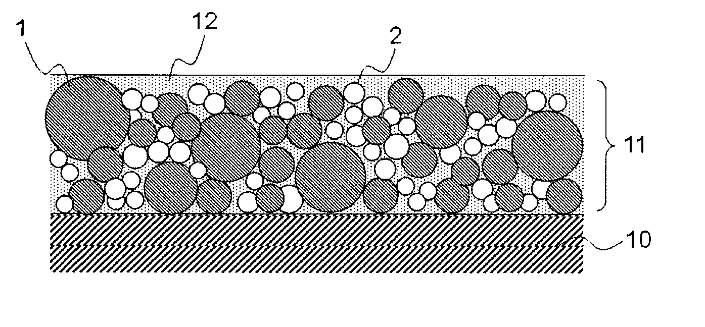
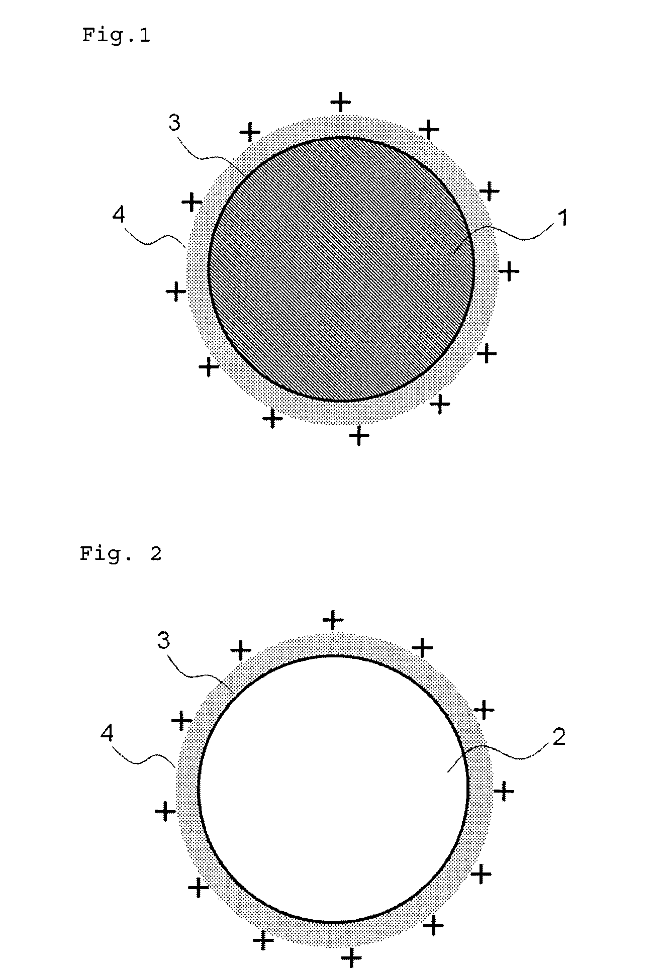
Image

Examples
example 1
Preparation of Suspension Containing Drug Particle, 1
[0170]1 g of probucol was dissolved in a mixture of 40 mL of acetone and 20 mL of ethanol which are a good solvent for probucol to prepare a solution thereof. Separately, to 100 mL of 2% (w / w) aqueous solution of polyvinyl alcohol (GOHSENOL EGOS, NIPPON GOHSEI) was added 15 g of 2 (w / w) aqueous solution of chitosan (CHITOSAN GH-400EF, NOF CORPORATION) to prepare a solution which is a poor solvent for probucol. To the solution at 40° C. with stirring at 400 rpm, the afore-prepared solution of probucol was added dropwise in a constant speed (20 mL / min) to give a suspension of crystalline drug particle (probucol), which was prepared by so-called “diffusion of a good solvent into a poor solvent”.
[0171]Consequently, the acetone and ethanol were removed in vacuo. The zeta potential on the particle surface was measured with an electrophoresis (Malvern Instruments, ZETASIZER Nano-Z). The result showed that the zeta potential on the drug p...
example 2
Preparation of Suspension Containing Drug Particle, 2
[0177]A drug particle of Example 2 was prepared in the same manner as Example 1, provided that the drug particle was not milled with a desktop-type ball mill. The mean particle size of the drug particle was 21.5 μm.
example 3
Preparation of Suspension Containing Drug Particle, 3
[0178]A drug particle of Example 3 was prepared in the same manner as Example 1, provided that the amount of 2% (w / w) aqueous solution of chitosan added to a solution equivalent to the poor solvent was 60 g. The mean particle size of the drug particle measured with a laser diffraction-scattering was 4.5 μm. The zeta potential was +45 mV, the content of probucol in the drug particle was 53% and the yield ratio was 65%.
PUM
| Property | Measurement | Unit |
|---|---|---|
| Particle size | aaaaa | aaaaa |
| Particle size | aaaaa | aaaaa |
| Particle size | aaaaa | aaaaa |
Abstract
Description
Claims
Application Information