Label-free sensing of pna-dna complexes using nanopores

a pnadna complex and nanopore technology, applied in the field of label-free sensing of pnadna complexes using nanopores, can solve the problems of not being parallelized, routineized, nor cost-effective enough to compete with other “next generation sequencing” methods, and not being able to distinguish a pool of same-sized ss nucleic acids and/or ds nucleic acids on the basis of sequence differences, so as to increas

Inactive Publication Date: 2012-11-01
TRUSTEES OF BOSTON UNIV
View PDF3 Cites 17 Cited by
  • Summary
  • Abstract
  • Description
  • Claims
  • Application Information

AI Technical Summary

Benefits of technology

[0021]In one embodiment, the ds biomolecule is a ds DNA. In one embodiment, the ds biomolecule is a RNA/DNA hybrid. In one embodiment, the at least one probe is a PNA. Other probes include RNA, DNA, and modified forms thereof. In another embodiment, the PNA is a bis-PNA. In another embodiment, the PNA is a gamma-PNA (γ-PNA). In one preferred embodiment, the γ-PNA can have a higher binding to affinity to DNA. For example, the γ-PNA has a modified nucleobase, guanidinium G-clamp (X) that replaces cytosine in the canonical G:C binding. The G-clamp results in increased thermal stability of matched duplexes due to formation of five hydrogen bonds with guanine. The probe's function is to hybridize to the ds biomolecule by complement base pairing to form a stable complex. Not the entire probe needs to hybridize to the

Problems solved by technology

However, one factor limiting the adaptability of this process to a greater number of applications is the large negative linear charge density inherent in nucleic acid strands, which significantly reduces the stability of nucleic acid complexes.
However, current literature indicates that nanopore sequencing is still at the proof-of-concept experimental stage, with some laboratory-based data to back up the different components of the sequencing method, but not yet parallelized, routineized, nor cost-effective enough to compete with other “next generation sequencing” methods.
The method does not facilitate distinguishing a pool of same-sized ss nucleic acid and/or ds nucleic acids on the basis of sequence differences.
The siz

Method used


Image

  • Label-free sensing of pna-dna complexes using nanopores
  • Label-free sensing of pna-dna complexes using nanopores
  • Label-free sensing of pna-dna complexes using nanopores

Examples


example 1

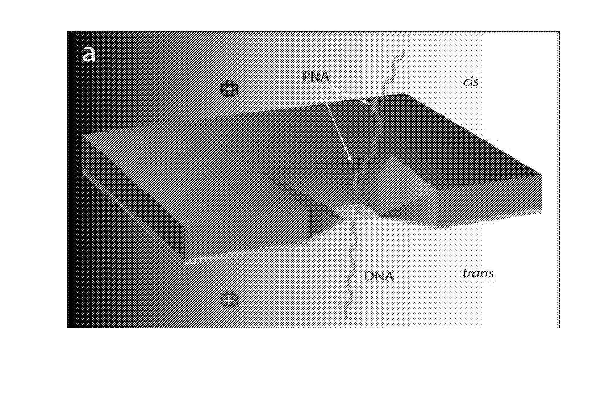
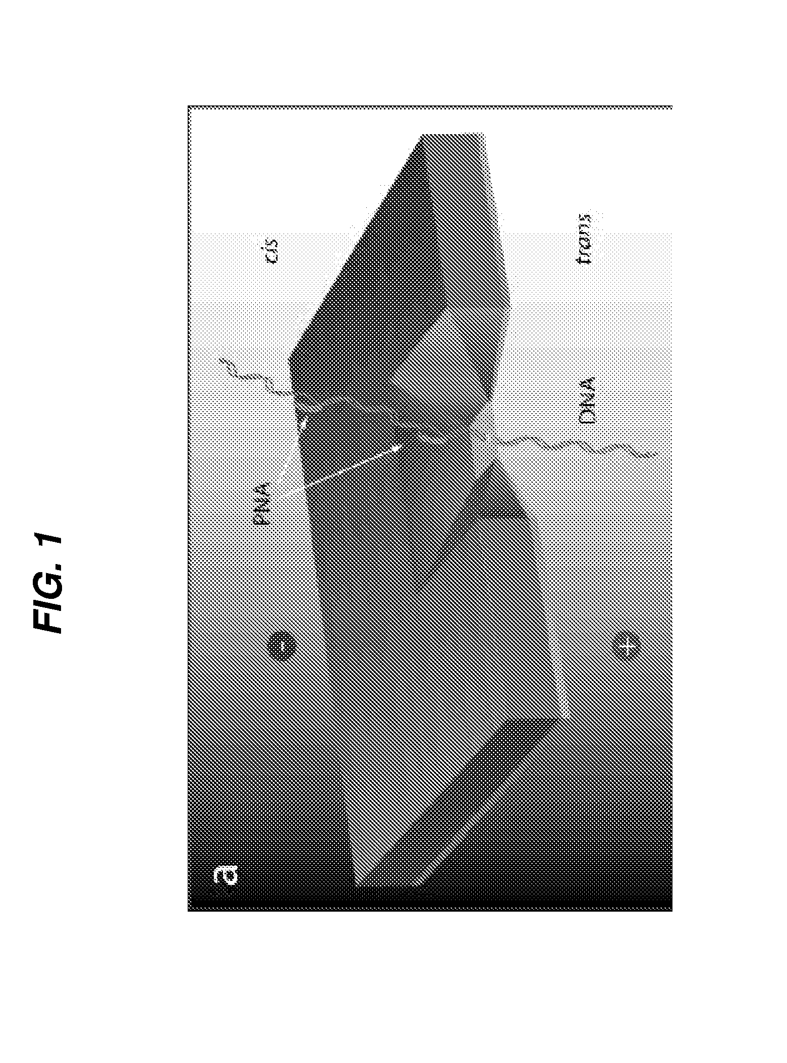
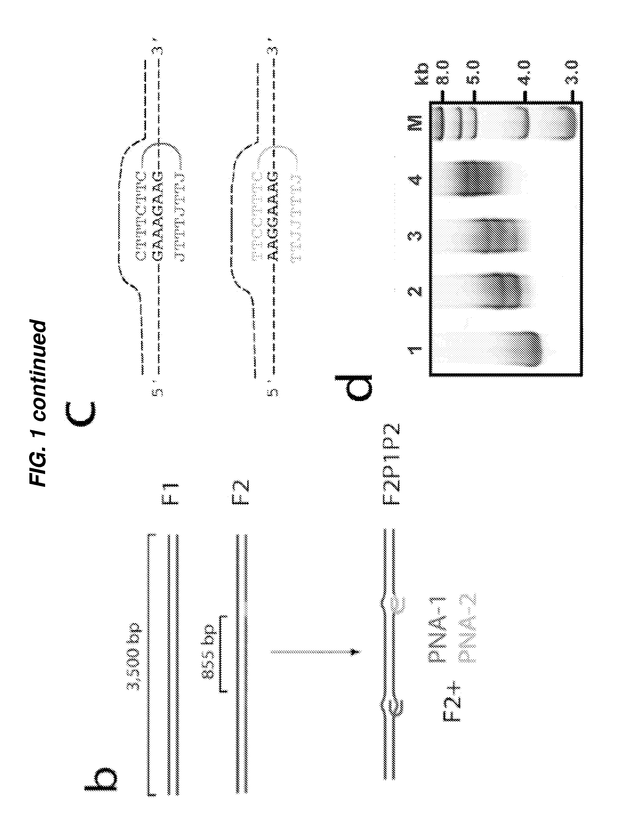
[0178]FIG. 1a shows a schematic of a nanopore system for the detection of PNA-DNA complexes in a ds DNA. A solitary 4-5 nm pore fabricated in a thin (˜20 nm) SiN membrane is assembled between two miniature fluid chambers (‘trans’ and ‘cis’), and hydrated using a 1M KCl buffered solution, as previously described (7). A voltage bias is then applied across the SiN membrane using a pair of AgCl electrodes. When the trans chamber is positively biased, DNA molecules in the cis chamber (4) thread through the nanopore into the trans chamber. To show that nanopores can discriminate among PNA-bound and PNA-free DNA samples, two DNA fragments (PCR-amplified from the -phage genome) of nearly equal lengths were have prepared (3,500 bp, see FIG. 1b). The first fragment (F1) serves as a negative control, which does not include target sequences for either of the two bis-PNA probes (PNA-1 and PNA-2). The positive sample (F2) contains two different binding sites for our bis-PNA probes (see sequences ...

example 2

[0182]In this example, the inventors show the use of two bis-PNA to create a P-loop and the detection of that P-loop. Two bisPNA probes were used to bind to two closely spaced binding sites on a linearized pUC19 plasmid. The two closely spaced binding sites represent the signature site in pathogen genome. As a result, an extended P-loop comprising of 19 nt in each strand was formed (FIG. 5). The target sequence was located approximately 500 bp from one end of the plasmid and 2200 bp from the other end, as shown in the figure. Complex formation was confirmed by nondenaturing gel electrophoresis, which showed a clear shift in the mobility of the DNA / PNA hybrid molecule as compared to the untagged DNA lane (data not shown).

[0183]A 4 nm diameter nanopore was used to compare the hybridized complex with a control of intact, double-stranded linear pUC19. FIG. 6 shows a typical DNA translocation events (FIG. 6a) of the 2700 bp plasmid control DNA, and typical translocation events of the DNA...

example 3

[0185]The nanopore based assays using γ-PNA probes are conducted in the same manner as for the nanopore based assays using bis-PNA except that the nanopore is 3.5 nm.

[0186]For this study, the inventors took a 1 kbp dsDNA molecule, with a single binding site located in the center (so ˜500 bp on either side of the γ-PNA binding site) and compared the readout signal from the nanopore based assay (FIG. 9). The parameters for the nanopore system are as described in Examples 1 and 2.

[0187]FIG. 9a shows an exemplary data obtained for the control experiment where no γ-PNA is added and mixed with the dsDNA, illustrating qualitatively an electrical current trace of a handful of single-molecule translocation events. Similar to FIGS. 2a and 6a in Examples 1 and 2 respectively, the single-molecule translocation events are seen as single block drop in the electrical current trace, which are identified quantitatively in an all-points histogram (FIG. 9a right side).

[0188]FIG. 9b shows an exemplary ...


PUM

PropertyMeasurementUnit
Diameteraaaaaaaaaa
Electric potential / voltageaaaaaaaaaa
Electrical conductanceaaaaaaaaaa

Abstract

Embodiments disclosed herein relate to a method of detecting specific DNA sequences and the application of this method in the detection of pathogens, viruses, drug-resistant pathogens, genomic variations associated with disease/disorder susceptibility etc. based on specific signature sequences unique to the pathogens, viruses, drug-resistant pathogens or genomic variations. The method can also be used to distinguish a pool of same-sized dsDNA on the basis of sequence differences. The method uses non-optically labeled bis-PNA and/or gamma-PNA probes to tag specific target sequences for identification by solid-state nanopores.

Description

CROSS REFERENCE TO RELATED APPLICATION[0001]This application claims benefit under 35 U.S.C. §119(e) of the U.S. Provisional Application No. 61 / 236,187 filed Aug. 24, 2009, the contents of which are incorporated herein by reference in its entirety.GOVERNMENT SUPPORT[0002]This invention was made with Government support under contract No. HG-004128 awarded by the National Institute of Health and contract No. PHY-0646637 awarded by the National Science Foundation. The Government has certain rights in the invention.BACKGROUND OF INVENTION[0003]The ability of nucleic acids to spontaneously form stable, sequence-specific complexes with other nucleic acids, which serve as molecular probes, has been exploited for a wide range of applications in life sciences, biotechnology, medicine, and forensics. Examples range from polymerase chain reaction (PCR) and fluorescence in situ hybridization (FISH) to DNA microarrays and sequencing by hybridization. Current methods for detection of nucleic acids...

Claims


Application Information

Patent Timeline
IPC IPC(8): G01N27/26B82Y5/00
CPCC12Q1/6839C12Q1/689G01N33/48721C12Q2525/107
Inventor MELLER, AMITFRANK-KAMENETSKII, MAXIMWANUNU, MENIKUHN, HEIKOSINGER, ALONMORRISON, WILL
Owner TRUSTEES OF BOSTON UNIV
Why Eureka
Try Eureka