
Methods and compositions for wound healing

a composition and wound healing technology, applied in the field of methods and compositions, can solve the problems of special challenges, insufficient function of wound healing components, impaired wound healing, etc., and achieve the effects of promoting and enhancing wound healing

Pending Publication Date: 2015-10-08
IMBED BIOSCI
View PDF4 Cites 44 Cited by
  • Summary
  • Abstract
  • Description
  • Claims
  • Application Information

AI Technical Summary

Benefits of technology

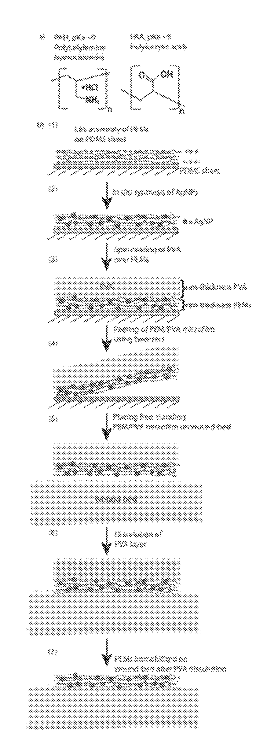
The present invention provides methods and compositions for promoting wound healing and enhancing the surface properties of biomedical devices and tissues by altering the chemical composition and physical attributes of the wound bed. The invention provides formulations that can deliver antimicrobial compounds, extracellular matrices, and other scaffolds and cytoactive agents to a wound. The invention also provides methods for incorporating nanoparticles and microparticles into the wound bed to engineer its physical characteristics and chemical composition. The invention can adjust key cellular elements and promote favorable outcomes such as vascularization, fibrosis, and epithelial coverage. The invention contemplates that the microsheet can be applied to a variety of wound surfaces without affecting their normal growth and proliferation.

Problems solved by technology

Wound healing is impaired when these components, either individually or as a whole, do not function properly.
Additionally, wounds that are extensive in size, regardless of the initiating cause, present special challenges due to the large surface area that must be re-epithelialized to re-establish surface integrity.
Delayed wound healing causes substantial morbidity in subjects with diabetes.
These pathological changes can ultimately lead to chronic ulceration, which may necessitate amputation.
Chronic wounds and wounds with pathological or dysregulated healing represent a major health burden and drain on health care resources.
However, in many patients, due to either the local wound environment or systemic disease or other factors, the wound healing processes can become asynchronous (i.e., loss of connectivity with triggering mechanisms associated with prior cellular events) and are unable to progress to closure, resulting in a chronic ulcer.
Wounds that do not readily heal can cause the subject considerable physical, emotional, and social distress as well as great financial expense (Richey et al., 1989, Annals of Plastic Surgery 23:159).
Indeed, wounds that fail to heal properly and become infected may require excision of the affected tissue.
Unfortunately, certain types of wounds (e.g., diabetic ulcers, pressure sores) and the wounds of certain subjects (e.g., recipients of exogenous corticosteroids) do not heal in a timely manner (or at all) with the use of such dressings.
Thus, except in very limited circumstances, the promotion of wound healing with these agents has met with little success.
All of these approaches fall short of promoting optimal healing conditions in many of the most challenging wounds.
It is likely that a major contributing factor to the failure of these traditional approaches is the fact that they do not alter the intrinsic chemistry / structure of the wound bed itself that has been shown in many cases to contribute significantly to their persistence.
Additionally, the historical use of a single factor or set of factors to treat all wounds often falls short due to the great heterogeneity found in wound beds themselves and the complex environment of the wound itself containing a community of signaling molecules that frequently modulate the activity of individual molecules.
However, currently available methods of applying silver as a bactericidal agent for wound treatment are inadequate.
However, while such formulations have a high concentration of silver, there is no residual activity, necessitating frequent applications (e.g., up to 12 times a day) which poses a severe logistical burden in clinical settings.
Additionally, nitrate is toxic to wounds and to mammalian cells.
However, such formulations also have limited residual activity and have to be applied twice a day.
Bacterial resistance does develop to these formulations, and, impaired re-epithelialization has also been observed.
Bone marrow toxicity has been observed with silver sulfadiazine, primarily due to its propylene glycol component.
However, dressings have to be impregnated with large amount of silver, which results in cytotoxicity to mammalian cells.
The complex nature of pathologic wounds, and the lack of significant clinical progress based on current therapies, indicates the urgent need for new and unconventional approaches.

Method used


Image

  • Methods and compositions for wound healing
  • Methods and compositions for wound healing
  • Methods and compositions for wound healing

Examples


example 1

Covalent Immobilization of a Protein to a Surface

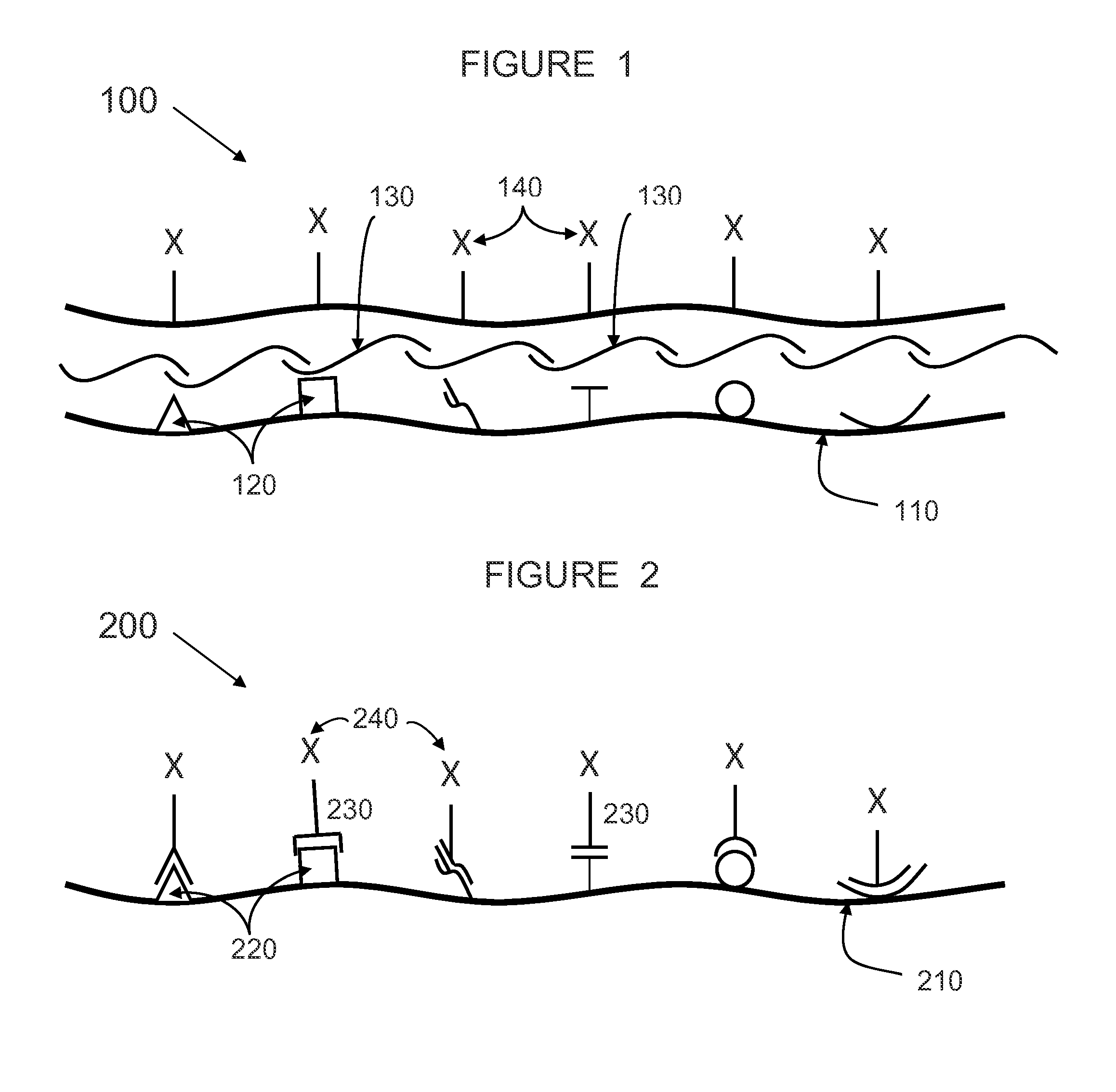
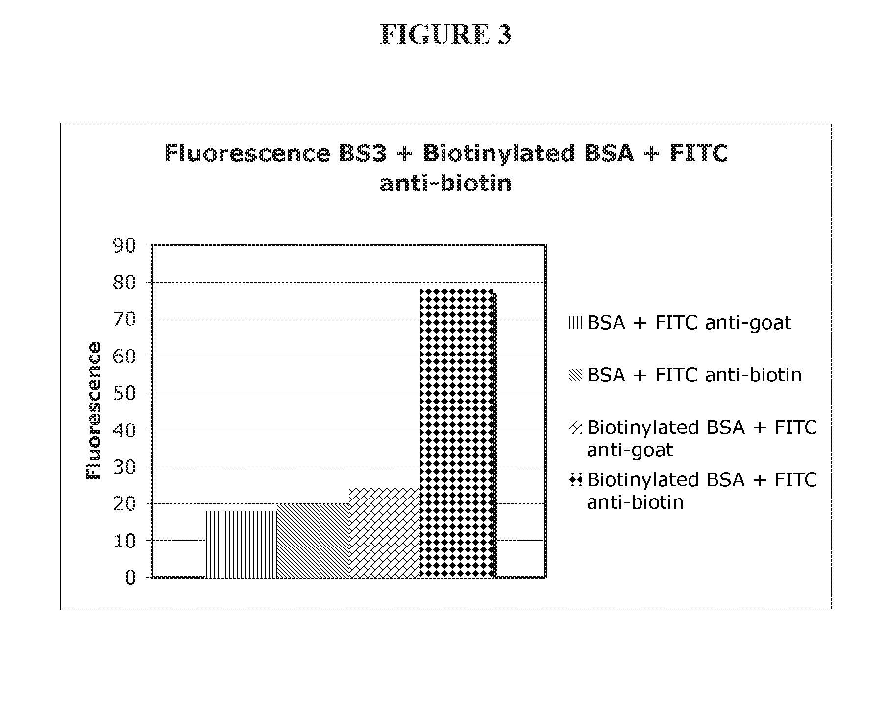
[0359]To demonstrate that proteins can be covalently immobilized on model surfaces, bovine serum albumin (BSA) was used as the protein in the model system. Bovine serum albumin was biotinylated (BSA used as control) and covalently attached to gamma-amino propyl silanes (GAPS)-treated glass surfaces using the homobifunctional bifunctional cross-linker BS3 (1 mM for 15 min.). BS3 contains an amine-reactive N-hydroxysulfosuccinimide (NHS) ester at each end of an 8-carbon spacer arm. The NHS esters react with primary amines at pH 7-9 to form stable amide bonds, along with release of the N-hydroxysulfosuccinimide leaving group. For detection purposes, the biotinylated BSA was labeled with FITC-labeled anti-biotin antibodies and the BSA control was probed using FITC-antigoat antibodies.

[0360]The large fluorescent signal seen in FIG. 3 from the surfaces treated with biotinylated BSA and FITC-antibiotin antibody (diamonds) relative to control...

example 2

Layer by Layer Deposition of Polyelectrolytes in Ex Vivo Wound Beds

[0361]Experiments depositing polyelectrolytes in wound bed explants from mice were performed. Cutaneous wounds from euthanized mice were harvested. The excised wounds were sequentially treated with aqueous solutions (0.5M NaCl, PBS at pH 7.0) containing polystyrene sulfonate (PSS, 1 mg / ml) and FITC-labeled poly allylamine hydrochloride (FITC-PAH, 1 mg / ml). Between adsorption steps, the wounds were treated rinsed with PBS. After each treatment with FITC-PAH, the intensity of fluorescence was recorded. As seen in FIG. 4, the growth in fluorescence after each treatment cycle with PSS and FITC-PAH demonstrates growth of a multilayered polyelectrolyte film on the wound bed, relative to control treatments.

example 3

Use of Mesoscopic Cross-Linkers in Ex Vivo Wound Beds

[0362]Skin wounds are created in tandem on diabetic mice using a surgical skin punch. After wounding, one of the wounds is treated by contact with activated 10-micrometer diameter polystyrene beads with surfaces terminated in carboxylic acid groups. The activation of the carboxylic acid groups is achieved by incubation in EDC / NHS solution (200 mM / 50 mM) in phosphate buffered saline (PBS) (10 mM phosphate, 120 mM NaCl, 2.7 mM KCl; pH 7.6) for 1 hour at room temperature. Following activation, the beads were pipetted into the wound beads and allowed to incubate within the wound beds for 1 hr. After incubation, the wound beds are washed exhaustively with PBS, and then incubated with collagen (10 micromolar in PBS). The remaining wound is used as a control and treated as described above, but without activation of the beads with NHS / EDC. The size of the wound is measured after 2, 4, 6, 8 and 10 days. It is observed that the treated woun...


PUM

PropertyMeasurementUnit
thickaaaaaaaaaa
thickaaaaaaaaaa
thickaaaaaaaaaa

Abstract

The present invention relates to methods and compositions for wound healing. In particular, the present invention relates to promoting and enhancing wound healing by utilizing cross-linker covalent modification molecules to attach and deliver wound active agents to a wound. In addition, the present invention provides methods and compositions utilizing oppositely charged polyelectrolytes to form a polyelectrolyte layer on a wound surface. The invention further relates to incorporating wound active agents into a polyelectrolyte layer for delivery to a wound.

Description

FIELD OF THE INVENTION[0001]The present invention relates to methods and compositions for the modulation of wound healing. In particular, the present invention relates to nanoscale polymer layers comprising bioactive agents and microsheets comprising a nanoscale polymer layer supported on a sacrificial polymer layer.BACKGROUND OF THE INVENTION[0002]The primary goal in the treatment of wounds is to achieve wound closure. Open cutaneous wounds represent one major category of wounds and include burn wounds, wounds resulting from chemical (especially alkali) burns, wounds from physical trauma, neuropathic ulcers, pressure sores, venous stasis ulcers, and diabetic ulcers. Open cutaneous wounds routinely heal by a process which comprises six major components: i) inflammation, ii) fibroblast proliferation, iii) blood vessel proliferation, iv) connective tissue synthesis, v) epithelialization, and vi) wound contraction. Wound healing is impaired when these components, either individually or...

Claims


Application Information

Patent Timeline
Patent Type & Authority Applications(United States)
IPC IPC(8): A61L15/42A61L15/24A61L15/26A61L15/44
CPCA61L15/42A61L15/44A61L15/24A61L15/26A61L2400/12A61L2300/404A61L2300/206A61L2300/102A61L2300/104A61F13/02A61L15/22A61L15/46A61P17/02
Inventor AGARWAL, ANKITABBOTT, NICHOLAS L.
Owner IMBED BIOSCI
Learn More