

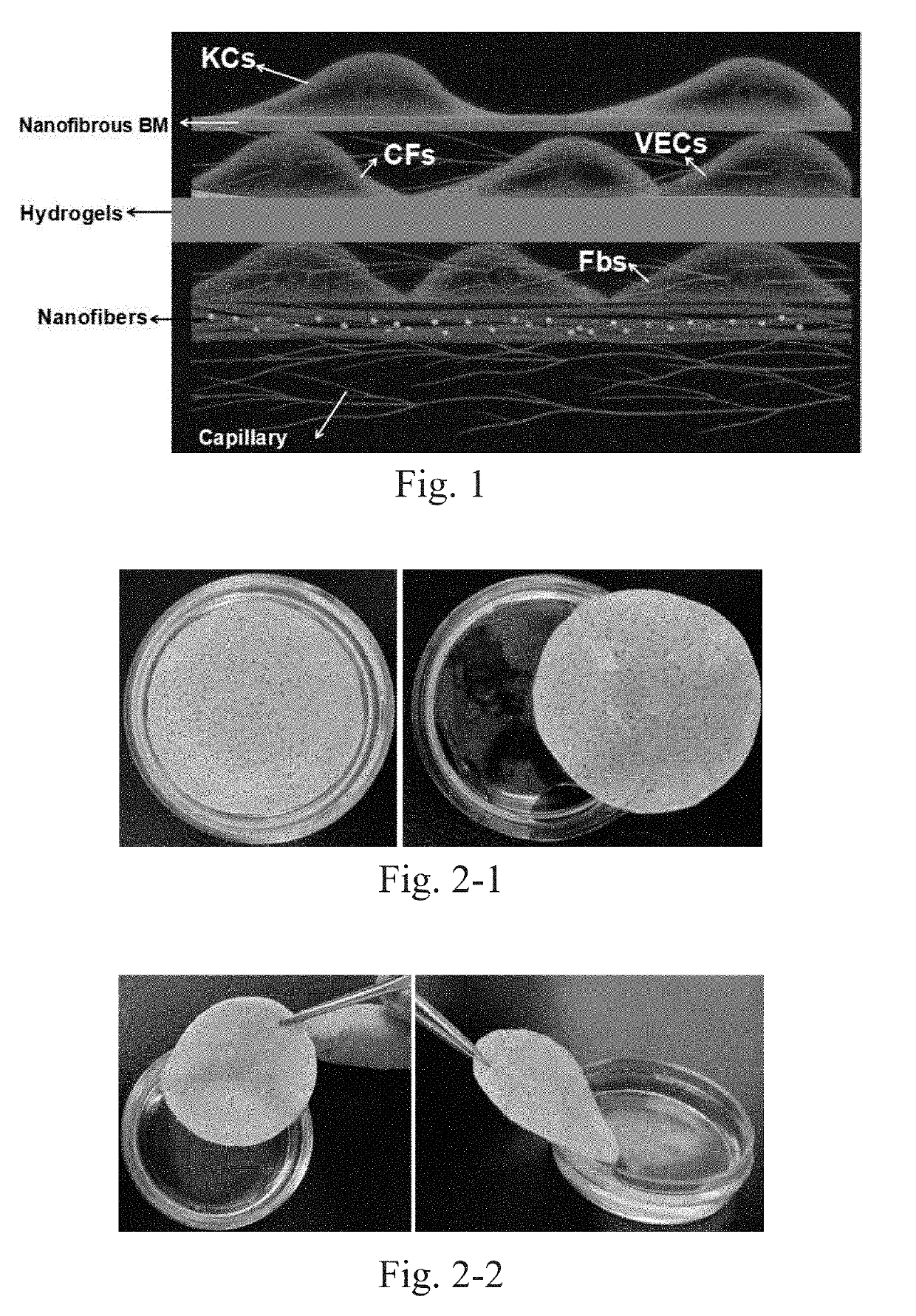
Vascularized full thickness tissue-engineered skin assembled by hydrogel, nanofibrous scaffolds and skin cell layers and preparation method thereof
a technology of nanofibrous scaffolds and skin cells, applied in the field of polymer materials and biomedical materials, can solve the problems of skin damage, difficult filling, and destruction of its integrity as a barrier, and achieve the effects of facilitating cell adhesion and growth, uniform distribution, and facilitating cultivation and function maintenan
- Summary
- Abstract
- Description
- Claims
- Application Information
AI Technical Summary
Benefits of technology
Problems solved by technology
Method used
Image

Examples
embodiment 1
nd Purification of Keratinocytes
[0051]The foreskin was clarified by repeated washing with sterile PBS several times until the PBS is clear. The foreskin is cut into strips after removing other tissues such as fascia; then Dispase is added before digestion overnight in a refrigerator at 4° C. The foreskin that had been digested overnight was rewarmed at 37° C. Then epidermis and dermis were separated, and the isolated epidermis was washed in PBS. The isolated epidermis was added to trypsin (containing EDTA) and digested at 37° C. The digested juice was filtered through a sieve, and the filtrate was collected and centrifuged. After washing with PBS, the cells were resuspended in a K-SFM medium, and the cells were inoculated at a cell density of 1×10⁵ / cm2 and cultured in a 37° C. incubator. After 2 days, the liquid was changed for the first time, and then the liquid was changed every 3 days.
embodiment 2
nd Purification of Circulating Fibroblasts
[0052]A lymphocyte separation solution was added to an L tube, and the mixture was centrifuged to the lower layer. Blood was diluted with PBS, and then was added to the L tube for centrifugation separation. The upper layer of plasma was discarded, and the leucorrhea layer was aspirated and added to a centrifuge tube to be diluted with PBS and centrifuged. The above procedures were repeated 3 times. Subsequently, the cells were counted after resuspending in PBS, and inoculated in a culture dish at a density of 1×10⁶ / mL.
embodiment 3
nd Purification of Vascular Endothelial Cells
[0053]The HUVEC cell line purchased from Sciencell was resuscitated at 37° C., and inoculated into a 100 mm culture dish. After the culture dish was full of cells, trypsin was used for passage digestion.
PUM
| Property | Measurement | Unit |
|---|---|---|
| Temperature | aaaaa | aaaaa |
| Fraction | aaaaa | aaaaa |
| Thickness | aaaaa | aaaaa |
Abstract
Description
Claims
Application Information