Inflamed plaques are most susceptible to ruptures and the formation of blood clots.
The rupture causes a large
thrombus (blood clot) to form on the inside of the
artery, which may completely occlude the
blood flow through the
artery, thereby injuring the heart or brain.
Frequently, however, injury to the vessel wall and disruption of the plaque core occur during restoration of vessel patency.
This leads to neointimization and remodeling of the vessel wall and
restenosis.
Blood clots may form as a result of the
spillage of plaque contents and due to triggering of the natural clot-forming
cascade of the blood, further contributing to
restenosis at the treatment site.
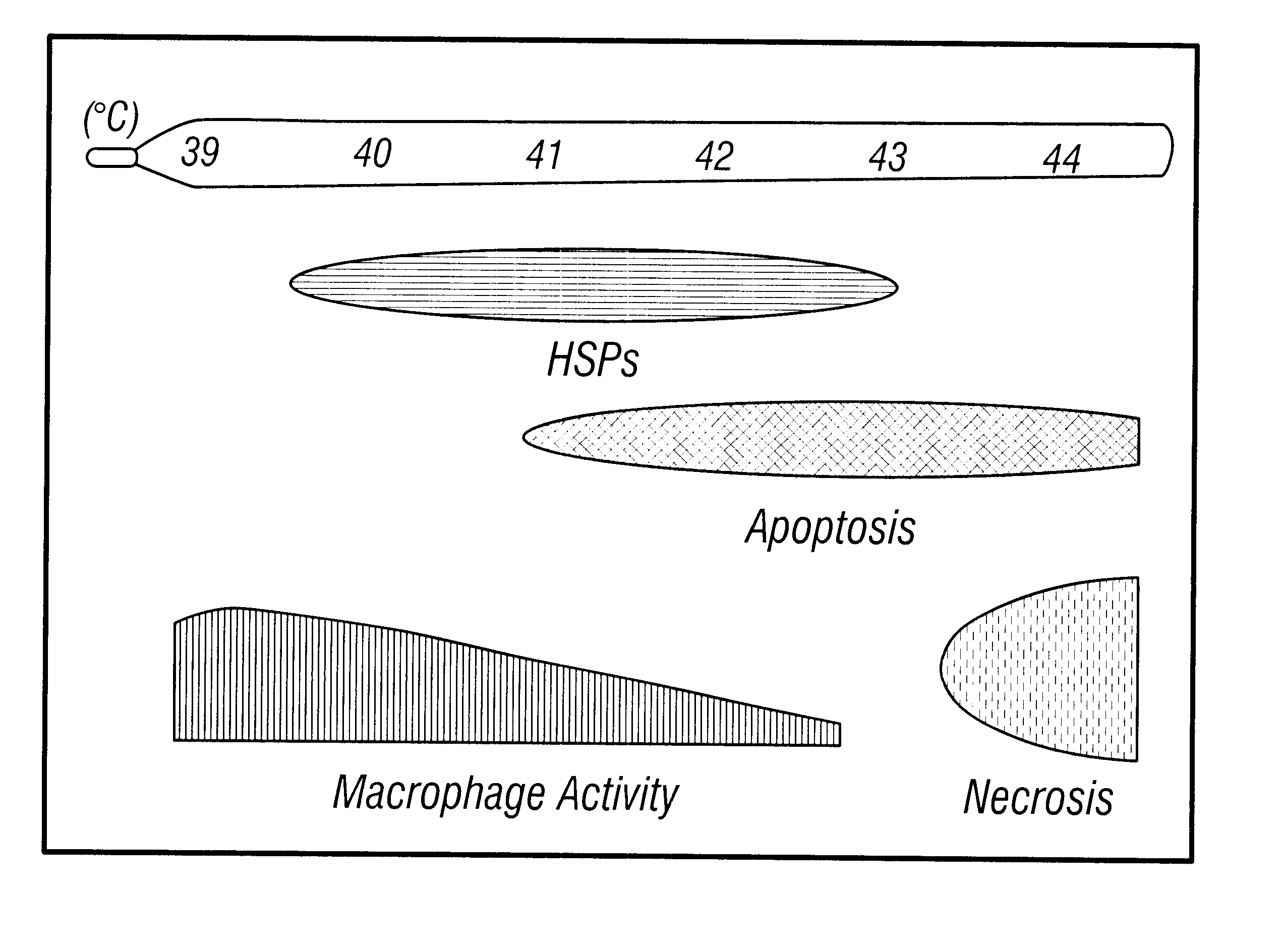
Previously no method was known for selectively inducing
apoptosis in macrophages or other inflammatory cells in a
blood vessel without also inducing
apoptosis in beneficial endothelial cells.
Such methods are not useful as therapeutic methods because of the risk that apoptosis will develop in
healthy tissue.
Preventing the synthesis of
prostaglandin, which serve as feedback inhibitors of macrophage function, limits the anti-inflammatory utility of indomethacin and presumably other inhibitors of
cyclooxygenase.
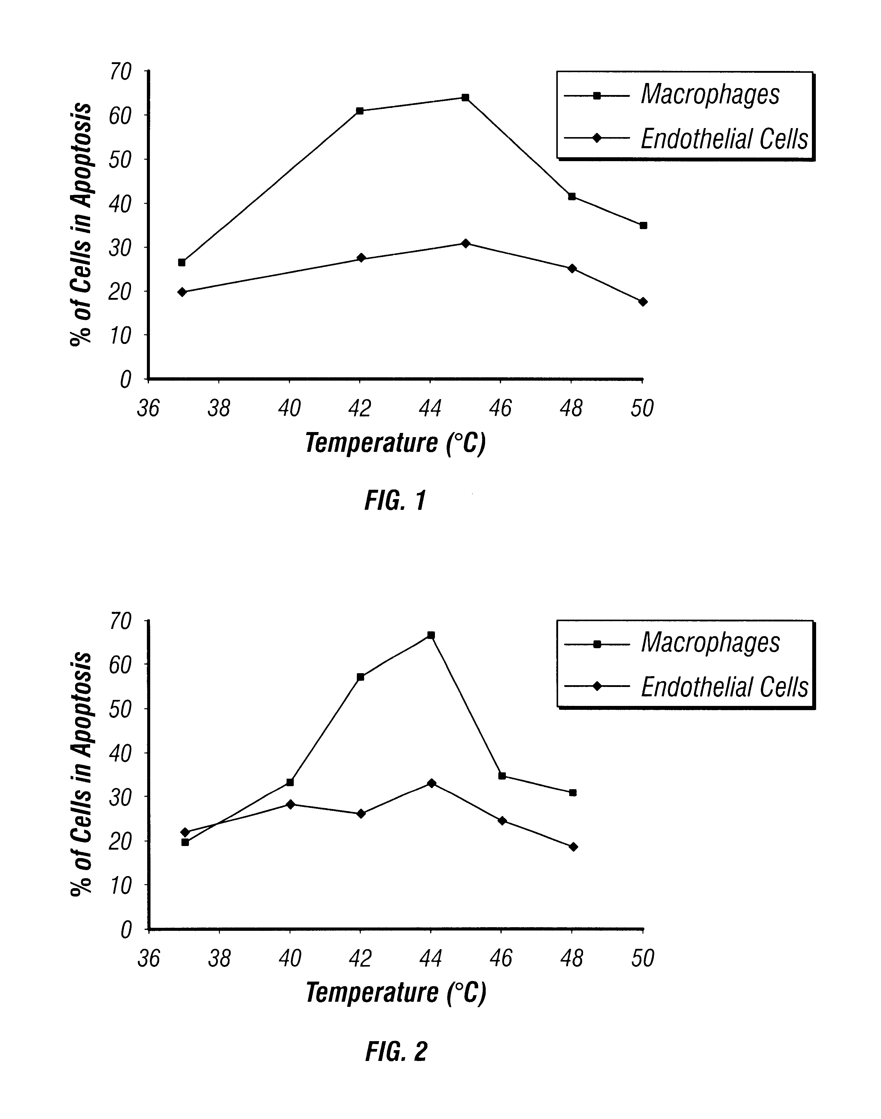
rance. These experiments could not determine whether
hyperthermia increased ATP utilization or inhibited its syn
Although the cellular phenomenon of apoptosis has been studied in some detail in
tissue culture, no studies have been directed toward developing that technique for the treatment of
inflammation.
While various
stent devices have almost always improved short-term results in vessel patency, at the present time it is not yet determined whether any of the many
stent designs and materials have significant advantages over the others.
For example, some stents penetrate the plaque, whereby a gruel-like material is extruded through the strut lattice, provoking an intense thrombotic and
inflammatory response.
Particular problems that have been associated with stents include
thrombus formation and cellular overgrowth.
During
stent placement the
blood vessel wall can be disturbed or injured, and
thrombosis often results at the injured site, causing
stenosis (narrowing) or complete blockage of the vessel.
As a result, the patient is placed at risk of a variety of complications, including heart
attack,
pulmonary embolism, and
stroke, depending on the placement of the stent.
It is well established that stainless steel implants such as pacemakers tend to release
chromium and
nickel ions, which can destroy or damage certain enzymes and proteins and can exacerbate allergies to these metals.
However, a potential problem with the sleeve or sheet type of stent is that blood may not adequately circulate to the vessel wall adjacent the stent.
Typically, these kinds of devices fail to distinguish normal vessel wall from atherosclerotic plaque, however.
For instance, other implanted metallic objects in the body, such as pacemakers, defibrillators or cardiovascular stents, may malfunction or heat up dangerously if exposed to a strong
electromagnetic field.
Additionally, the effects of high magnetic fields on
biological tissue are still not completely understood.
Such fields may cause
ionization of some biomolecules and cellular damage.
Another
disadvantage of techniques requiring high-energy
magnetic field production is that they require facilities that are costly to maintain and are not widely available to the medical
community.
Other methods of heating vessels or other annular organs, such as resistive heating of a fluid-filled
balloon catheter, for example, which require passage of
electricity to the body are inherently difficult to control and can also present a
hazard to the patient.
Moreover, all of the conventional intravascular heating methods are invasive and
catheter-based.
Ultrasound, particularly pulsed
high intensity ultrasound, can quickly cause excessive heating and even burning of
soft tissue in contact with bone or another acoustically reflective object, such as a conventional
metal stent, unless the conditions are carefully controlled.
Even though
ultrasound has been in use both diagnostically and therapeutically for over a decade, and its beneficial results have been attributed to both thermal and non-thermal effects, there has been little research on the effect of treatment dosage on the extent of
tissue heating.
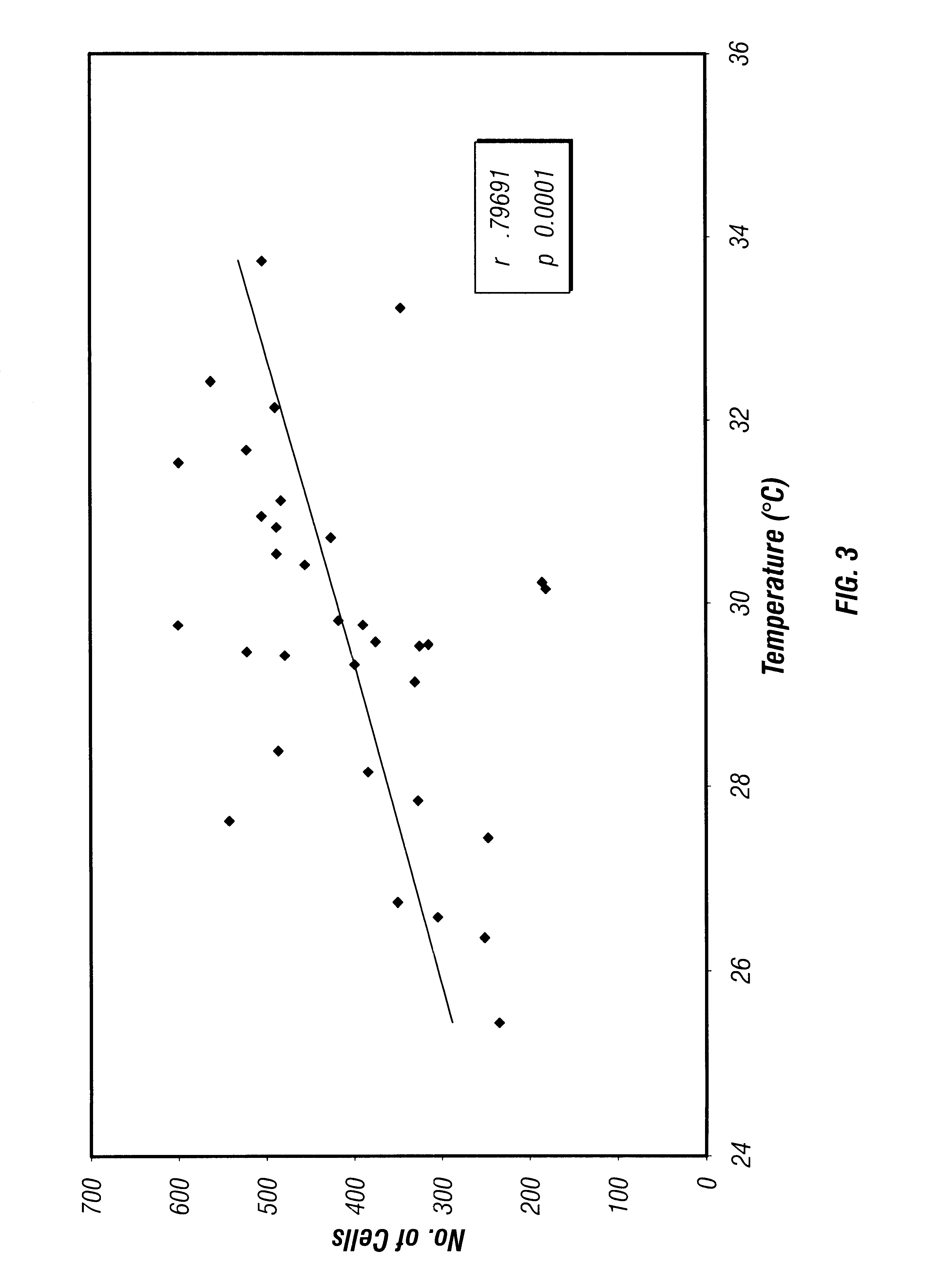
Thermal therapy devices have particular difficulty in establishing and maintaining the desired therapeutic temperature in highly perfused tissues.
Also, blood flowing through an artery makes it very difficult to heat a region of the vessel to a desired temperature.
Oftentimes, considerable damage to intervening tissue also occurs as a consequence of the higher energy
ultrasound required with existing ultrasound
thermal treatment methods.
Conventional
metal stents, when exposed to a focused
high intensity ultrasound beam may cause damage or burning of the surrounding cardiovascular tissue due to reflectance by the metallic surface of the stent.
Catheters that emit heat above about 45.degree. C. are not preferred because the use of such elevated temperatures may damage endothelial cells and produce a secondary
inflammatory response.
Blood flowing through an artery makes it very difficult to heat a region of artery wall to a desired temperature.

