Use of multiphoton excitation through optical fibers for fluorescence spectroscopy in conjunction with optical biopsy needles and endoscopes

a multi-photon excitation, optical fiber technology, applied in the direction of fluorescence/phosphorescence, spectroscopy, instruments, etc., can solve the problems of insufficient volume observed, insufficient angular spreading, and insufficient spatial resolution, etc., to achieve sufficient spatial resolution, convenient resolution, and use the effect of spatial resolution

Inactive Publication Date: 2005-01-04
CORNELL RES FOUNDATION INC
View PDF29 Cites 64 Cited by
  • Summary
  • Abstract
  • Description
  • Claims
  • Application Information

AI Technical Summary

Benefits of technology

An analogous advantage applies to fluorescence multiphoton excited by laser light transmitted through an optical fiber as in application to endoscopic tissue fluorescence. Although the illumination intensity fans out just as for one-photon excitation with roughly equal total power at each value of radius from the end of the fiber or focal plane of any focusing lens, the excited fluorescence does not follow this fanned out illumination. In the case of two-photon excitation through a single mode optical fiber, the distribution of fluorescence is localized near the fiber tip in a shape resembling a candle flame beginning at the fiber tip. This effective focal volume is defined by the spatial distribution of the square of the illumination intensity for two-photon excitation. The square of the illumination intensity falls off roughly as the reciprocal fourth power of the distance from the fiber tip so that the fluorescence excitation is localized. Significant fluorescence is emitted only from this limited volume where the square of the excitation intensity is large, as illustrated in FIG. 3. Lenses, prisms, etc. can be used to shift the effective focal volume beyond the end of the optical fiber and/or to provide a side-looking orientation without losing the advantages of multiphoton excitation.
This effect provides a well-defined focal volume for the fluorescence excitati

Problems solved by technology

Unfortunately, these clues are frequently not sufficient, especially for detection of the early onset of disease.
A first problem is that this ultraviolet light is strongly absorbed by hemoglobin and oxyhemoglobin in the blood, which are not fluorescent, so that penetration of the illumination into the tissue depends on their concentration and distribution.
A second problem is that the illumination exiting the optical fibers into tissue fans out at an included angle determined by the numerical aperture (NA)

Method used


Image

  • Use of multiphoton excitation through optical fibers for fluorescence spectroscopy in conjunction with optical biopsy needles and endoscopes
  • Use of multiphoton excitation through optical fibers for fluorescence spectroscopy in conjunction with optical biopsy needles and endoscopes
  • Use of multiphoton excitation through optical fibers for fluorescence spectroscopy in conjunction with optical biopsy needles and endoscopes

Examples


Embodiment Construction

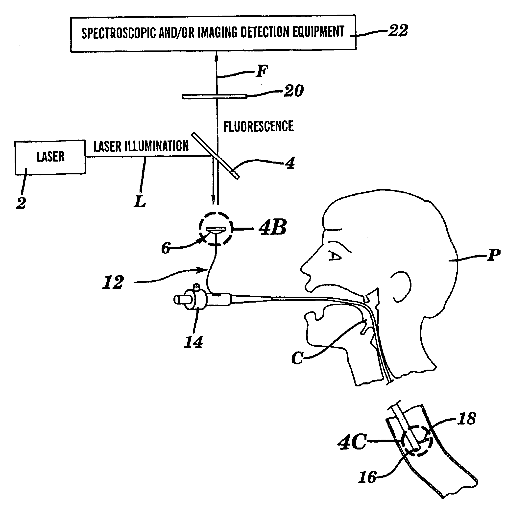
One aspect of the present invention relates to a method of detecting disease within a particular tissue of a plant or animal. This method involves activating the particular plant or animal tissue by application of radiation through at least one optical fiber under conditions effective to promote a simultaneous multiphoton excitation of the particular plant or animal tissue and to emit an intrinsic fluorescence characteristic. The intrinsic fluorescence characteristic is compared to fluorescence emitted by exciting healthy tissue of the particular plant or animal under the same conditions used to carry out the activating step. The particular tissue of a plant or animal where the intrinsic fluorescence characteristic differs from the fluorescence, emitted by exciting healthy tissue of the particular plant or animal under the same conditions used to carry out such activity, is then identified as potentially diseased.

Another aspect of the present invention involves a method of producing...


PUM


Abstract

The present invention is directed to a method of applying radiation through an optical fiber for detecting disease within a plant or animal or imaging a particular tissue of a plant or animal. In addition, fluorescence can be detected and localized within a subject by such application of radiation through an optical fiber. The radiation is effective to promote simultaneous multiphoton excitation. The optical fibers are used alone to examine internal regions of tissue, in conjunction with an optical biopsy needle to evaluate sub-surface tissue, or with an endoscope to evaluate tissue within body cavities.

Description

FIELD OF THE INVENTIONThe present invention relates to the use of multiphoton excitation in conjunction with optical biopsy needles and endoscopes.BACKGROUND OF THE INVENTIONMany crucial biological functions are mediated or accomplished by biomolecules and tissue structures that are intrinsically fluorescent. As a result, there is an opportunity to diagnose and study important biological events by measuring and localizing the spectra and tissue fluorescence emission. To investigate in vivo internal processes and structures in large organisms, such as human beings and agricultural animals, endoscopic procedures which penetrates body cavities or even solid tissue may be required.Endoscopy video imaging in body cavities ordinarily utilizes back-scattered white light applied through the endoscope to form a low-resolution color image of the internal surfaces of these cavities. Physicians often use the changes in shapes and changes in local apparent color (which are often due to changes i...

Claims


Application Information

Patent Timeline
IPC IPC(8): A61B5/00G01N21/64A61B1/06A61B1/07A61B1/00G02B23/26
CPCA61B5/0071A61B5/0075A61B5/0084A61B5/0086G01N21/64G01N21/645G01N21/6486A61B5/6848A61B1/043A61B1/07G01N2021/6484G01N21/6458G01N2021/6417
Inventor WEBB, WATT W.
Owner CORNELL RES FOUNDATION INC
Why Eureka
Try Eureka