Rapid cell block embedding method and apparatus

a cell block and cell block technology, applied in the field of rapid cell block embedding method and apparatus, can solve the problems of inefficient showing diagnostic cells in the final microtome section, inconvenient processing, and high cost, and achieves the effect of reducing the amount of cellular sample required, maximizing cell recovery and extraction efficiency, and minimizing processing tim

Active Publication Date: 2005-07-05
MASSACHUSETTS UNIV OF
View PDF19 Cites 97 Cited by
  • Summary
  • Abstract
  • Description
  • Claims
  • Application Information

AI Technical Summary

Benefits of technology

[0015]The present invention provides a method and apparatus that utilizes a flow-through processing and embedding technique for rapidly embedding cells. This flow-through processing maximizes the efficiency of cell recovery and of extractions during embedding, thereby decreasing the amount of cellular sample required, minimizing the amount of time for processing, and minimizing the amount of reagents needed for embedding. The method and appar...

Problems solved by technology

However, the collodion technique is substantially more dangerous than the clot technique due to the flammable nature of collodion and its ether solvent.
Currently available cell block preparation techniques such as the ones previously described suffer from a number of problems that make them cumbersome, costly, susceptible to contamination or mislabeling, and inefficient for showing diagnostic cells in the final microtome sections.
This manual transfer to the tissue cassette takes a considerable amount of time, as does the step of transferring the cells from a sample tube into a tissue processor.
Many of the cell fragments are wasted during the transfer and/or embedding steps.
In the past, techniques attempting to improve the concentration of cells within the sectionable material and avoid cell waste have proven less than ideal, because the techniques necessarily involve dilution of the microtome section with carrier substances such that relatively few cells are present, or do not decrease cell loss in pre-embedding steps.
Current cell block techniques typically take anywhere from 8 to 16 hours to complete because the extraction of water, alcohol, fats, and xylene in the tissue processor is time consuming.
While there are methods to speed up the tissue processing step (step 6 above), such as by using microwave radiation, vacuum pressure, elevated temperatures, and more rapidly diffusing chemicals, these methods suffer from their own set of problems.
These techniques only modestly decrease the tissue processing time (step 6 above) but are relatively tedious to establish in the laboratory.
Furthermore, these techniques do not enhance the efficiency of the other processing steps.
Another drawback with the cell block techniques described above are that they are also susceptible to mislabeling of a sample since the sample has to be manually moved between the sample tube, lens paper (or collodion, and other carrier substances), tissue cassette, and tissue mold.
Si...

Method used


Image

  • Rapid cell block embedding method and apparatus
  • Rapid cell block embedding method and apparatus
  • Rapid cell block embedding method and apparatus

Examples


Embodiment Construction

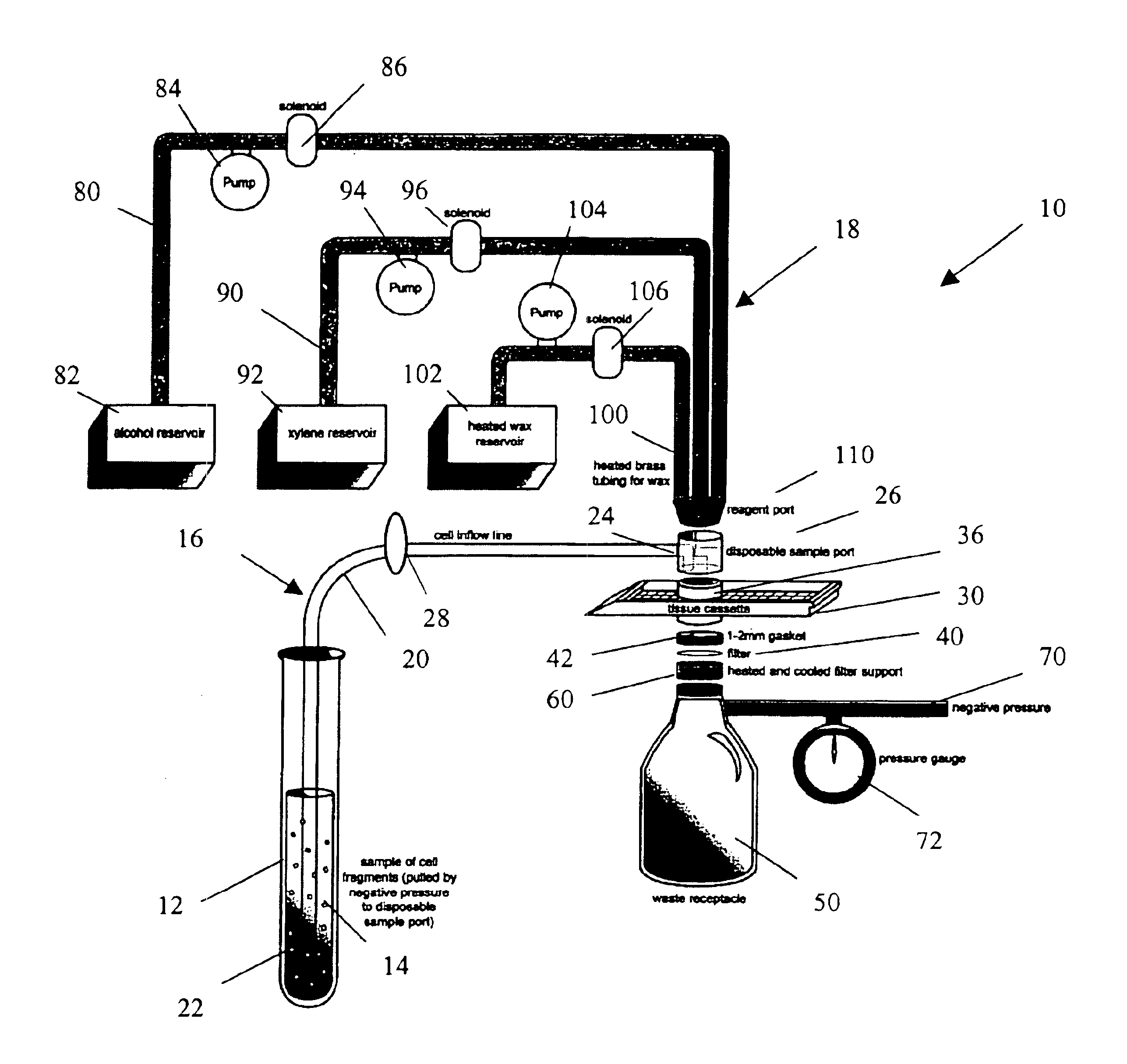
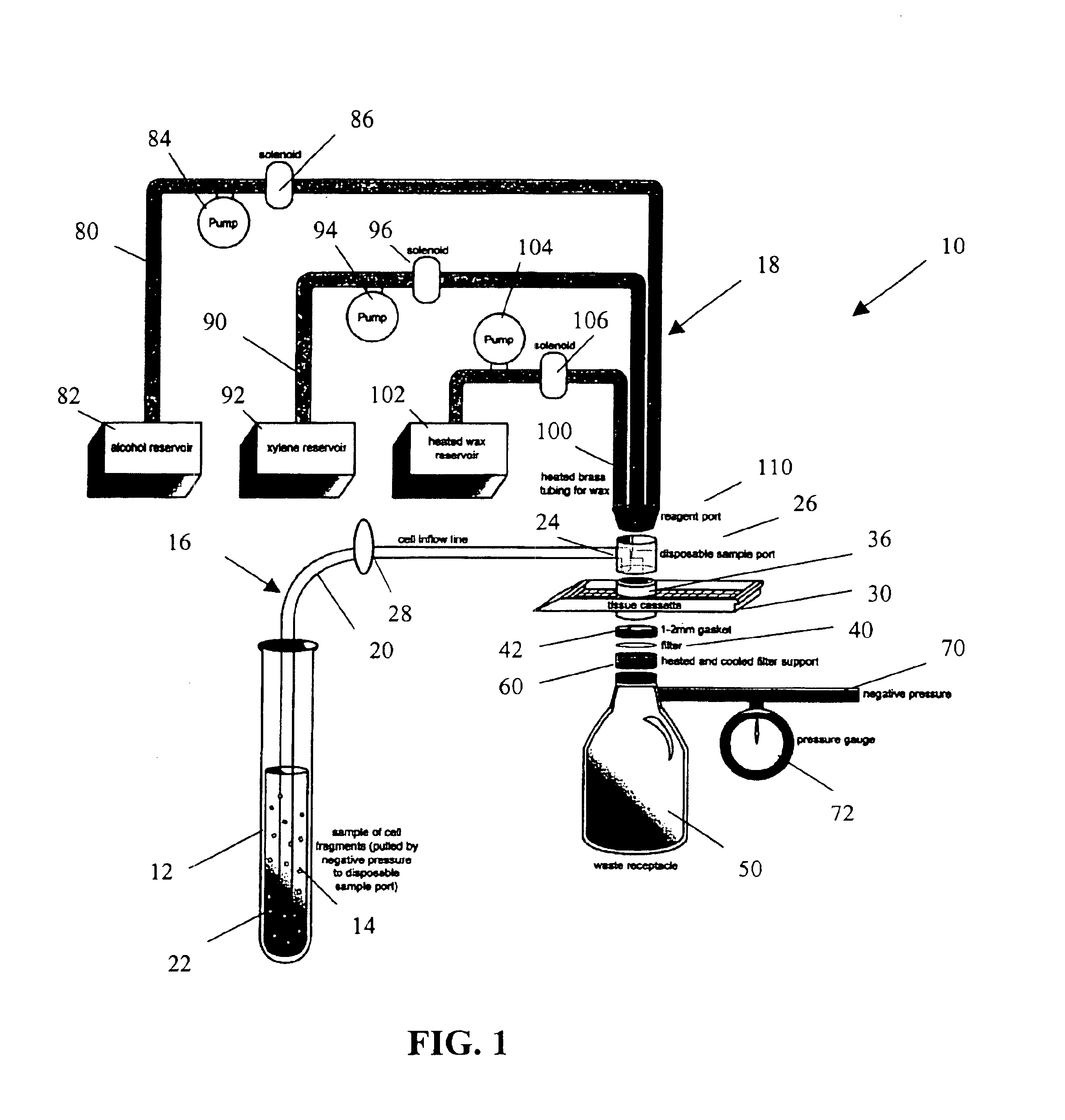
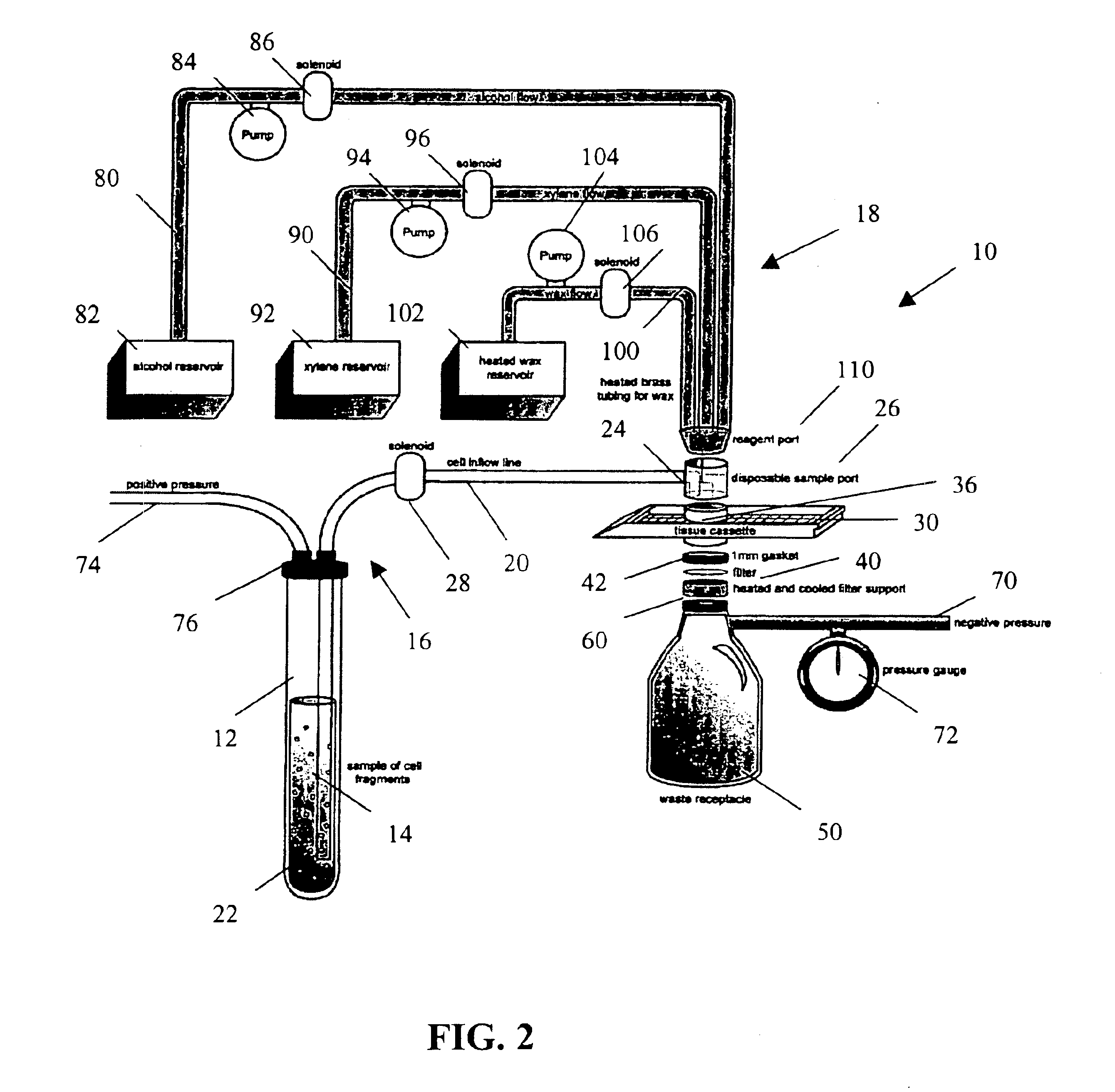
[0025]The present invention provides a method and apparatus for rapidly preparing a cell block for sectioning with a microtome. The cell block embedding apparatus 10 of the present invention is designed as a flow-through system. As shown in detail in FIG. 1, the apparatus 10 comprises a cell flow pathway 16 defined by an inflow tube 20 which has an inlet end 22 configured for placement within a container 12 holding a cell sample 14 therein and an outlet end 24 which removably connects to a disposable sample port 26. A solenoid tube clamp 28 can be disposed along the inflow tube 20 for regulating the flow of fluid through the tube 20. The container 12 can comprise any suitable shape, form, or size. For example, and as illustrated, the container 12 can be a standard 50 ml disposable centrifuge tube. The cell sample 14 can be in suspension in a physiologic saline solution such as Hanks buffered saline, or in a liquid preservative such as 50% aqueous alcohol. The cell suspension can be ...


PUM


Abstract

A method and apparatus for embedding cells that utilizes a flow-through embedding technique maximizes the efficiency of extractions and decreases time for embedding the cell fragments, minimizes cell loss, and automatically positions cell samples at the position in which a microtome blade will section them. The apparatus includes a cell flow pathway defined by an inflow tube for delivering cell fragments from a cell sample to a sample port. The sample port is in fluid communication with a tissue cassette having attached thereto a filter. The cell flow pathway is in communication with a reagent flow pathway for delivering the reagents through the sample port to the cassette. The apparatus is configured such that the application of pressure directs the cell fragments from the cell sample through the cell flow pathway, and effects delivery of the reagents through the reagent flow pathway. The apparatus produces an embedded cell block having concentrated cells near the plane of the block to be sectioned in a quick and efficient manner.

Description

CROSS-REFERENCE TO RELATED APPLICATIONS[0001]This application is based on U.S. provisional application No. 60 / 422,768 entitled “Automated Cell Block Embedding Apparatus” and filed on Oct. 31, 2002, of which priority is claimed.FIELD OF THE INVENTION[0002]The present invention relates to methods and devices for preparing cells for microscopic examination, and for archiving the cells for molecular and immunologic tests. More particularly, the invention relates to a method and apparatus for embedding cells and tissue fragments within a solid substrate, such as paraffin, to enable them to be cut with a microtome for microscopic evaluation, and to be stored in a stable state.BACKGROUND[0003]Many disease processes can only be diagnosed on the basis of histologic or cytologic examination using a light microscope. For instance, while the presence of a tumor can be detected using radiological devices, the determination of whether a tumor is benign or malignant still requires a pathologist's ...

Claims


Application Information

Patent Timeline
IPC IPC(8): C12M1/34C12M1/12G01N1/30G01N33/48B01L3/00G01N1/31G01N1/36
CPCB01L3/502G01N1/36B01L2300/0681B01L2300/0877B01L2400/0487G01N2001/315
Inventor FISCHER, ANDREW H.
Owner MASSACHUSETTS UNIV OF
Why Eureka
Try Eureka