Yeast cell surface display of proteins and uses thereof

Inactive Publication Date: 2008-12-16
THE BOARD OF TRUSTEES OF THE UNIV OF ILLINOIS
View PDF85 Cites 39 Cited by
  • Summary
  • Abstract
  • Description
  • Claims
  • Application Information

AI Technical Summary

Benefits of technology

[0049]FIG. 20 shows the flow cytometry profiles of antibody binding to yeast that have been transformed with double mutants from the selection shown in FIG. 19. Cells were stained for flow cytometry as described in FIG. 19. Double

Problems solved by technology

Although panning of antibody phage display libraries is a powerful technology, it possesses several intrinsic difficulties that limit its wide-spread successful application.
First, very high affinity antibodies (KD≦1 nM) are difficult to isolate by panning, since the elution conditions required to break a very strong antibody-antigen interaction are generally harsh enough (e.g., low pH, high salt) to denature the phage particle sufficiently to render it non-infective.
Secondly, the requirement for physical immobilization of an antigen to a solid surface produces many artifactual difficulties.
Also, physical tethering reduces the translational and rotational entropy of the antigen, resulting in a smaller ΔS upon antibody binding and a resultant overestimate of binding affinity relative to that for soluble antigen and large effects from variability in mixing and washing procedures lead to difficulties with reproducibility.
Thirdly, the presence of only one to a few antibodies per phage particle introduces substantial stochastic variation, and discrimination between antibodies of similar affinity becomes impossible.
Display of antibodies on the surface of Escherichia coli has been developed as an alternative methodology solving several of the problems associated with phage display (Francisco, et al., '93), but introduces new

Method used


Image

  • Yeast cell surface display of proteins and uses thereof
  • Yeast cell surface display of proteins and uses thereof
  • Yeast cell surface display of proteins and uses thereof

Examples


example 1

Media / Buffers

[0094]The following media / buffers were used herein:

E. Coli

[0095]LB media (1×): Bacto tryptone (Difco, Detroit, Mich.): 10.0 g; Bacto yeast extract (Difco): 5.0 g; NaCl:10.0 g. Make up to 1 L, autoclave. For plates, add 15 cg / L Agar and autoclave.

Ampicillin

[0096]Stock: 25 mg / ml of sodium salt of ampicillin in water. Filter sterilize and store in aliquots of 4 mls at −20° C. Working [ ] 35-50 μg / ml; 4 ml of aliquot in 1 L->100 μg / ml; 2 ml of aliquot in 1 L->50 μg / ml. Add to autoclaved LB only after it has cooled to ˜55° C.

[0097]

SOC media100 mL

[0098]2% Bacto tryptone: 2.0 g; 0.5% Yeast Extract (Difco):0.5 g; 10 mM NaCl: 0.2 ml 5 M; 10 mM MgCl2: 1.0 ml 1 M; 10 mM MgSO4: 1.0 ml 1 M; 20 mM Dextrose: 0.36 g. Autoclave or filter sterilize.

[0099]

YEASTSynthetic Minimal + Casamino acids (SD-CAA)500mLDextrose (Glucose)10.00gYeast Nitrogen Base w / o Amino Acids (Difco)3.35gNa2HPO4•7H2O5.1gNaH2PO4•H2O4.28gCasamino Acids (Trp-, Ura-) (Difco)2.5g

[0100]Add dH2O to final volume. Filter s...

example 2

Protocol: Replica Plating

[0110]1. Choose a material suitable for colony lifts, making sure it is washed, dried and sterile.

[0111]2. Mark the bottom of each fresh replica plate with an arrow to line up the plate. Also mark the starting plate.

[0112]3. Take the top off the starting plate, turn it upside down and line up the arrow on the bottom with the mark on the colony lift material. Lay the plate down onto the surface of the material and gently put pressure on the entire plate. Make sure the plate doesn't move around after it has touched the material. Remove the plate and replace the lid. Portions of the colonies that transferred to the material can be seen.

[0113]4. Repeat this procedure with one of the fresh replica plates. Make sure the arrow lines up with the mark also to make an exact replica. Hold up to the light to see the tiny colonies that transferred.

[0114]5. Repeat the entire procedure for each replica plate to be made.

[0115]6. Incubate the replica plates at the appropriat...

example 3

Protocol: Electrotransformation of Yeast

[0116]The cell preparation procedure was as follows: Step 1. Inoculate 50 ml of YPD with an overnight culture to an OD of 0.1. Step 2. Grow cells at 30° C. with vigorous shaking to an OD600 of 1.3 to 1.5 (approximately 6 hours). Step 3. Harvest in cold rotor at 3500 rpm for 5 minutes at 4° C. Discard supernatant. Step 4. Thoroughly wash the cells by resuspending in 50 ml cold sterile water. Centrifuge as above and discard supernatant. Step 5. Repeat step 4 with 25 ml cold water. Step 6. Resuspend in 2 ml of ice-cold sterile 1 M sorbitol. Centrifuge as above and discard supernatant. Step 7. Resuspend in 50 ml ice-cold 1 M sorbitol. Final volume of cells is about 150 ml (enough for 3 transformations).

Electrotransformation:

[0117]1. Place 0.2 cm cuvettes and white slide chamber on ice.

[0118]2. In an eppendorf tube, add 50 ml of yeast suspension and gently mix in <5 ml (0.1 mg) of plasmid DNA in TE. Make sure to add DNA to yeast already in eppendor...


PUM

PropertyMeasurementUnit
Fractionaaaaaaaaaa
Fractionaaaaaaaaaa
Fractionaaaaaaaaaa

Abstract

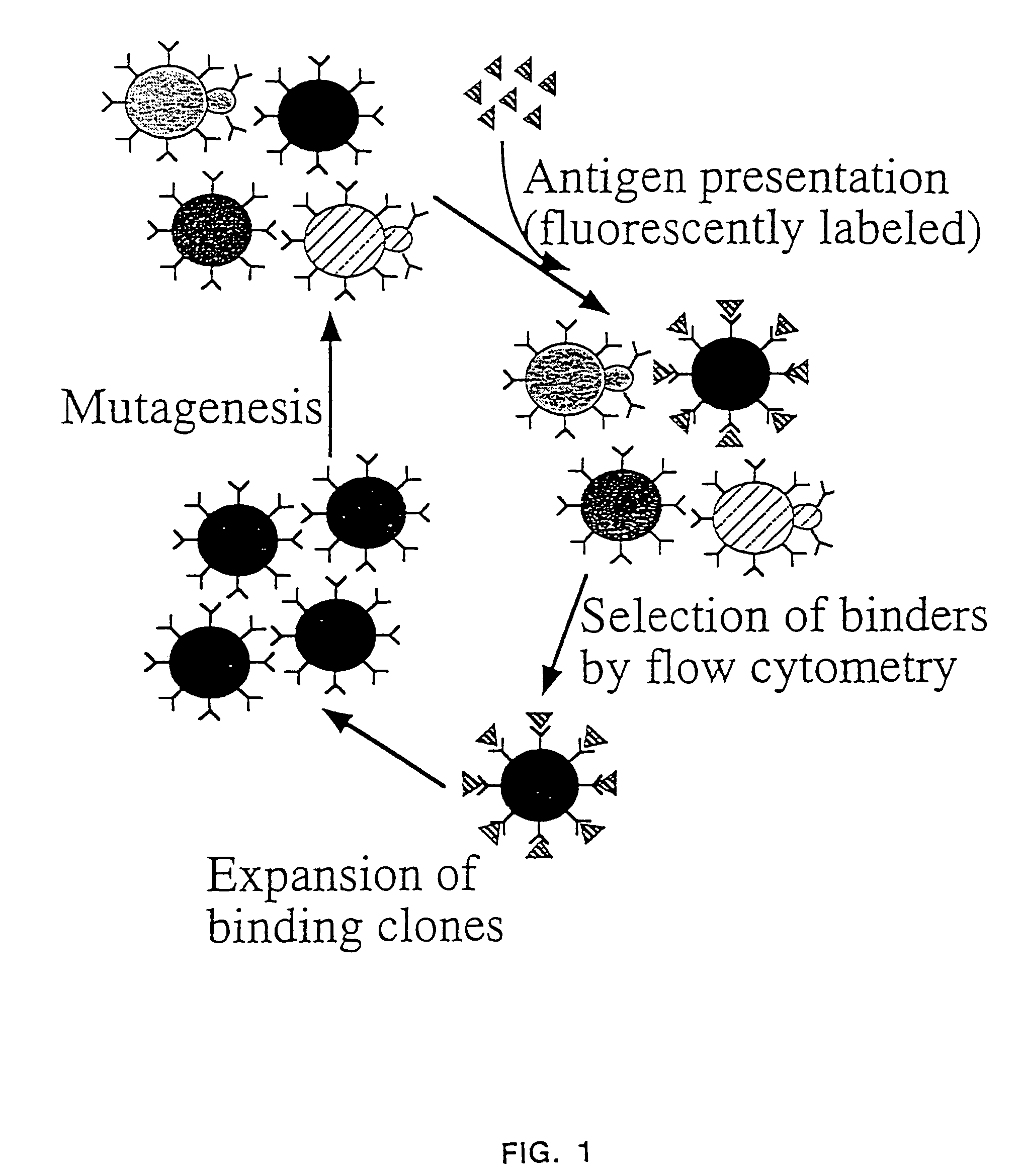
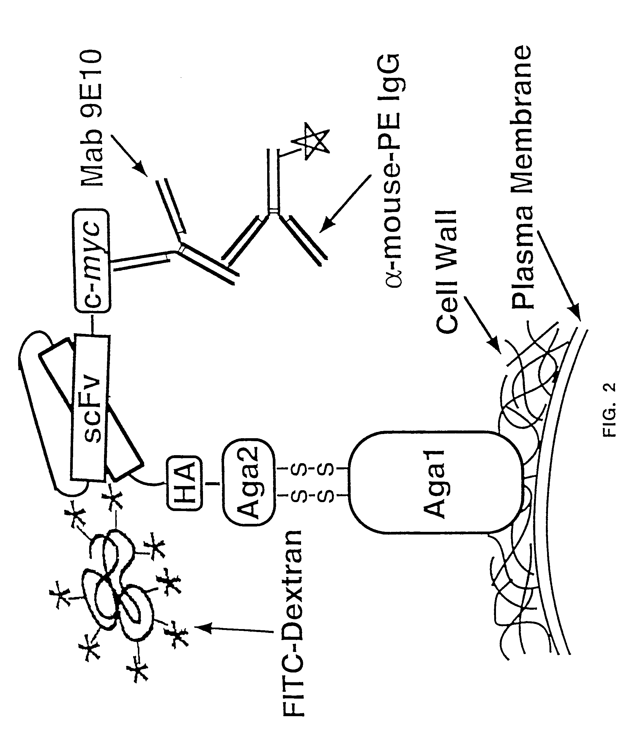
The present invention provides a genetic method for tethering polypeptides to the yeast cell wall in a form accessible for binding to macromolecules. Combining this method with fluorescence-activated cell sorting provides a means of selecting proteins with increased or decreased affinity for another molecule, altered specificity, or conditional binding. Also provided is a method for genetic fusion of the N terminus of a polypeptide of interest to the C-terminus of the yeast Aga2p cell wall protein. The outer wall of each yeast cell can display approximately 10 protein agglutinins. The native agglutinins serve as specific adhesion contacts to fuse yeast cells of opposite mating type during mating. In effect, yeast has evolved a platform for protein-protein binding without steric hindrance from cell wall components.

Description

CROSS-REFERENCE TO RELATED APPLICATIONS[0001]This application is a divisional of U.S. Ser. No. 09 / 724,108, filed Nov. 28, 2000, which is a continuation of U.S. Ser. No. 09 / 009,388, filed Jan. 20, 1998, which is a continuation-in-part of U.S. Ser. No. 08 / 866,398, filed May 30, 1997, now abandoned, which claims benefit of 60 / 018,741, filed May 31, 1996.FEDERAL FUNDING LEGEND[0002]This invention was produced in part using funds obtained through a grant from the National Institutes of Health grant R01AI35990. Consequently, the federal government has certain rights in this invention.BACKGROUND OF THE INVENTION[0003]1. Field of the Invention[0004]The present invention relates generally to the fields of immunology and protein chemistry. More specifically, the present invention relates to the display of peptides and proteins on the yeast cell surface for selection of sequences with desirable binding properties from combinatorial libraries.[0005]2. Description of the Related Art[0006]Antibod...

Claims


Application Information

Patent Timeline
IPC IPC(8): C07K14/37C07K14/39C07K16/28C07K16/46C07K17/00C07K17/02C07K17/06C12N1/19C12N15/63C12N15/64C12Q1/02C12Q1/68C12N1/15C12N15/10C12N15/81C40B40/02G01N33/68
CPCC12N15/1037C40B40/02G01N33/68G01N33/6854C07K14/705G01N2333/39
Inventor WITTRUP, K. DANEKRANZ, DAVID M.KIEKE, MICHELEBODER, ERIC T.
Owner THE BOARD OF TRUSTEES OF THE UNIV OF ILLINOIS
Why Eureka
Try Eureka