Tissue engineered osteochondral composite stent and preparation method thereof

A technology of tissue engineering bone and composite scaffold, applied in bone implants, medical science, prosthesis, etc., can solve the problem of low hyaline cartilage ratio, insufficient hydrophilicity and compatibility of osteocartilage scaffold, fibrin glue, etc. Weak mechanical properties and other problems, to achieve the effect of increasing the proportion of hyaline cartilage, improving hydrophilicity and collagen distribution uniformity, and increasing the density and depth of cell planting

Active Publication Date: 2013-01-09
TSINGHUA UNIV
View PDF4 Cites 20 Cited by
  • Summary
  • Abstract
  • Description
  • Claims
  • Application Information

AI Technical Summary

Problems solved by technology

[0008] 1. When using tissue engineering to simply repair articular cartilage defects, the healing interface between the graft body and the transplantation bed is the cartilage-cartilage and cartilage-bone interface. The integration of these two interfaces is slow, and the new tissue is formed on the surface of the transplantation bed during the repair process. Unstable, prone to repair failure
[0009] 2. At present, in the tissue engineering repair process, the proportion of hyaline cartilage in the cartilage tissue is relatively small, and the physiological functions and mechanical properties of the composite tissue cannot be maintained for a long time. The materials and construction methods of the cartilage scaffold are not bionic enough. The proportion of hyaline cartilage in cartilage tissue is small, and the physiological function and mechanical properties of the composite tissue cannot be maintained for a long time
[0010] 3. The hydrophilicity and compatibility of osteochondral scaffolds are not ideal enough. At present, the met

Method used

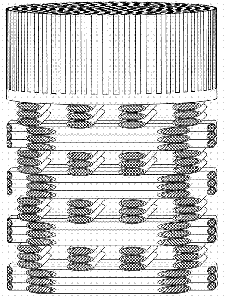
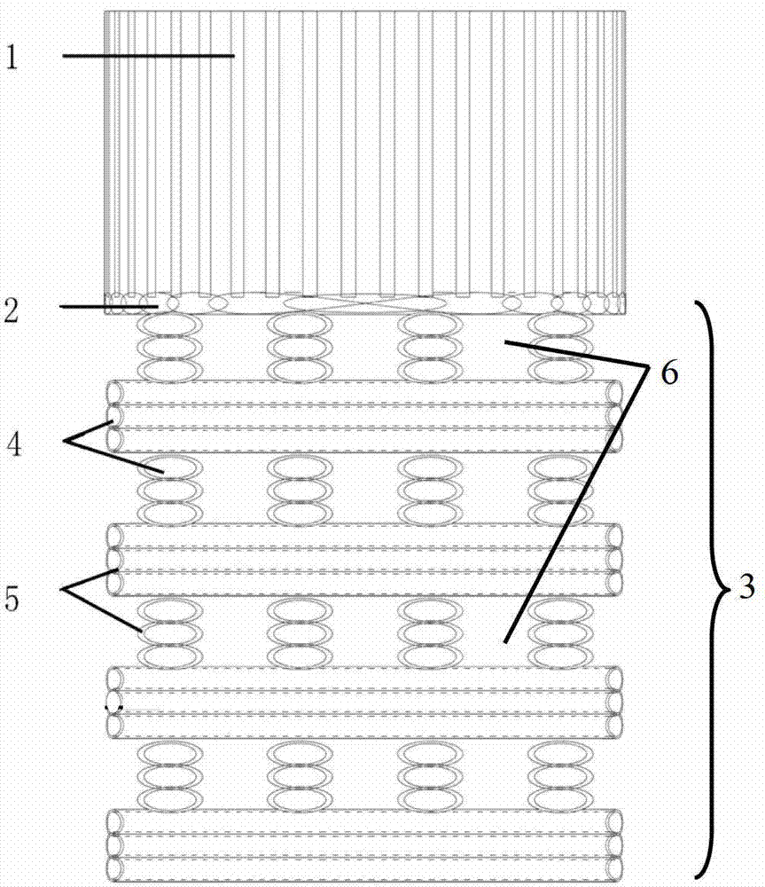
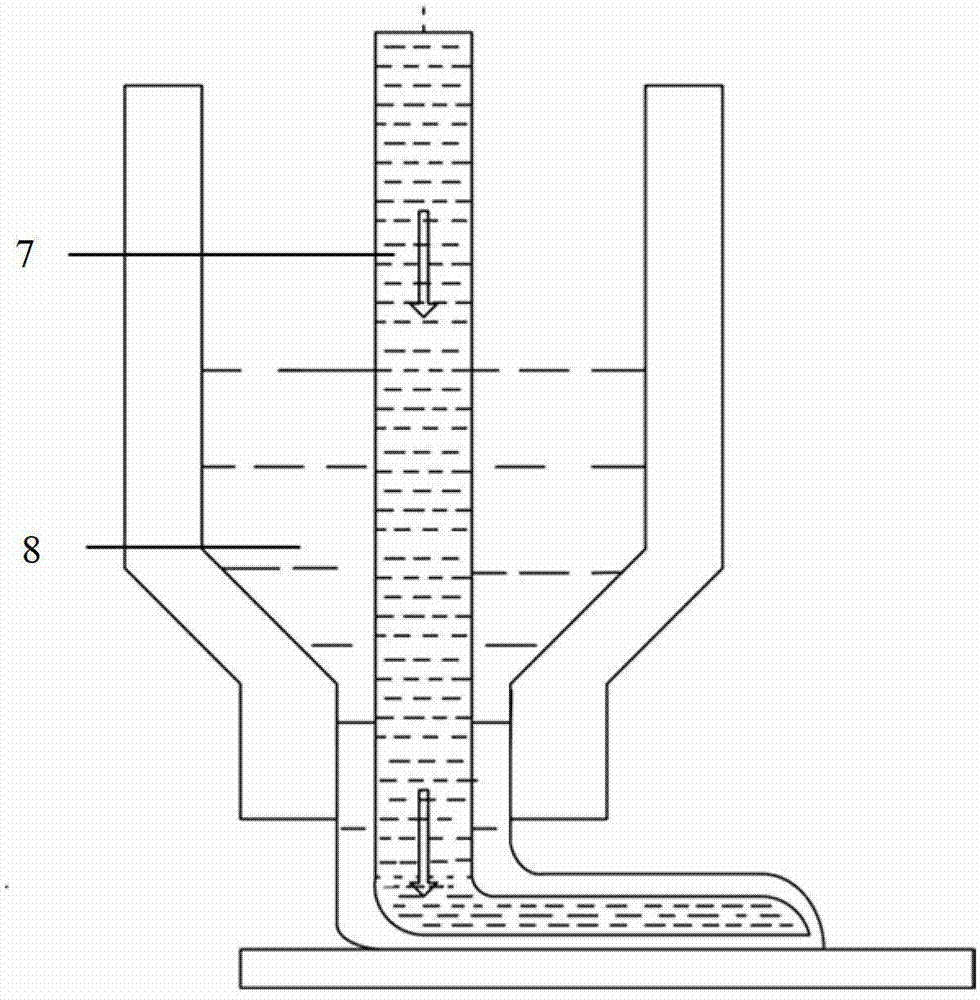

Image

  • Tissue engineered osteochondral composite stent and preparation method thereof
  • Tissue engineered osteochondral composite stent and preparation method thereof
  • Tissue engineered osteochondral composite stent and preparation method thereof

Examples


Embodiment 1

[0046] Example 1: Construction of Tissue Engineering Osteochondral Scaffold

[0047] The cartilage matrix material and chitosan extracted from the bovine bone source are dissolved in 0.5% acetic acid solution to prepare a cartilage matrix material-chitosan mixed solution with a mass volume concentration of 10mg / ml. In the solution, the cartilage matrix material and chitosan The mass ratio is 0.5:1. Put the mixed solution into the mold and build a temperature gradient of 10°C / mm in a low temperature environment of -200°C. The mixed solution was solidified and then freeze-dried for 24 hours to obtain a cartilage scaffold with an oriented structure. The scaffold was cross-linked with a genipin solution and adjusted to Neutral, ready for use after freeze-drying. Mix PLGA / tricalcium phosphate at a mass ratio of 5:1 and dissolve in 1,4-dioxane to form a solution with a concentration of 500mg / ml, and dissolve type I collagen in 0.5% acetic acid solution to form a concentration of 2m...

Embodiment 2

[0048] Example 2: Construction of Tissue Engineering Osteochondral Scaffold

[0049] The cartilage matrix material and chitosan extracted from bovine bone are dissolved in 3% acetic acid solution to prepare a cartilage matrix material-chitosan mixed solution with a concentration of 50 mg / ml. The mass ratio of cartilage matrix material to sodium alginate in the solution is It is 5:1. Put the mixed solution into the mold and build a temperature gradient of 10°C / mm in a low temperature environment of -80°C. After the mixed solution is solidified and freeze-dried for 24 hours to obtain a cartilage scaffold with an oriented structure, the scaffold is cross-linked and adjusted with sodium polyphosphate solution It is neutral and ready for use after freeze-drying. PLGA / tricalcium phosphate was mixed and dissolved in chloroform at a mass ratio of 10:1 to form a solution with a concentration of 50 mg / ml, and type I collagen was dissolved in 0.5% acetic acid solution to form a collagen...

Embodiment 3

[0050] Example 3: Tissue-engineered osteochondral composite scaffold used for defect repair of rabbit articular cartilage

[0051] The cartilage matrix material and chitosan extracted from bovine bone are dissolved in 0.5% acetic acid solution to prepare a cartilage matrix material-chitosan mixed solution with a concentration of 20mg / ml. The mass ratio of cartilage matrix material and gelatin in the solution is 1 :1. Put the mixed solution into the mold and build a temperature gradient of 10°C / mm in a low temperature environment of -200°C. The mixed solution was solidified and then freeze-dried for 24 hours to obtain a cartilage scaffold with an oriented structure. The scaffold was cross-linked with a genipin solution and adjusted to Neutral, ready for use after freeze-drying. Mix PLGA / tricalcium phosphate at a mass ratio of 5:1 and dissolve in 1,4-dioxane to form a solution with a concentration of 200mg / ml, and dissolve type I collagen in 0.5% acetic acid solution to form a ...


PUM

PropertyMeasurementUnit
Diameteraaaaaaaaaa
Diameteraaaaaaaaaa
Diameteraaaaaaaaaa

Abstract

A tissue engineered osteochondral cartilage composite stent and a preparation method thereof belong to the technical field of tissue engineering and bio-manufacturing. The preparation method comprises the following steps of: simulating natural materials and structures of osteochondral cartilage; preparing a cartilage stent with an oriented structure by applying the principle of oriented crystallization; preparing a core-spun structural stent by applying a low-temperature deposition manufacturing process based on the rapid forming technique and designing a special extrusion and injection device with a core-spun sprayer; and connecting the oriented structural stent and the core-spun structural stent in a dissolution-adhesion method so as to construct the tissue engineered osteochondral cartilage composite stent with the structure of oriented structural cartilage and core-spun structural bone. According to the tissue engineered osteochondral cartilage composite stent and the preparation method thereof, with respect to the defect treatment of articular cartilages, a material with biological activity is adopted and the osteochondral cartilage composite stent with a special bionic structure is manufactured; the cell implanting density and depth of the stent can be improved, and the simultaneous regeneration of bones and cartilage tissues can be promoted with respect to the different functional regional characteristics of the osteochondral cartilage, so that the repairing effect of osteochondral cartilage defects is improved.

Description

technical field [0001] The invention relates to a preparation method of a tissue-engineered osteochondral composite bracket, which belongs to the technical field of tissue engineering and biomanufacturing. Background technique [0002] Articular cartilage is a specialized connective tissue with biophysical properties such as toughness, good elasticity, and low coefficient of friction. Trauma and joint inflammation can easily lead to articular cartilage defects and affect the stability of the joints, resulting in the occurrence of osteoarthritis and joint degenerative lesions. Such lesions can not only make people lose their ability to work, but have even become one of the main causes of physical disabilities. one. [0003] The osteochondral complex tissue at the joint end of adult can be divided into hyaline cartilage layer, calcified layer and subchondral bone from the depth direction of the joint. Each layer is connected with a unique composition and structure and perform...

Claims


Application Information

Patent Timeline
IPC IPC(8): A61F2/28A61L27/36A61L27/26A61L27/12
CPCA61F2/30756A61F2002/30001
Inventor 熊卓张婷刘建孟国林张腊全林峰孙伟李丹
Owner TSINGHUA UNIV
Why Eureka
Try Eureka