Caspase 3-sensitive fluorescence enhancement nano developer and preparation method thereof

A caspase and fluorescence enhancement technology, applied in fluorescence/phosphorescence, material excitation analysis, peptide, etc., can solve problems such as poor stability, short excitation light wavelength, complex preparation, etc., and achieve good hydrophilicity and high sensitivity , good biocompatibility

Inactive Publication Date: 2013-10-09
UNIV OF SCI & TECH OF CHINA
View PDF2 Cites 9 Cited by
  • Summary
  • Abstract
  • Description
  • Claims
  • Application Information

AI Technical Summary

Problems solved by technology

[0004] The purpose of the present invention is to propose a fluorescence-enhanced nano-imaging agent sensitive to caspase 3 and its preparation method, so as to obtain a fluorescence-enhanced nano-imaging agent that can be used to detect the specific enzyme activity of apoptosis and the apoptotic state of l...

Method used


Image

  • Caspase 3-sensitive fluorescence enhancement nano developer and preparation method thereof
  • Caspase 3-sensitive fluorescence enhancement nano developer and preparation method thereof
  • Caspase 3-sensitive fluorescence enhancement nano developer and preparation method thereof

Examples


Embodiment 1

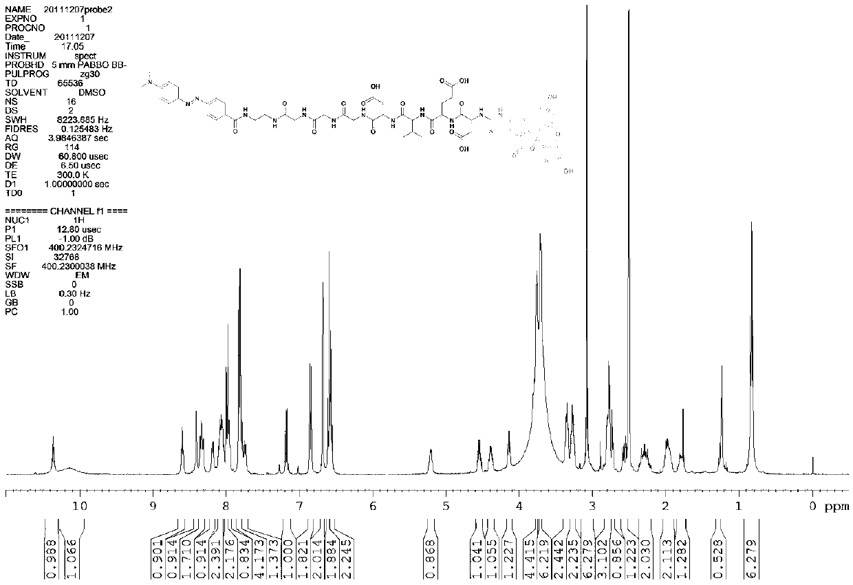
[0031] First, the oligopeptide fragment glycine-glycine-glycine-aspartic acid-valine-glutamic acid-aspartic acid-fluorenylmethoxycarbonyl is synthesized by solid-phase peptide synthesis.

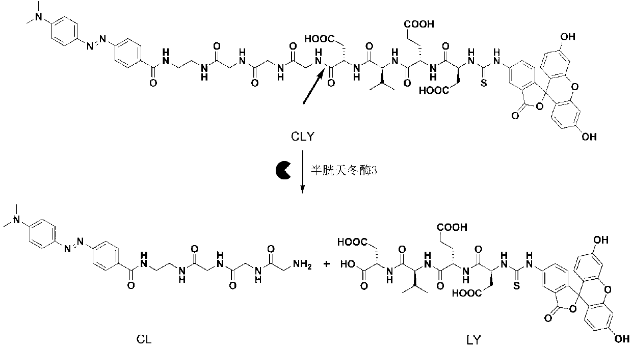
[0032] The synthesis route of the final compound CLY (Dabcyl-EDA-Gly-Gly-Gly-Gly-Asp-Val-Glu-Asp-FITC) in this example is as follows:

[0033]

[0034] .

[0035] First use the solid-phase peptide synthesis method to synthesize the oligopeptide sequence glycine-glycine-glycine-aspartic acid-valine-glutamic acid-aspartic acid-fluorenyl moxycarbonyl, press: 1 mmol 2-chlorotris Benzyl chloride resin was swollen in 2-3 ml of N, N-dimethylformamide for 4-8 minutes, and then 2 mmol of N-fluorenylmethoxycarbonyl-glycine was added, followed by 2 mmol of N, N-dimethylformamide. Isopropylethylamine, after reacting for 2-3 hours, react with 100 microliters of methanol for 5-10 minutes, add the mixed solution 4- 5 ml, react for 4-8 minutes, cut off the fluorenyl moxyprotecting group of glycine, Kai...

Embodiment 2

[0041] Embodiment 2: In vitro enzyme activity detection experiment

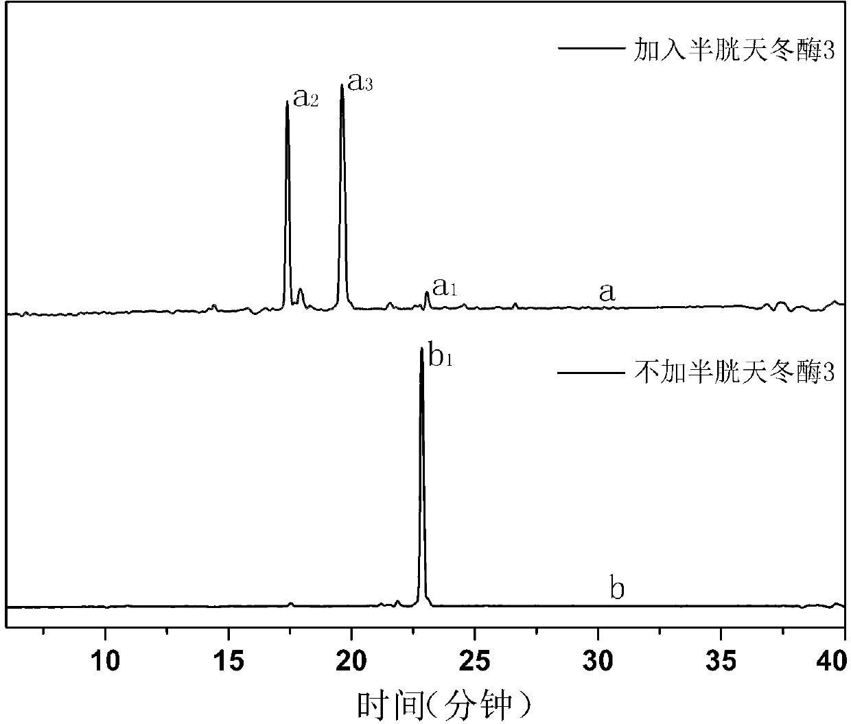
[0042] In the in vitro experiment of this embodiment, the final compound CLY with a concentration of 0.26 micromoles per liter was used, and the concentration was 50 millimoles per liter of 4-hydroxyethylpiperazineethanesulfonic acid, 0.1% of 3-[(3- cholesterylaminopropyl)dimethylamino]-1-propanesulfonic acid, 50 mmol per liter of sodium chloride, 10 mmol per liter of EDTA, and 5 cysteine ​​with a unit volume of 50 microliters Aspartase 3 was incubated in a solution with a total volume of 100 microliters and incubated at 37°C for 80 minutes to complete the identification and cleavage of the enzyme. At the same time, the fluorescence emission spectrum was scanned to obtain the curve of the fluorescence emission spectrum changing with time. The detection of enzyme activity in the apoptotic cell lysate is carried out in the following way: 2 The human liver cancer cell line HepG2 was cultured in a 37°C incubator...

Embodiment 3

[0045] Example 3: Western blot experiment and apoptotic cell imaging experiment of UV-induced apoptotic cells

[0046] Firstly, the culture of human hepatoma cell HepG2 was carried out: at a volume concentration of 5% CO 2 The human liver cancer cell line HepG2 was cultured in a 37°C incubator in an air environment using DMEM medium containing 10% bovine serum albumin by volume; the cells in the logarithmic growth phase were washed three times with 0.01 moles per liter of sterile PBS buffer , and then digested with trypsin at a mass volume concentration of 0.25%; the sub-disc cells were rinsed with DMEM, then counted, and the cell concentration was diluted to 3×10 per ml 4 cells.

[0047] Then, test the changes of apoptosis-related proteases in human liver cancer cells HepG2 after ultraviolet induction and final compound CLY, and verify the influence of ultraviolet induction and final compound CLY on cells: human liver cancer cells HepG2 were divided into five groups, except ...


PUM


Abstract

The invention discloses a caspase 3-sensitive fluorescence enhancement nano developer and a preparation method thereof. The preparation method is characterized by comprising the following steps: obtaining a connection having an appropriate length through simple solid-phase polypeptide synthesis; and obtaining the developer through conventional liquid-phase carboxylic acid/ammonia condensation reaction and fluorescent molecular marker reaction. Under physiologic conditions, the developer has favorable hydrophilicity and is easy to prepare; and meanwhile, the molecule of the fluorescence enhancement nano developer contains an oligopeptide sequence which can be specifically recognized by caspase 3, so that the oligopeptide sequence can be cut by the caspase 3 produced in the cell apoptosis process, thus indicating whether the cell enters an apoptosis state. Compared with the existing fluorescence enhancement apoptosis diagnosis kit, the preparation method disclosed by the invention is simple; the enzyme-sensitive fluorescence enhancement nano developer disclosed by the invention has favorable biocompatibility; the activity detection of a specific enzyme related to apoptosis can be completed in an in-vitro environment and a cell cracking solution through the detection of fluorescent signals; and the cell apoptosis state can be monitored on a living cell level.

Description

technical field [0001] The invention belongs to the technical field of nano-imaging agents, and in particular relates to a nano-imaging agent for fluorescence-enhanced imaging of cell apoptosis and a preparation method thereof. Background technique [0002] Fluorescent nano-imaging agents that use the phenomenon of fluorescence enhancement in biological systems to detect the state of cell apoptosis have been found in the journal of the American Chemical Society "Journal of the American Chemical Society" (J.Am.Chem.Soc., 2012, 134, 17972- 17981) reported that the fluorescent group with aggregation-induced emission characteristics was used to integrate the biological process with the aggregation process, but the excitation light wavelength required by the imaging agent in the detection of the apoptosis state of living cells is in the ultraviolet region, which is The excitation light of this wavelength is harmful to biological systems such as cells. The research on the applica...

Claims


Application Information

Patent Timeline
IPC IPC(8): C07K7/06G01N21/64
Inventor 梁高林唐安明
Owner UNIV OF SCI & TECH OF CHINA
Why Eureka
Try Eureka