

Method used for preparing microcarrier/polymer composite scaffold by electro-deposition
A composite scaffold and microcarrier technology, applied in prosthetics, medical science, etc., can solve the problems of inactivation of bioactive components, load of bioactive components, affecting biological performance, etc., and achieve the effect of short preparation time and mild conditions
- Summary
- Abstract
- Description
- Claims
- Application Information
AI Technical Summary
Problems solved by technology
Method used
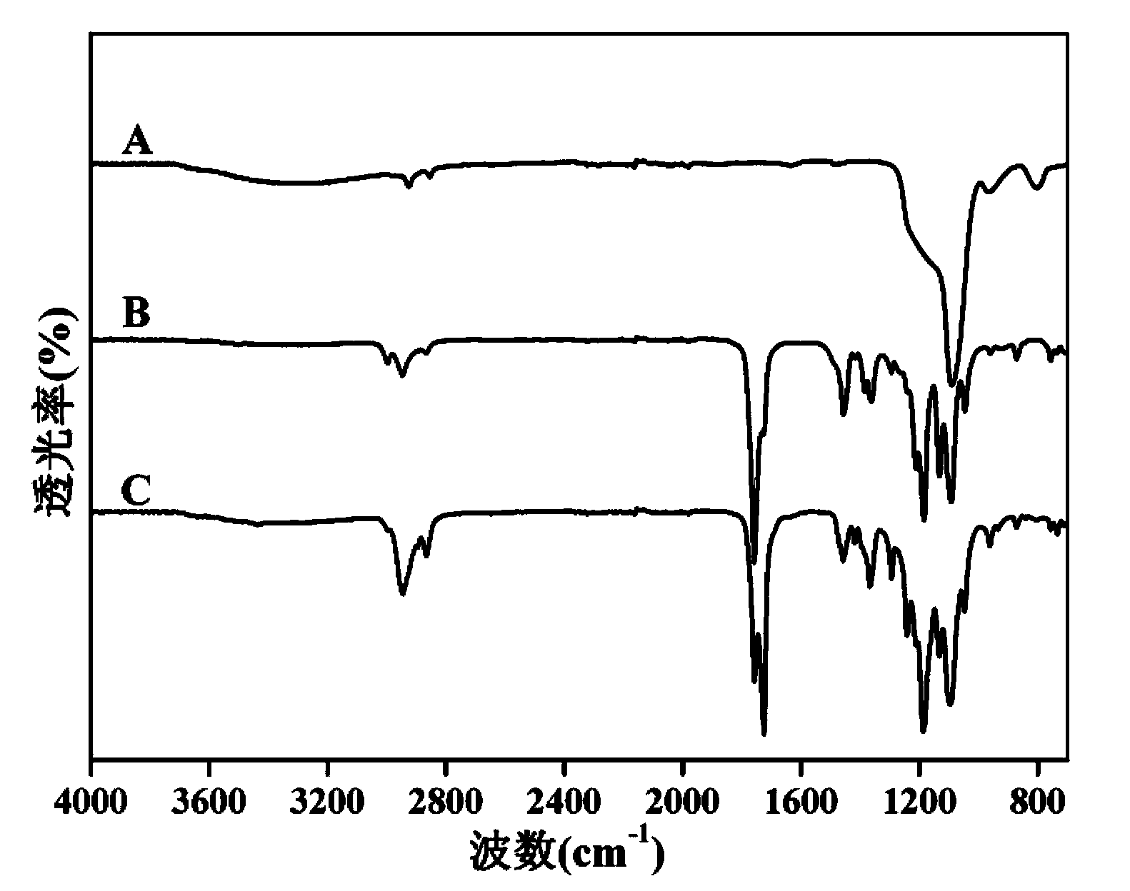
Image

Examples
Embodiment 1
[0027] (1) Preparation of mesoporous silica nanoparticles, which are aminated and loaded with dexamethasone;
[0028] (2) PLLA / PCL70:30 three-dimensional porous nanofibrous scaffold was prepared by thermal phase separation technology: PLLA:PCL was dissolved in tetrahydrofuran with a mass ratio of 70:30, heated and stirred to form a homogeneous solution with a concentration of 10wt%; Then inject it into the syringe, and place it at -80°C quickly to cause the phase separation process to occur overnight; after taking it out, return the syringe, soak the gelled polymer in deionized ice water for 2 days, and change the solvent every day Three times of deionized ice water; after freeze-drying, a three-dimensional nanofiber scaffold with a porous structure was obtained.
[0029] (3) Weigh 50 mg of the mesoporous silicon loaded with dexamethasone prepared in (1), dissolve it in 200 mL of absolute ethanol, ultrasonicate and stir to obtain a uniform electrolyte;
[0030] (4) Fix the th...
Embodiment 2
[0032] (1) Prepare chitosan microspheres and load bone morphogenetic protein;
[0033] (2) Preparation of PLLA nanofiber membrane by electrospinning technology: PLLA was dissolved in a mixed solution of dichloromethane and N,N-dimethylformamide with a volume of 9:1 to obtain a 10wt% electrospinning solution; 18G The injection needle is a spinneret, and the nanofiber membrane is received under the conditions of a voltage of 10kV, a receiving distance of 15cm, and a liquid supply rate of 1mL / h.
[0034] (3) Weigh 20 mg of chitosan microspheres loaded with bone morphogenetic protein prepared in (1), dissolve in 200 mL of absolute ethanol, ultrasonicate and stir to obtain a uniform electrolyte;
[0035] (4) Fix the nanofiber membrane scaffold prepared in (2) on the stainless steel electrode as the working electrode, the blank platinum electrode as the counter electrode, and the calomel electrode as the reference electrode, and place them together in the electrolyte solution in (3)...
Embodiment 3
[0037] (1) Prepare PLGA microspheres, use chitosan for surface modification, and load sphingosine phosphate;
[0038] (2) Collagen nanofiber membrane was prepared by electrospinning technology; collagen was dissolved in hexafluoroisopropanol to obtain 8wt% electrospinning solution; 18G injection needle was used as the spinneret, and the voltage was 16-18kV, accepted The nanofibrous membrane was received under the condition that the distance was 15cm and the liquid supply rate was 0.8mL / h.
[0039] (3) Weigh 30 mg of PLGA microspheres loaded with sphingosine phosphate prepared in (1), dissolve in 200 mL of isopropanol, ultrasonicate and stir to obtain a uniform electrolyte;
[0040] (4) Fix the nanofiber membrane support prepared in (2) on the stainless steel electrode as the working electrode, the blank titanium electrode as the counter electrode, and the calomel electrode as the reference electrode, and place them together in the electrolyte in (3). Electrochemical depositio...
PUM
| Property | Measurement | Unit |
|---|---|---|
| Concentration | aaaaa | aaaaa |
Abstract
Description
Claims
Application Information