
Novel three-dimensional tendon biological patch, and preparation method and applications thereof

A biological patch and three-dimensional biological technology, applied in prosthetics, medical science, etc., can solve the problems of hindering wound healing, difficult fusion, poor fixation effect of biological patch and tendon stump, etc., to achieve rapid regeneration and early repair, not easy to rub and damage each other, and beneficial to interpenetration and fusion

Pending Publication Date: 2019-05-07
上海白衣缘生物工程有限公司
View PDF10 Cites 0 Cited by
  • Summary
  • Abstract
  • Description
  • Claims
  • Application Information

AI Technical Summary

Problems solved by technology

[0015] Due to the lack of mechanical strength of the acellular material patch in practical application, chemical cross-linking agents such as epoxy or glutaraldehyde need to be introduced in the process of processing, or it is used together with synthetic materials, which has the following disadvantages: Potential cell Toxicity, slow degradation rate, mismatch with tissue regeneration, can lead to fibrosis, chronic inflammation and other reactions
If there is a deviation in the method or inappropriate material selection when treating the wound of the tendon stump, the tendon stump is likely to be shifted or the force on both sides is obviously uneven, which will delay the growth of new tissue and hinder The recovery of the wound will heal, and it will cause secondary injury, and even the bad situation of partial or crooked after healing
[0018] In the prior art, the planar tendon biological patch and the tendon stump are simply sutured, and the suture is the key joint. Because of the simple suture, it is easy to shift and shake inside the suture, especially the stability of the tendon stump. Poor; resulting in poor fixation between the biological patch and the tendon stump, difficult to fuse; in the case of active force or passive force, the muscle fiber layer of the stump is still prone to slipping or dislocation, which affects the formation of new tissue, Delays wound healing and functional recovery

Method used


Image

  • Novel three-dimensional tendon biological patch, and preparation method and applications thereof
  • Novel three-dimensional tendon biological patch, and preparation method and applications thereof
  • Novel three-dimensional tendon biological patch, and preparation method and applications thereof

Examples


Embodiment 1

[0047] The preparation process of the three-dimensional tendon biological patch can be found in figure 1 ,details as follows:

[0048] (1) Pretreatment: Take the fresh pig small intestine tissue from the slaughterhouse, immediately wash it repeatedly with water until it is completely clean, soak it in 0.5% acetic acid solution for 60 minutes, the weight ratio of pig small intestine to acetic acid solution is 1:5, and use physical scraping method The mucous layer, muscular layer, serosa layer and lymph nodes of the porcine small intestine and jejunum were removed, and the submucosa was separated, washed at least 3 times with purified water to obtain the tendon biological repair material, that is, the small intestinal submucosa, hereinafter referred to as SIS material.

[0049] (2) Virus inactivation: use a mixed aqueous solution containing 1.0% peracetic acid and 15% ethanol, the ratio of SIS material to the mixed aqueous solution is 1:10, soak at room temperature for 100min un...

Embodiment 2

[0059] Preparation of planar tendon biopatch

[0060] Steps 1-4 are similar to Example 1.

[0061] (5) freeze-drying, processing and shaping, sterilization

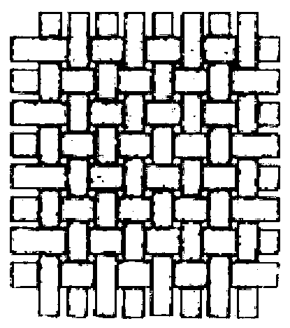
[0062] Superimpose and fix the submucosa processed by the above steps on the mold in 6 layers, after freeze-drying for 24 hours, cut into thin strips evenly in the longitudinal direction, twist them into threads, and weave them into figure 2 The planar mesh-like tendon bio-patch, and then the raw material of the bio-patch was cut into rectangles or squares of appropriate size, packed in PET bags, and finally sterilized by irradiation.

Embodiment 3

[0064] In order to ensure the safety of the biological patch, the products prepared in Examples 1 and 2 were tested for immunogenic substances.

[0065] (1) Detection method of residual cells: fixed with 10% neutral formalin, embedded in paraffin, cut into thin slices of 0.4 micron, dewaxed with xylene, dehydrated with serial alcohol, stained with hematoxylin-eosin, and microscope Next, observe the cell residue and matrix fiber structure.

[0066] (2) DNA content detection method: according to YY / T0606.25-2014 "Determination of DNA residues in biological materials of animal origin: fluorescent staining method".

[0067] (3) α-Gal antigen content detection method: After the sample was fixed with paraformaldehyde, it was routinely embedded in paraffin and sectioned, with a thickness of 4 microns. Immunohistochemical reaction was carried out by using the specific affinity between biotin-labeled BSI-B4 and α-Gal antigen. Judgment of staining results: dark brown-yellow particles ...


PUM

PropertyMeasurementUnit
Concentrationaaaaaaaaaa

Abstract

The invention discloses a novel three-dimensional tendon biological patch, and relates to the woven three-dimensional tendon biological patch having an opening. Based on the obtaining of larger mechanical properties, tendon stumps can be placed in the biological patch; and therefore, problems of poor mechanical strength of planar biological patches, poor fixation effects at suture and easy occurrence of peristalsis between stumps and the biological patches can be solved. On the basis of ensuring mechanical properties, the opening end of the biological patch can perform omnidirectional packaging on the tendon stumps, so that better fixing effects can be realized; firm attachment with each other can be achieved, uneasy moving can be realized, so that the internals of the tendon stumps can berelatively more stable; a better local small environment can be provided for the tendon stumps by adopting the biological patch, mutual infiltration and fusion can be conveniently achieved, the growth of new wound cells and the formation of new tissues can be realized, so that the structures and functions of damaged tissues can be repaired more rapidly; and the biological patch can be completelydegraded and absorbed, so that safety can be fully guaranteed.

Description

technical field [0001] The invention relates to the field of biological materials, in particular to a novel biological patch for tendon injury surgery. Background technique [0002] Tendon is a cord-like or membranous dense connective tissue at both ends of the muscle belly, which facilitates muscle attachment and fixation. Tendons are viscoelastic tissues that transmit forces generated by muscles to bones, causing movement of the limb. The biological properties of tendons affect the contractility and sports performance of muscles to a certain extent. Tendon injury is a common disease, common or more common in the elderly and certain groups of people. It is an abnormality in the sense of anatomical structure. The underlying cause is damage to the tendon, partial or complete tear, or even complete rupture. . Repeated strenuous exercise often results in abrasion or tearing of delicate soft tissues; injury to these tendons can cause pain and dysfunction. For the treatment o...

Claims


Application Information

Patent Timeline
IPC IPC(8): A61F2/00A61L27/36
Inventor 韩韦红葛翠兰钱锵张国强
Owner 上海白衣缘生物工程有限公司
Learn More