
Correlative light-and transmission electron microscopy sample treatment reagent and CLEM detection method

A transmission electron microscope and sample processing technology, applied in the detection field, can solve the problems of weak fluorescent signal in ultra-thin sheets, difficulty in compatibility with fluorescent signal and sample cell structure preservation, and unsatisfactory structural information, achieving high consistency and simple and effective method Effect

Active Publication Date: 2019-06-18
ZHEJIANG UNIV
View PDF13 Cites 2 Cited by
  • Summary
  • Abstract
  • Description
  • Claims
  • Application Information

AI Technical Summary

Problems solved by technology

[0003] In the light microscope-electron microscope combined technology scheme, the method of fluorescence observation after embedding first fixes, dehydrates, infiltrates, and embeds the sample, and then observes the fluorescence signal and electron microscope image of the completely consistent sample, which has a higher consistency , can obtain more accurate results, but on the one hand, the sample preparation technology is difficult to be compatible with the preservation of the fluorescence signal and the sample cell structure, and the ultra-thin sheet fluorescence signal is very weak when the sample signal is obtained after embedding, and the structural information is not ideal; on the other hand Integrated light-electron microscopy equipment is not yet popular

Method used


Image

  • Correlative light-and transmission electron microscopy sample treatment reagent and CLEM detection method
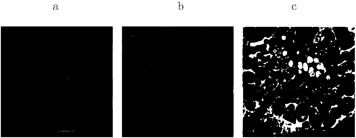
  • Correlative light-and transmission electron microscopy sample treatment reagent and CLEM detection method
  • Correlative light-and transmission electron microscopy sample treatment reagent and CLEM detection method

Examples


Embodiment 1

[0061] Embodiment 1 A kind of light microscopy-transmission electron microscopy combined sample processing reagent

[0062] A light microscope-transmission electron microscope sample processing reagent, including fixative: 2.5% glutaraldehyde, embedding agent: hydrophilic basic resin GMA, 0.5% sodium borohydride, nuclear fluorescent dye DAPI, transmission electron microscope dye Uranyl acetate and lead citrate.

[0063] The fixing agent is 2.5% glutaraldehyde or a fixing agent mixed with 4% paraformaldehyde and 0.5% glutaraldehyde, wherein 2.5% glutaraldehyde is preferred. When 2.5% glutaraldehyde was used as a fixative, the effect of preserving the ultrastructure of the sample cells was the best.

[0064] The nuclear fluorescent dye is acridine orange (Acridine Orange, AO), ethidium bromide (Ethidium Bromide, EB), propidium iodide (Propidium Iodide, PI), Diaminophenylindole (DAPI), Hoechst dye, EthD III, 7- One of AAD or RedDot2.

[0065] The TEM dye is uranyl acetate or l...

Embodiment 2

[0066] Embodiment 2: the culture of the cell that has expressed EYFP-Mito molecule

[0067] (1) Plasmid construction of EYFP-Mito fusion gene:

[0068] Digest pAcGFP1-Mito with restriction endonuclease BamH I / Not I, and recover a 4.1kb fragment after electrophoresis; digest pEYFP-N1 with BamH I / Not I, electrophoresis, and recover a 0.7kb fragment with a DNA gel recovery kit PCR product, T4DNA ligase, ligated the recovered two fragments, transformed Escherichia coli DH5α, selected a single clone and amplified it in a small amount, and sequenced the obtained clone to confirm that the sequence was correct. Extract the plasmid and measure its concentration with an ultraviolet spectrophotometer;

[0069] (2) Cell culture and transfection:

[0070] 37 degrees Celsius CO 2 In a cell culture incubator, culture human cervical cancer Hela cells in RPMI 1640 medium containing 10% fetal bovine serum (FBS). When the cells grow to 70-80% density, mix serum-free RPMI1640 containing 12ug ...

Embodiment 3

[0071] Embodiment 3: the sample processing of the cell that has expressed EYFP-Mito molecule

[0072] (1) Sample processing:

[0073] After confirming the expression of EYFP-Mito protein, discard the supernatant of the culture medium, digest the cells with trypsin, and collect by centrifugation at 2000rpm after neutralizing the digestion solution in the culture medium, discard the supernatant, lightly wash the cells with PBS, and divide into 3 equal parts, After removing PBS, add 2.5% glutaraldehyde prepared in PBS, or 4% paraformaldehyde prepared in PBS, or a mixed fixative (4% paraformaldehyde+0.5% glutaraldehyde prepared in PBS), and fix at 4°C Overnight, samples were treated with 0.5% sodium borohydride in PBS for 5 min at 4°C, followed by dehydration infiltration.

[0074] The samples were dehydrated with 50%, 70%, and 95% ethanol at 4°C for 10 minutes, and then the samples were treated with 70%, 85%, and 100% GMA at -20°C for 10 minutes each; after replacing with new 10...


PUM


Abstract

The invention provides a correlative light-and transmission electron microscopy sample treatment reagent and a CLEM detection method using the provided correlative light-and transmission electron microscopy sample treatment reagent. The reagent comprises a fixing agent, an embedding agent-hydrophilic alkaline resin GMA, sodium borohydride, nuclear fluorescent dye and transmission electron microscopy dye. The CLEM detection method comprises steps of fixing a sample, removing background fluorescence, dehydrating the sample, permeating, embedding, polymerizing, ultrathin slicing, laser confocal microscope imaging, acquiring a transmission electron microscopy image, and processing the image in later period. The method overcomes the difficulty of being compatible with a fluorescent signal and solves a problem of storage of a sample cell structure in the prior art. The sample has a strong fluorescent signal and complete ultramicrostructure information can be retained; the method adopts a simple but effective image normalization method, jointly uses existing fluorescent microscope and transmission electron microscopy so as to acquire highly consistent correlative light-and electron microscopy images. The method combines positioning information of a target molecule and ultramicrostructure information, thereby being very practical.

Description

technical field [0001] The invention belongs to the technical field of detection, and in particular relates to a light microscope-transmission electron microscope combined sample processing reagent and a CLEM detection method using the reagent. Background technique [0002] Knowing the precise structural information of target cells and organelles, as well as the location of target proteins in cells and their impact on structure, can provide necessary information for the study of growth, development, reproduction, learning and memory, signal transduction, immune response, metabolism and other life activities. information. Fluorescence microscopy alone cannot obtain the structural information of the sample, and the immunocolloidal gold labeling electron microscopy technique for studying the precise positioning of proteins is limited by the difficulty of preserving epitopes, the difficulty of antibodies entering the resin, the low labeling success rate, and the high non-specifi...

Claims


Application Information

Patent Timeline
IPC IPC(8): G01N1/30G01N1/36
Inventor 王贝贝
Owner ZHEJIANG UNIV
Learn More