
Mycoplasma pneumoniae antigen and application thereof in simultaneous quantitative detection of Mycoplasma pneumoniae IgG and IgM content in peripheral blood

A detection technology for mycoplasma pneumoniae and antibodies, which is applied in the field of molecular biology and biological detection, can solve the problems of easy quenching, large amount of reagent samples, long incubation time, etc., and achieve the effect of reducing the misdiagnosis rate

Active Publication Date: 2020-04-07
NANJING VAZYME MEDICAL TECH CO LTD
View PDF5 Cites 3 Cited by
  • Summary
  • Abstract
  • Description
  • Claims
  • Application Information

AI Technical Summary

Problems solved by technology

However, considering its specificity, it is prone to false positives
Although real-time fluorescent quantitative polymerase chain reaction (FQ-PCR) has high sensitivity and specificity, it has high nutritional requirements and takes a long time. The judgment of some results takes nearly one month. For the early diagnosis of Mp has produced certain obstacles
However, when the colloidal gold method is used to diagnose Mycoplasma pneumoniae, a large amount of reagent samples and a large amount of blood collection are used.
Although the detection of Mycoplasma pneumoniae antigen or NAT can be used for early diagnosis of infection, due to sample contamination or non-pathogenic bacterial colonization, the diagnostic false positive rate is high and the cost is expensive
Although the culture of Mycoplasma pneumoniae is considered as the gold standard for the diagnosis of MP, it has obvious methodological limitations, such as long culture time and low sensitivity.
The detection of Mycoplasma pneumoniae antibody can distinguish between colonized bacteria and pathogenic bacteria, but the markers used to detect antibodies at home and abroad are mostly conventional markers such as fluorescein isothiocyanate (FITC), which have a short fluorescence lifetime, are easy to quench, and emit light. Instability, small Stokes shift, and monochromatic excitation of a single light source are the limitations of FITC. Therefore, the positive rate of antibodies labeled with common fluorescent agents such as FITC is low and it is easy to miss the diagnosis.

Method used


Image

  • Mycoplasma pneumoniae antigen and application thereof in simultaneous quantitative detection of Mycoplasma pneumoniae IgG and IgM content in peripheral blood
  • Mycoplasma pneumoniae antigen and application thereof in simultaneous quantitative detection of Mycoplasma pneumoniae IgG and IgM content in peripheral blood
  • Mycoplasma pneumoniae antigen and application thereof in simultaneous quantitative detection of Mycoplasma pneumoniae IgG and IgM content in peripheral blood

Examples


Embodiment 1

[0027] Embodiment 1 New Mycoplasma pneumoniae P80 antigen preparation

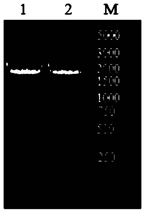
[0028] The epitope-rich region sequence (nucleotide sequence such as SEQ ID NO.3) and corresponding antibody sequence (amino acid sequence such as SEQ ID NO.4) of Mycoplasma pneumoniae outer membrane adhesion protein is known, and its binding is predicted by Pymol biological software Site-related amino acid residues are subjected to site-directed mutation according to conventional methods in the art to obtain a mutant Mycoplasma pneumoniae antigen P80, the amino acid sequence is shown in SEQ ID NO.1, and the nucleotide sequence is shown in SEQ ID NO.2 . The mutant sequence was integrated into the pET28a plasmid through the routine operation of genetic engineering, transformed into DH5α, and then transformed into E. coli to construct a prokaryotic expression system to express the mutant antigen P80 exogenously. Purified by Ni column, the purity was greater than 90%. From figure 1 It can be seen that the s...

Embodiment 2

[0029] Example 2 New Mycoplasma pneumoniae antigen P80 reactivity test

[0030] In order to study the immunoreactivity of the novel Mycoplasma pneumoniae antigen P80, it was diluted to 100mg / L, and 20% bovine serum was added to the protein stock solution to simulate serum samples. The relative immunoreactivity of P80 was determined with FUJIREBIO reagent on the Siemens BNII system. The calculated actual effective concentrations of P80 and natural Mycoplasma pneumoniae antigen (nucleic acid sequence such as SEQ ID NO.3) are 91 mg / L and 40.7 mg / L, respectively. The measurement results are shown in Table 1, indicating that P80 has good immunoreactivity, and the measured concentration can reach 94.9% of the actual concentration after excluding the matrix effect, which is 34.6% higher than the natural antigen.

[0031] Table 1 Comparison of reactivity between recombinant antigen and natural antigen

[0032]

[0033] Actual concentration = relative molecular mass of natural / rec...

Embodiment 3

[0034] Embodiment 3 detects the preparation of Mycoplasma pneumoniae IgG, IgM quantum dot immunochromatography kit

[0035]The test strip adopts the principle of double-antibody sandwich method. Add 50 μL of the quantum dot stock solution into the EP tube containing the activation buffer, and place it in a vortex mixer to mix and activate for 0.5 h. Add 450 μL of coupling buffer and 75 μg of mouse anti-human IgM and mouse anti-human IgG antibodies, and place in a vortex mixer to shake and mix for 0.5 h. After centrifuging to remove the supernatant, block with 10% ethanolamine solution and 5% Casein solution, and mix with a vertical mixer for 45 minutes. Resuspend and remove the supernatant to prepare water-soluble quantum dot conjugates coated with mouse anti-human IgM and mouse anti-human IgG antibodies, add buffer and store in the dark at 2-8°C.

[0036] Take a marker pad with a width of 8 mm, use a gold sprayer to dilute the quantum dot-labeled mouse anti-human IgM and T...


PUM


Abstract

The invention discloses a Mycoplasma pneumoniae antigen and its application in simultaneous quantitative detection of Mycoplasma pneumoniae IgG and IgM content in peripheral blood. The amino acid sequence of the mutant Mycoplasma pneumoniae antigen P80 of the invention is as shown in SEQ ID NO.1. The immunoreactivity of the Mycoplasma pneumoniae antigen p80 and the antibody is about 34.6% higher than that of the natural antigen. By combining the Mycoplasma pneumoniae antigen P80 with the quantum dot immunochromatography technology, the shortcoming that existing detection methods of Mycoplasmapneumoniae antibody, such as immunochromatography assay (ICA) and enzyme-linked immunosorbent assay (ELISA), can only detect qualitatively or semi-quantitatively, and the MP-IgM and MP-IgG detection must be carried out twice is solved; and the detection of MP-IgM and MP-IgG can be realized at the same time, so as to distinguish the patients with recent infection or previous infection and reduce the misdiagnosis rate.

Description

technical field [0001] The invention belongs to the technical field of molecular biology and biological detection. Specifically, the invention relates to the development of a mycoplasma pneumoniae mutant and a fingertip blood sampling method to detect mycoplasma pneumoniae quantum dot immunochromatography kit. Background technique [0002] Mycoplasma pneumoniae (Mycoplasma Pneumoniae, Mp) is one of the most important pathogens causing community acquired pneumonia (Community Acquired Pneumonia, CAP). It is an ultrafilterable pathogenic microorganism between bacteria and viruses. According to reports, pneumonia caused by Mp infection accounts for 10-40% of CAP, and the incidence trend is still on the rise in recent years. Clinically, Mp infection mainly manifests as symptoms of bronchitis and upper respiratory tract infection. Its main features are slow onset, headache, fever, sore throat and discomfort, and the most typical dry cough and other symptoms. [0003] Usually, the...

Claims


Application Information

Patent Timeline
Patent Type & Authority Applications(China)
IPC IPC(8): C07K14/30C12N15/31G01N33/569
CPCC07K14/30G01N33/56933
Inventor 曹林唐波徐晓昱赖迪文
Owner NANJING VAZYME MEDICAL TECH CO LTD
Learn More