Covered implantable medical device and preparation method thereof

A technology for implanting medical devices and coatings, applied in the field of implanted medical devices and their preparation, can solve problems such as increasing platelet attachment, activating the thrombin system, poor blood compatibility, and poor prognosis of patients, so as to promote chemical Effects of cross-linking, good anticoagulant properties, and good economic value

Active Publication Date: 2020-10-13
WUHAN YONGSEN BIOTECH
View PDF7 Cites 2 Cited by
  • Summary
  • Abstract
  • Description
  • Claims
  • Application Information

AI Technical Summary

Problems solved by technology

However, this method can only reduce the risk of voids at the interface between the polymer and the framework material, and the firmness of the interface between the polymer and the framework material does not change significantly
[0005] In addition, implanted medical devices such as artificial blood vessels and stent grafts that are in direct

Method used


Image

  • Covered implantable medical device and preparation method thereof
  • Covered implantable medical device and preparation method thereof
  • Covered implantable medical device and preparation method thereof

Examples


Embodiment 1

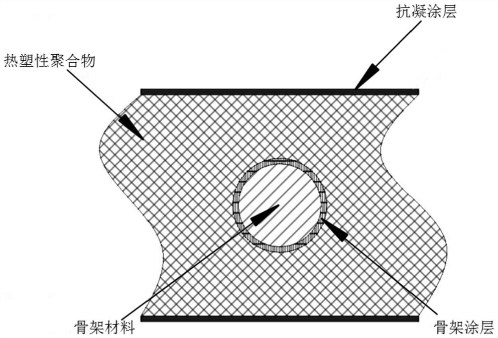
[0034] A covered artificial blood vessel, wherein the skeleton material is a polyester thread, the coating is a copolymer of hexamethylene diamine and pimelic acid, the thickness is 5 nm, the thermoplastic polymer is polyurethane, and the anticoagulant material is VA64.

[0035] The preparation method is specifically as follows:

[0036] Step 1: Dissolve the thermoplastic polymer polyurethane in the DMAc organic solvent to prepare a 20wt% thermoplastic polymer solution, defoam, attach the solution to the mold surface by electrostatic spinning, and dry to remove the organic solvent;

[0037] Step 2. After pretreatment with 5wt% sodium hydroxide solution for 8 hours, the surface molecular chains are partially hydrolyzed to form active carboxyl functional groups and then introduced into the coating. The coating is composed of hexamethylene diamine and pimelic acid in the skeleton through amidation reaction. The surface is polymerized in the form of ABAB head-to-tail bonding.

[0038] Ste...

Embodiment 2

[0043] A covered artificial blood vessel, wherein the coating is octamethylene diamine and tridecanedioic acid copolymer, the thickness is 5nm, the thermoplastic polymer is polyurethane, and the anticoagulant compound is low molecular heparin.

[0044] The preparation method is specifically as follows:

[0045] Steps together Example 1

[0046] Step 2: After pretreatment with 5wt% sodium hydroxide solution for 8 hours, the surface molecular chain is partially hydrolyzed to form active carboxyl functional groups and then introduced into the coating, the coating is octamethylene diamine and tridecanedioic acid through amidation reaction It is polymerized in the form of ABAB head-to-tail bonding on the surface of the framework.

[0047] Step three and step four are the same as in Example 1

[0048] Step 5: In 0.1wt% of 1-(3-dimethylaminopropyl)-3-ethylcarbodiimide aqueous solution, add 2wt% of polyethyleneimine (Mw=600Da) to make it and polyurethane For the reaction, the quaternization r...

Embodiment 3

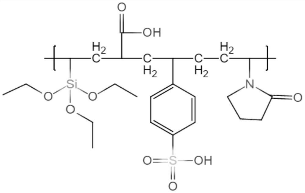
[0052] A covered artificial blood vessel, wherein the coating is: a copolymer of decamethylene diamine and eicosandioic acid, the thickness is: 5nm, the thermoplastic polymer is polyurethane, and the anticoagulant compound is a heparin-like copolymer, wherein Heparinoid copolymer is made by addition polymerization of vinyl triethoxy silane, N-vinyl pyrrolidone and sodium p-styrene sulfonate.

[0053] The preparation method is specifically as follows:

[0054] Steps together Example 1

[0055] Step 2. After pretreatment with 5wt% sodium hydroxide solution for 8 hours, the surface molecular chains are partially hydrolyzed to form active carboxyl functional groups and then introduced into the coating. The coating is hexamethylene diamine and adipic acid through an amidation reaction on the surface of the skeleton with ABAB head It is polymerized in the form of tail bonding.

[0056] Step three and step four are the same as in Example 1

[0057] Step five is specifically:

[0058] Take 5.5...


PUM

PropertyMeasurementUnit
Thicknessaaaaaaaaaa

Abstract

The invention belongs to the technical field of medical devices, and discloses a covered implantable medical device and a preparation method thereof. The implantable medical device comprises a skeleton material, the surface of the skeleton material is sequentially covered with a coating and a thermoplastic polymer film, the surface of the thermoplastic polymer material film is grafted with an anticoagulant compound; the coating is formed by condensation polymerization of diamine A and dibasic acid B in the form of ABAB head-to-tail bonding on the skeleton surface through amidation reaction; the anticoagulant compound has carboxyl or sulfonic acid groups, specifically one or more of VA64, low molecular weight heparin, heparinoid copolymer and diosgenin. Bonding strength between the polymermaterial and the skeleton material is improved by arranging the coating on the surface of the skeleton material, anticoagulation modification is performed on the covered skeleton material, and the problem about blood compatibility of the implantable device is solved.

Description

Technical field [0001] The present invention relates to the technical field of medical devices, in particular to an implanted medical device with specific functions formed by partially or completely wrapping a skeleton material by a thermoplastic polymer and a preparation method thereof. Background technique [0002] The prevalence and mortality of cardiovascular diseases in China are still on the rise. Cardiovascular deaths account for more than 40% of the residents’ disease deaths, ranking first, higher than tumors and other diseases. Implanted medical devices for the treatment of cardiovascular diseases have also been increasingly developed. Among them, artificial blood vessels and covered stents are one of the most commonly used cardiovascular implant devices. At present, the main materials used in the two types of products are polyester, polytetrafluoroethylene and other traditional materials that have been used for decades, with strong hydrophobicity and general clinical b...

Claims


Application Information

Patent Timeline
IPC IPC(8): A61L27/50A61L27/34A61L27/54A61L33/04A61L33/06A61L33/10C08F226/10C08F230/08C08F212/14
CPCA61L27/507A61L27/34A61L27/54A61L33/0011A61L33/0076A61L33/0088A61L33/04A61L33/064A61L33/08C08F226/10A61L2300/232A61L2300/216A61L2300/42A61L2300/606A61L2420/02A61L2420/06C08L5/10C08L39/06C08L75/04C08F230/085C08F212/30
Inventor 欧阳晨曦徐天成焦培龙行吴瑶刘思诗
Owner WUHAN YONGSEN BIOTECH
Why Eureka
Try Eureka