Photo-caged fluorescent molecules

a fluorescent molecule and photocage technology, applied in the field of fluorescent molecules, can solve the problems of cell membrane damage, method is associated with a number of major limitations, and molecules are not ideally suited for use as photocaged fluorescent molecules

Inactive Publication Date: 2005-02-24
BOARD OF RGT THE UNIV OF TEXAS SYST
View PDF4 Cites 12 Cited by
  • Summary
  • Abstract
  • Description
  • Claims
  • Application Information

AI Technical Summary

Benefits of technology

Because the current caged fluorophores can be uncaged in a localized area, are cell permeable, and can cross gap junctions, they have even wider applications in cell biology research. First, they do not have to be delivered into cells through invasive techniques such as microinjection or electroporation. Second, they make it feasible to obtain paired data under both control and test conditions and to perform a set of experiments on the same coupled cells over a period of time. Table 1 below summarizes the advantages of using localized photo-uncaging of fluorophores to study the permeability of gap junctions, versus the traditional techniques of microinjection, dual whole cell patch clamping, and FRAP. TABLE 1LocalizedMicroinjectionDual whole celluncaging ofof tracersFRAPpatch clampingpermeable dyesInvasivenessInvasiveIntenseInvasiveNon-invasiveillumination &photo-damageCell integrityCompromisedMaintained (ifCompromisedMaintaineddyes are cellpermeable)TemporalLowGoodVery highGoodresolutionSimplicity ofSpecialRequire highTechnicallyEasy, applicableset-up andtechnique,power laserdemanding &to cellexecutionlimited cellstime consumingpopulations ortissuesQuantificationReliable only ifReliable if noConductance doesReliable & allowof molecularcells recoverphoto-damagenot always reflectmultipletransfer ratesvery rapidlymolecularmeasurements intransfer,coupled cell pairsproblematic inwell-coupled cellsfrom theinterference ofseries resistanceMulti-colorFeasibleDifficultFeasibleFeasibleimaging
Thus, localized uncaging of the fluorophores can be used as a powerful and convenient method for studying intercellular communication in intact cell populations. The caged and cell permeable fluorophores are first loaded into fully intact cells. Localized uncaging of the fluorophores can then be performed in order to selectively mark individual cells amongst coupled cells. Subsequent fluorescent imaging and data analysis then provide quantitative information on the molecular transfer rates across gap junctions of coupled cells.

Problems solved by technology

Thus, these molecules are not ideally suited for use as photo-caged fluorescent molecules.
However, the method is associated with a number of major limitations.
First, cell membranes are usually damaged during the operation, so special skill and extra care are needed to minimize cell injury.
Also, only a limited number of cells can be injected at a time, so it is not convenient to study intercellular communications in cell populations or tissue preparations.
Finally, the microinjection process may disrupt the extracellular and intracellular concentrations of other molecules affecting gap junction permeability.
It is very difficult to maintain the intracellular Ca2+ concentration at a normal physiological level during microinjection.
Thus, studies of fluctuations in concentration of molecules such as Ca2+ in relation to gap junction permeability may be adversely affected.
The technique known as “scrape loading” suffers from similar drawbacks (el-Fouly, et al., 1987).
However, the accuracy of junctional current measurement is affected by the relative values of uncompensated electrode resistances, nonjunctional resistances, junctional resistance and seal resistances (Harris, 2001).
While the technique offers some abilities to alter cytoplasmic constituents, it can also cause problems when concentrations of cellular ions, such as Ca2+, must be accurately controlled, or when certain important cytoplasmic factors should be prevented from being diluted out in the pipette.
This intense laser illumination may damage cells.
Moreover, the GJ-FRAP technique maybe incompatible with multi-color imaging when other biochemical changes inside cells need to be studied by fluorescent sensors.

Method used


Image

  • Photo-caged fluorescent molecules
  • Photo-caged fluorescent molecules
  • Photo-caged fluorescent molecules

Examples


example 1

General Synthesis

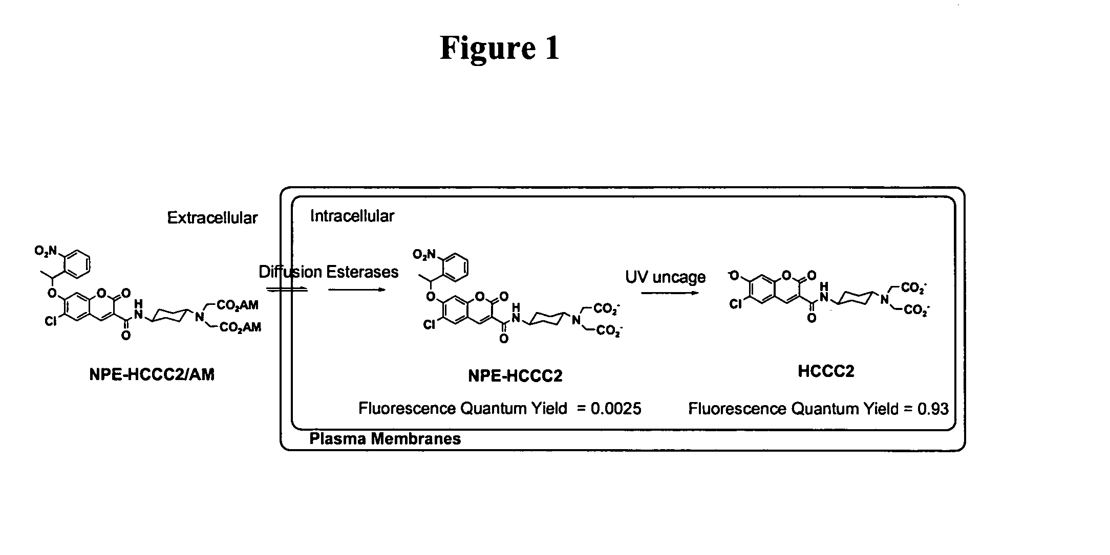
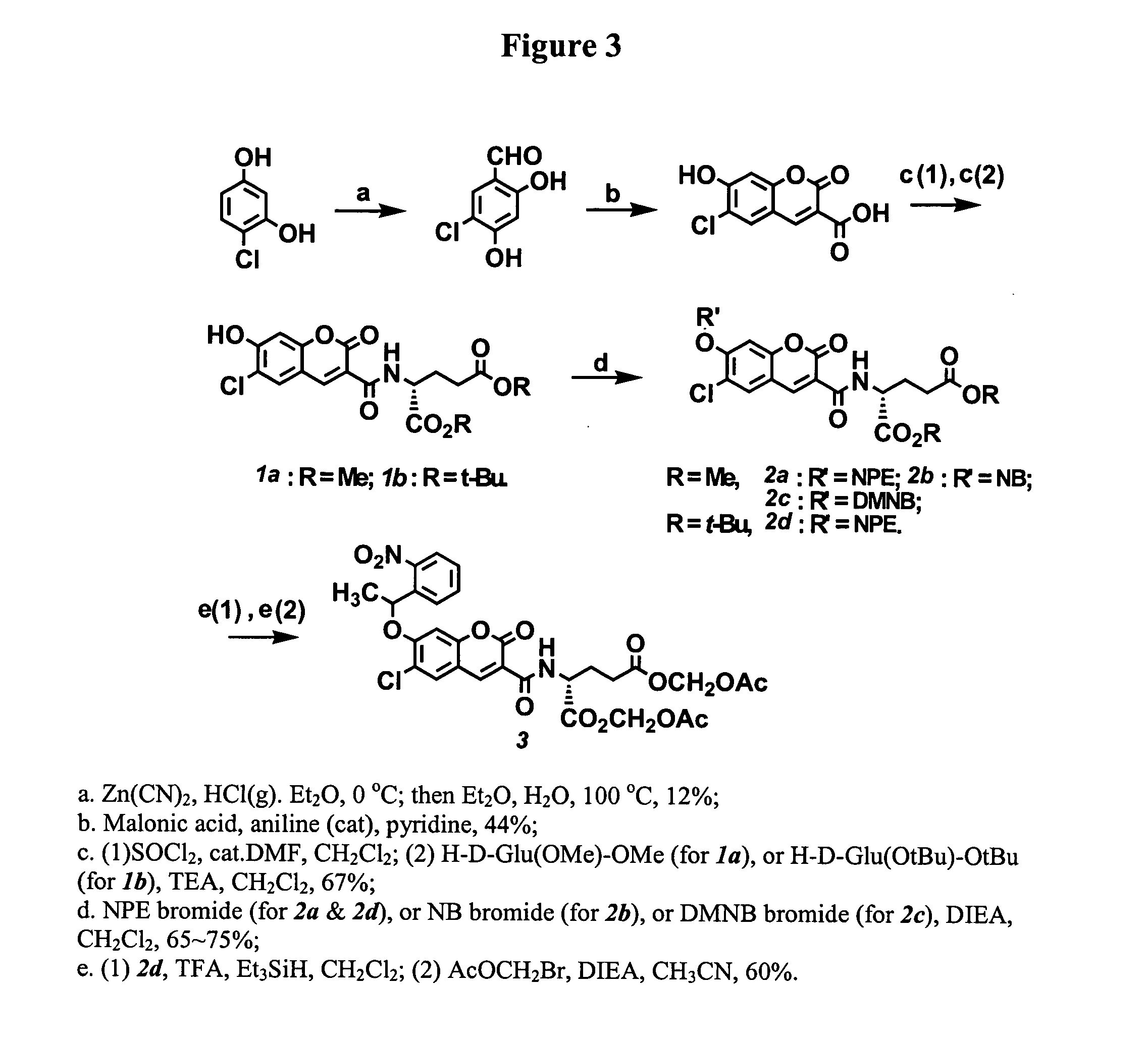
6-Chloro-7-hydroxy-coumarin 3-carboxylate was prepared in two steps starting with 4-chloro-resorcinol. Coupling coumarin 3-carboxylate with the methyl ester of D-glutamate provided coumarin 3-carboxamide (1a, FIG. 3). The dye absorbs maximally at 408 nm with an extinction coefficient of 44,000. The fluorescent quantum yield of the molecule is 0.93 measured in an aqueous buffer (100 mM KCl, 20 mM Mops, pH 7.35). Three caged derivatives of this fluorophore were synthesized by masking the 7-hydroxyl group of coumarin with the following caging groups: 1-(2-nitrophenyl)ethyl (“NPE,” 2a, FIG. 3), 2-nitrobenzyl (“NB,” 2b, FIG. 3), and 4,5-dimethoxy-2-nitrobenzyl (“DMNB,” 2c, FIG. 3).

example 2

Photoactivation and Excitation

The three caged coumarins prepared in Example 1 (2a, 2b, 2c, FIG. 3) were dissolved in aqueous buffer (pH 7.35) containing 100 mM KCl and 20 mM Mops to final concentrations of 2 μM. The solutions were photolyzed with 365 nm light from a filtered mercury lamp. Fluorescence emission spectra at 408 nm of the solutions were taken with a Fluorolog spectrometer (Jobin-Yvon) after different UV exposures. FIG. 4 shows the spectra of NPE-caged coumarin (2a, FIG. 3).

All three caged fluorophores prepared in Example 1 are essentially non-fluorescent, as shown by their negligible fluorescence quantum yields in Table 2 below. UV illumination (365 nm) removed the caging group and progressively generated highly fluorescent coumarin 1a, as shown in FIG. 5. The fluorescence enhancements after uncaging ranged from 200 times (2b) to nearly 800 times (2c). This robust change in fluorescence quantum yields is important for generating high contrast optical signals with a ...

example 3

Esterification

Protection of the two carboxylates of the D-glutamate moiety with acetoxymethyl (“AM”) ester groups is useful for cellular imaging applications. AM esters are stable in physiological solutions but are susceptible to enzymatic hydrolysis by cellular esterases. The enzyme hydrolysis regenerates negatively charged species which cannot diffuise out of cells (Grynkiewicz, et al., 1985). This irreversible trapping process has been applied to enrich various ion indicators, fluorescent probes, and second messengers into cells to higher concentrations (Li, et al., 1998). With the current caged coumarin derivatives, hydrolysis of two methyl esters of compound 2a under a number of conditions was accompanied by the concomitant hydrolysis of the amide bond of coumarin 3-carboaximide. Thus, protecting two carboxylates of glutamate with t-butyl groups is a more feasible synthetic route. Acid treatments of the intermediate 2d (prepared from 1b, FIG. 3) followed by esterification wit...


PUM

PropertyMeasurementUnit
wavelengthsaaaaaaaaaa
non-fluorescentaaaaaaaaaa
fluorescenceaaaaaaaaaa

Abstract

A class of photo-caged and cell permeable fluorescent molecules having high uncaging cross sections, robust fluorescence enhancement, and flexible chemistry for bioconjugation. Some of the photo-caged fluorescent molecules are derived from 6-chloro-7-hydroxy-coumarin 3-carboxamide. The fluorescent molecules are useful for cellular imaging applications and particularly for tracing the molecular transfer between cellular gap junctions. The fluorescent molecules also have an emission wavelength that spectrally complements with the emission wavelength of other fluorophores, enabling simultaneous multi-color imaging.

Description

BACKGROUND The present invention is generally related to fluorescent molecules for cellular imaging applications and, in particular, to photo-caged fluorescent molecules which are cell permeable and useful for the study of cellular imaging applications and tracing molecular transfer in cellular gap junctions. Photo-caged fluorescent dyes have wide applications in tracking the spatiotemporal dynamics of molecular movements in biological systems. These caged tracers are weakly or non-fluorescent when key functional groups of fluorophores are masked by photo-labile protecting groups, or cages. Photo-activation “uncages” the molecule by removing the protecting group and abruptly switches on the fluorescence of the parent dyes. By selectively photoactivating labeled molecules present in a certain area of a cell, the progression of the fluorescent signal can then be tracked over time as it moves away from the “uncaging” site into the dark surrounding area. (Adams, et al., 1993; Politz, ...

Claims


Application Information

Patent Timeline
Patent Type & Authority Applications(United States)
IPC IPC(8): C07D215/54C07D311/02C07D311/16C07D405/12C12Q1/68
CPCC07D215/54C07D311/16C07D405/12C12Q1/6816C12Q2563/107
Inventor LI, WEN-HONGZHAO, YURUI
Owner BOARD OF RGT THE UNIV OF TEXAS SYST
Why Eureka
Try Eureka