With anatomic
MR imaging, the presence of moving
biological tissue can be highly problematic because it can produce image artifacts, obscure the detection of lesions, and more generally complicate the interpretation of
MR images.
The time scale for acquiring diagnostic MRI typically ranges from several seconds to several minutes, which can yield significant postural, cardiac, respiratory, and
blood flow image artifacts that can confound the ability to detect
pathology.
For example,
motion artifacts due to normal or abnormal
respiratory movements can degrade
image quality in MR scans where the patient is either allowed to breathe freely, breathes inadvertently, or if the MR study requires scan times in excess of a patient's ability to hold their breath.
Although this has been used to correct for motion-related artifacts in functional
neuroimaging studies, such a method cannot monitor diaphragmatic motion where a projection profile includes moving structures (liver,
stomach, etc.) and slightly moving structures (
lung, shoulder).
In the case of MR
neuroimaging, the inability of the subject simply to remain still during the examination period may significantly compromise MR scan quality.
However, repeated breath holding may not be feasible for many coronary patients and navigation techniques to-date have not generally provided a robust method which works over a range of different
breathing patterns in a variety of patients.
Another drawback to these approaches is that success or failure is usually not apparent for some time after the start of imaging, and many times not until the imaging has been completed.
This then requires a manual registration and analysis of the
perfusion images, which is cumbersome and time-consuming because the user must carefully arrange each image to compensate for the
respiratory motion before proceeding to a
region of interest time-intensity analysis.
The absence of beam-hardening artifacts from bone allows complex approaches to anatomic regions that may be difficult or impossible with other imaging techniques such as conventional CT.
However, both the stereotactic and the frameless techniques are typically limited to the use of rigid devices like needles or
biopsy forceps, since their adequate operation requires either mechanical attachments or line-of-
sight between the light sources and the sensors.
However, the application of this technology to MRI is problematic due to the simultaneous use of RF signals by the MR scanning.
Potential difficulties are the heating of the receiving antenna in the device by the
high amplitude excitation RF transmissions of the MRI
scanner and artifacts in the MR image.
However, this method may be subject to heating of the coil, and requires time to implement that reduces the
temporal resolution available for repeated MRI acquisitions.
However, each invention also has significant inherent limitations.
In addition, as BOLD imaging is typically coupled with a repetitive behavioral task (e.g., passive sensory, cognitive, or sensorimotor task) to localize BOLD signals in the vicinity of neurons of interest, there is significant potential for fMRI to be confounded by the presence of
small head motions.
Random head motion decreases the statistical power with which brain activity can be inferred, whereas task-correlated motion cannot be easily separated from the fMRI
signal due to neuronal activity, resulting in spurious and inaccurate images of
brain activation.
In addition, head motion can cause mis-registration between neuroanatomical MR and fMR images that are acquired in the same examination session.
An analogous problem exists for aligning anatomical and functional
MR images performed on different days.
It is well known that motion between images acquired with MRI greatly reduces their utility and effectiveness.
To date, however, no generally acceptable solution has been reported.
The simplest approach is to average
imaging data repetitively, although this reduces spatial resolution.
Prior art attempts at tracking motion using cross-correlation and other simple
distance measurement techniques have not been highly effective where
signal intensities vary either within images, between images, or both.
However, unlike the present invention, the system disclosed by Nevo is not capable of
position tracking when imaging gradients are inactive, nor is it capable of measurements outside the sensitive volume of the imaging gradients (i.e., significantly outside the
magnet bore in the static fringe
magnetic field of the MRI system, or even outside the
magnet room entirely).
As in anatomical MRI, these schemes remain an incomplete solution to the problem and the search for improved motion suppression continues.
However, it is still possible to achieve poor activation
image quality if patients exhibit task-correlated motion on the order of 1
millimeter.
This problem is particularly manifest in specific patient populations (e.g.
dementia, immediate post-acute phase of
stroke).
Furthermore, image-based coregistration algorithms suffer from methodological limitations.
Consequently, the resulting co-registered images still can suffer from residual motion
contamination that impairs the ability to interpret brain activity.
However, it increases attentional demands and consequently modulates fMRI signals of brain activity, and may therefore not be broadly applicable across patient populations.
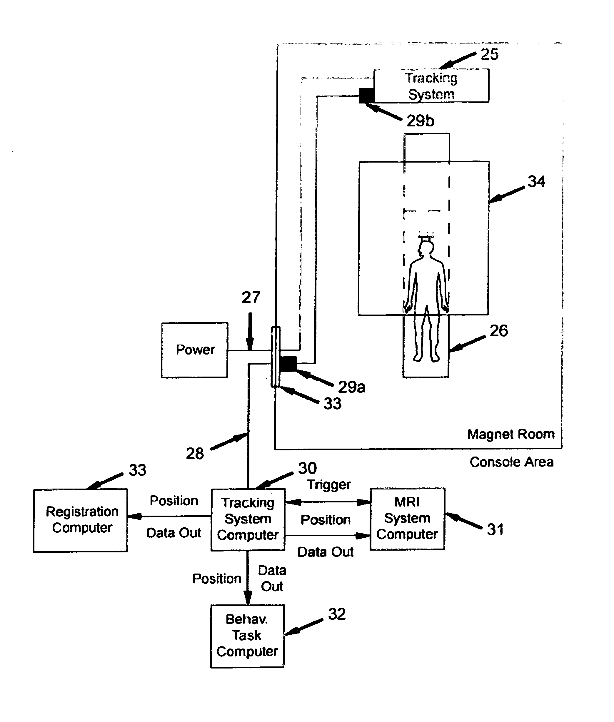
There are also several drawbacks to the use of an external, MRI-compatible position-tracking device.
Such measurements are inherently limited to sensing the motion at the surface of an object, not the interior.
Another limitation is the necessity to transform the position data into the co-ordinate system of MR
image acquisition.

