Blood test prototypes and methods for the detection of circulating tumor and endothelial cells

a technology of endothelial cells and blood test prototypes, which is applied in the field of blood test prototypes and methods for the detection of circulating tumors and endothelial cells, can solve the problems of difficult purification of cells for analysis, negative prediction of cardiovascular diseases risk, and limited detection of exfoliated abnormal cells by routine cytopathology, so as to improve the anti-tumor immune response and dramatic utility in management

Inactive Publication Date: 2005-11-03
CHEN WEN TIEN +2
View PDF6 Cites 77 Cited by
  • Summary
  • Abstract
  • Description
  • Claims
  • Application Information

AI Technical Summary

Benefits of technology

[0041] Cytotoxic lymphocytes enriched by the CAM methods from blood of cancer patients may be valuable in their own right: careful comparison of their gene expression profile in comparison to non-tumor associated lymphocytes may yield valuable information concerning the type of ongoing immune reaction and inflammation that are being mounted against the metastatic tumor cells. Moreover, another valuable therapy approach may be to expand these cells in vitro, for example, using IL-2, and then reintroduce them into the patients to augment their anti-tumor immune response. This approach may have dramatic utility in the management of melanoma and other tumors.
[0042] Embodiments of the present invention would be useful both for diagnostic and therapeutic purposes in providing the ability to separate, for example, the small fraction of CTC that are metastatic from the large number of other circulating cells in a patient's body.
[0043] Embodiments of the present invention: (1) can isolate specifically viable target cells such as tumor and endothelial cells but leave alone unrelated or damaged cells; (2) can achieve an enrichment of over one hundred target cells such as tumor or endothelial cells, from over five billion cells in whole blood; (3) can identify target cells such as “cancer cells” or “endothelial progenitor cells” from normal blood cells readily in the same assay format; (4) can enrich cells from background normal blood cells that are useful in diagnosis and treatment of patients suffering with a disease such as metastatic cancers and cardiovascular diseases.

Problems solved by technology

Thus, CEC may be a negative predictor of the risk of cardiovascular diseases.
These cells can be hard to purify for analysis.
Therefore, the detection of exfoliated abnormal cells by routine cytopathology is often limited.
Compounding this heterogeneicity problem, the frequency of neoplastic cells present in each clinical specimen is variable, which biases and complicates the quantification of differential gene expression in randomized mixed population.
These cells do not contain high quality RNA and thus present technical problems for molecular analyses (Karczewski et al., 1994).
This method typically has several limitations: (1) the subsequent sample processing is complicated, (2) cell viability cannot readily be established, and (3) selection of the cells to be dissected is based mainly on morphological criteria, which has a high frequency of giving rise to false-positive results.
However, these methods may not effectively enrich viable tumor cells from normal cells.
That is, 500-1,000 fold cell enrichment is often found to be relatively modest enrichment which generates substantial background noise adversely affecting further molecular analysis.
In addition, enrichment methods based on physical separation techniques are often cumbersome, lengthy, and involve steps (e.g. more than 2-3 rounds of centrifugation) that can result in cellular damage.
However, cells that exist in clusters or clumps are discarded during the FACS process, and in some instances, for example, ovarian cancer, most of the cells are present as aggregates, making FACS CTC or CEC detection highly ineffective.
Despite its great power for enrichment, there are also inherent limitations associated with all of the antibody-based cell separation methods.
The most serious one is that cancer cells usually express putative tumor-specific antigens to variable degrees (Sabile et al., 1999); hence it is easy to lose a large and potentially non-random subset of tumor cells during the collection.
Overall, such antibody-based cell separation methods have a higher than desired false-negative rate.
Therefore, it is still a challenging task to detect the presence of thousands of cancer or endothelial cells in one mL of blood (Gulati and Acaba, 1993).
Invadopodia are not found on differentiated normal blood cells or on primary tumor cells, and they do not function effectively on dead or dying cells.

Method used


Image

  • Blood test prototypes and methods for the detection of circulating tumor and endothelial cells
  • Blood test prototypes and methods for the detection of circulating tumor and endothelial cells
  • Blood test prototypes and methods for the detection of circulating tumor and endothelial cells

Examples


example 1

CTC and CEC from Blood

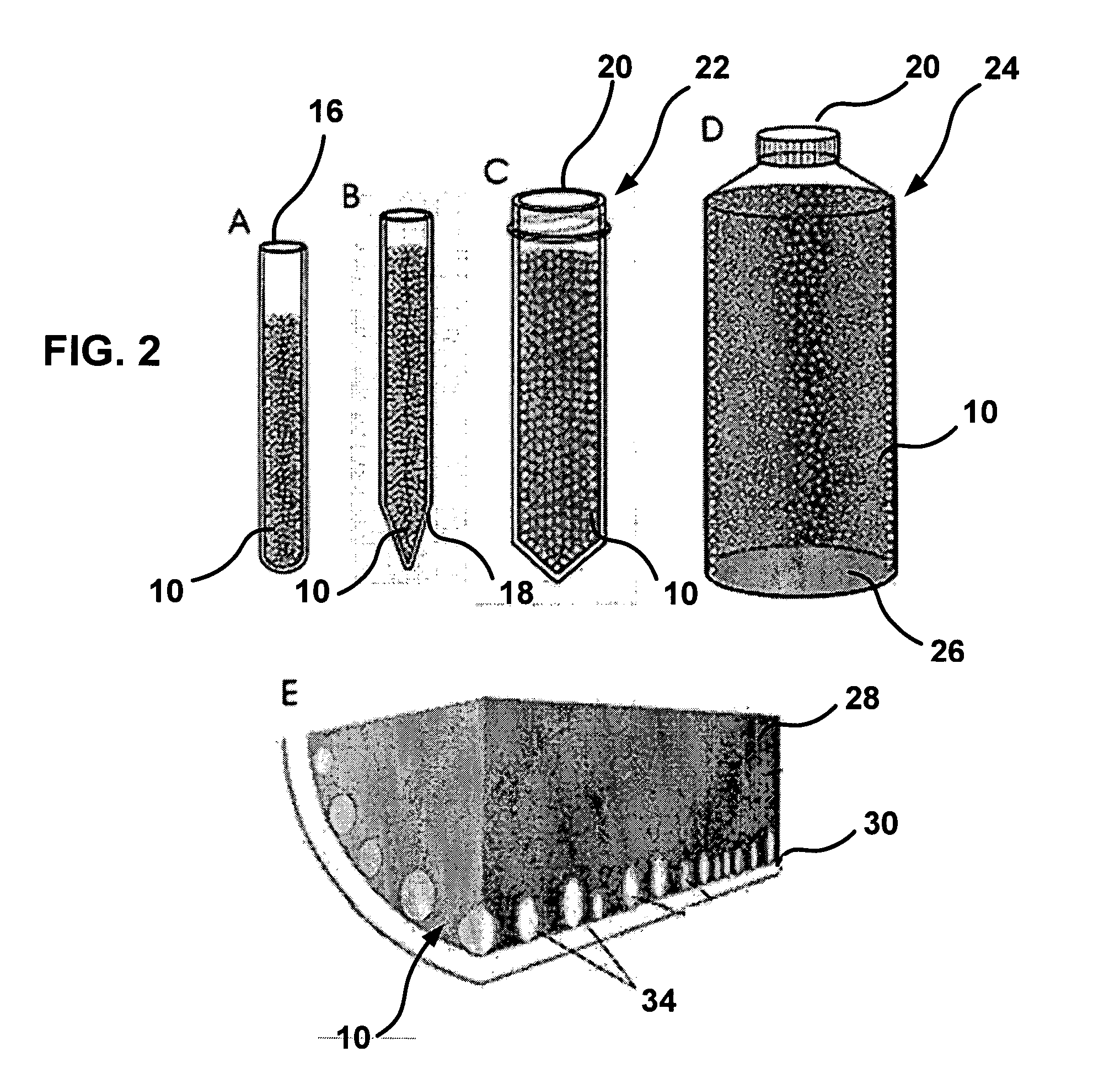
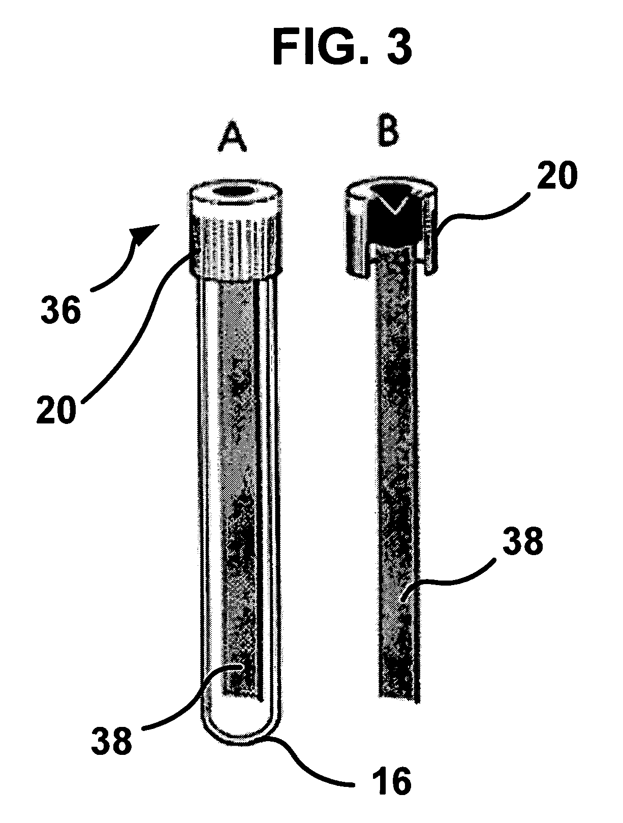
[0072] Whole blood may be placed in a CAM blood collection unit, such as a blood collection tube (FIGS. 2 and 3). The tube may be incubated at about 37° C. and rotated to imitate blood flow so as to increase contact between cells and CAM. Blood may be collected in the presence of anticoagulants, i.e., Anticoagulant Citrate Dextrose solution USP (ACD, Baxter Healthcare Corporation, Deerfield, Ill.) plus 50 units of lithium heparin per mE, to prevent clotting in the CAM blood test unit. The sealed CAM-blood tube may be placed on a roller and rotated at 5-30 cycles per minute at about 37° C., and then incubated for 1-3 hours for cell attachment to occur.

example 2

Specificity and Sensitivity Control

[0073] Human tumor cell lines of different tumor origins may be chosen for use in performing specificity and sensitivity control experiments. For examples, the human colon tumor cell line SW-480, human gastric tumor cell line RF-48, several breast tumor cell lines, human malignant melanoma line LOX, and several ovarian tumor cell lines may be used. Tumor cell lines may be purchased from American Type Culture Collection (Manassas, Va.). All cell lines should be confirmed to be negative for Mycoplasma infection. The tumor cell lines should be examined for: (a) high affinity binding to CAM within one hour after plating; (b) high proliferation rate; and (c) the tumor cell lines should be readily and stably (100%) fluorescently labeled with red or green fluorescent dyes prior to use or transformed with an expression plasmid for green fluorescent protein (GFP) in order to be able to visualize the tumor cells directly at the end of the enrichment procedu...

example 3

Determination of Cell Viability in a CAM-Blood Filtration Unit v. Blood Collection Tube

[0076] Another problem is the cell viability of the blood samples, which may vary during transportation to the research laboratory. Increasing the time of storage may be expected to damage cells in the blood. To determine if tumor cells in the CAM blood unit can stay viable during shipping, 3,000 GFP-tumor cells were spiked into 3 mL of cord blood and control medium containing 15% human serum (Sigma). Each aliquot was stored at 4° C. for series of time (4, 6, 8, 12, 16, 24, 36 and 48 hours). Each aliquot was then captured by CAM and the percent recovery of GFP-tumor cells by CAM determined. For each time point, four duplicate experiments were performed, and percent recoveries determined. The results showed that CAM-captured tumor cells survived better than suspended cells in blood.

[0077] CAM-enriched cells may be counted by any means known to those of ordinary skill in the art, including microsc...


PUM

PropertyMeasurementUnit
diameteraaaaaaaaaa
timeaaaaaaaaaa
timeaaaaaaaaaa

Abstract

Methods and devices for isolating and diagnosing disease with a cell adhesion matrix system, mimicking a metastatic, cardiovascular or placental environment, are disclosed. The cell adhesion matrix facilitates the enrichment of target cells such as metastatic tumor cells, fetal cells and endothelial progenitor cells from a fluid sample such as blood for diagnostic and therapeutic applications in treating patients afflicted with disease, such as cancerous, cardiovascular and fetal diseases, as well as for research applications in molecular analysis of metastatic, and cardiovascular and fetal diseases. Blood test prototypes and methods for the cell enrichment and detection of circulating tumor and endothelial cells using multiplex molecular analysis are described herein. In addition, methods and compositions for determining host immunity to tumor in subjects with risk of cancer progression and methods for isolating an enriched fraction of fetal cells from pregnant females for prenatal diagnosis are also described herein.

Description

CROSS REFERENCE TO RELATED APPLICATIONS [0001] This application claims benefit of priority from U.S. Provisional Patent Application Ser. No. 60 / 516,571, filed on Oct. 31, 2003 and is a continuation-in-part application of U.S. patent application Ser. No. 10 / 122,268, filed on Apr. 11, 2002, which claims benefit of priority from U.S. Provisional Patent Application Ser. No. 60 / 332,408, filed on Nov. 16, 2001, and is further a continuation-in-part of U.S. patent application Ser. No. 10 / 220,347 filed on Aug. 28, 2001 which claims benefit of priority from U.S. Provisional Application Ser. No. 60 / 231,517, filed on Sep. 9, 2000, all of which are herein incorporated by reference in their entirety.BACKGROUND OF THE INVENTION [0002] 1. Field of the Invention [0003] The present invention generally relates to an improved cell adhesion matrix (“CAM”) and an improved cell isolation device for separating target cells such as tumor, fetal and angiogenic cells from blood or other tissue fluid samples ...

Claims


Application Information

Patent Timeline
Patent Type & Authority Applications(United States)
IPC IPC(8): C12M1/34C12Q1/68G01N1/30G01N33/48G01N33/50
CPCG01N1/30G01N33/574G01N33/5091
Inventor CHEN, WEN-TIENCHEN, LEECHEN, CHE
Owner CHEN WEN TIEN
Why Eureka
Try Eureka