Methods for identifying drug pharmacology and toxicology

Inactive Publication Date: 2006-12-07
RIBONOMICS
View PDF3 Cites 12 Cited by
  • Summary
  • Abstract
  • Description
  • Claims
  • Application Information

AI Technical Summary

Benefits of technology

[0017] The invention provides a ex vivo methods and compositions for identifying mechanistic biomarkers and for elucidating potential toxicity and pharmacology of chemical compounds and their underlying mechanisms and pathways. The methods of the invention provide a means for separating and characterizing the pharmacology and toxicity of drug candidates, for example, thiazolidinediones (TZDs), and provide specific screens and biomarkers that allow for population or patient stratification. The methods of the invention thus have significant utility across the drug discovery and development process. In an embodiment, the methods combine a microarray with a cell-based screen of test compounds. The gene content of the microarray focuses on regulators of human gene expression, including regulators of mRNA production (transcription), regulators of mRNA utilization (po

Problems solved by technology
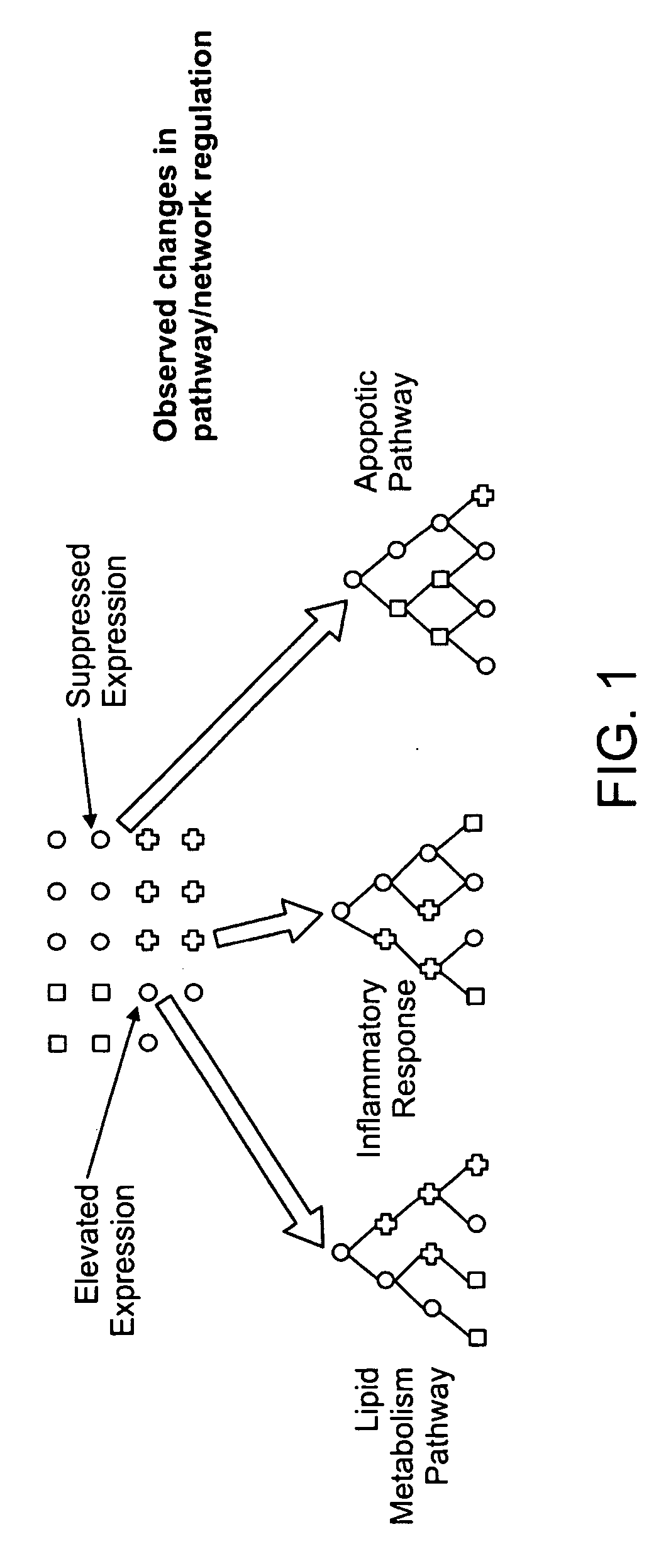
These studies can take years to complete at significant cost to the industry and are often poor indicators of the actual efficacy and safety of drugs in humans.
One of the most significant problems in drug discovery and development is the attrition of compounds.
The attrition rates in earlier stages of development are significantly worse, leading to fewer than 1 in 10,000 early stage candidates making it to market.
Estimates are that the development cost of every drug includes ˜$70 million US dollars for candidates that fail to make it to market.
However many of these assays have proven difficult to scale and therefore have limited scope in terms of the number of compounds that can be assessed.
Although these examples provide some useful information, biomarker discovery, patient stratification and development of personalized medicines is still in its infancy.
However, the datasets generated using these tools are extremely large making them difficult to manage and analyze.
Adding to these challenges, microarray data sets from large survey arrays such as these have proven to be extremely noisy and poorly reproducible.
This makes detection of low abundance transcripts and detection of modest, but biologically significant changes in gene expression extremely challenging using these tools.
Importantly, many regulatory molecules, including certain transcription factors, are expressed at low levels and modest changes in their expression level can signal or result in significant biological consequences.
These factors combine to make elucidation of biological mechanisms extremely challenging using existing tools.
Although these tools are attractive complement

Method used


Image

  • Methods for identifying drug pharmacology and toxicology
  • Methods for identifying drug pharmacology and toxicology
  • Methods for identifying drug pharmacology and toxicology

Examples


Example

Example 1

Determining the Cytotoxicity of Test Compounds

[0063] HepG2 cells were obtained from American Type Culture Collection (ATCC, Manassas, Va., cat. no. HB-8065). Cells were maintained as recommended in Minimal Essential Medium (MEM) (Gibco-BRL, a Division of Invitrogen, Carlsbad, Calif.) with 10% fetal bovine serum (FBS, HyClone, Logan, Utah) supplemented with antibiotics in p150 plates at 37° C., 5% CO2. Cells were split 1:5 and fresh media added every 3 days.

[0064] Cytotoxicity was assessed using the Alamar Blue-based CellTiter™ Blue Cell Viability Assay (Promega, Madison, Wis.) to determine the viable cell fraction that remained following a 72 hour treatment period. Cells (˜8,000 cells / well) were plated in 96 well BioCoat collagen coated plates (Becton Dickinson, Franklin Lakes, N.J.) using standard media. This allowed untreated control samples (0.25% DMSO) to be in late log phase (˜70% confluent) at completion of the study. Cells were then allowed to recover for 24 hours...

Example

Example 2

Determining the Apoptosis in Response to Test Compounds

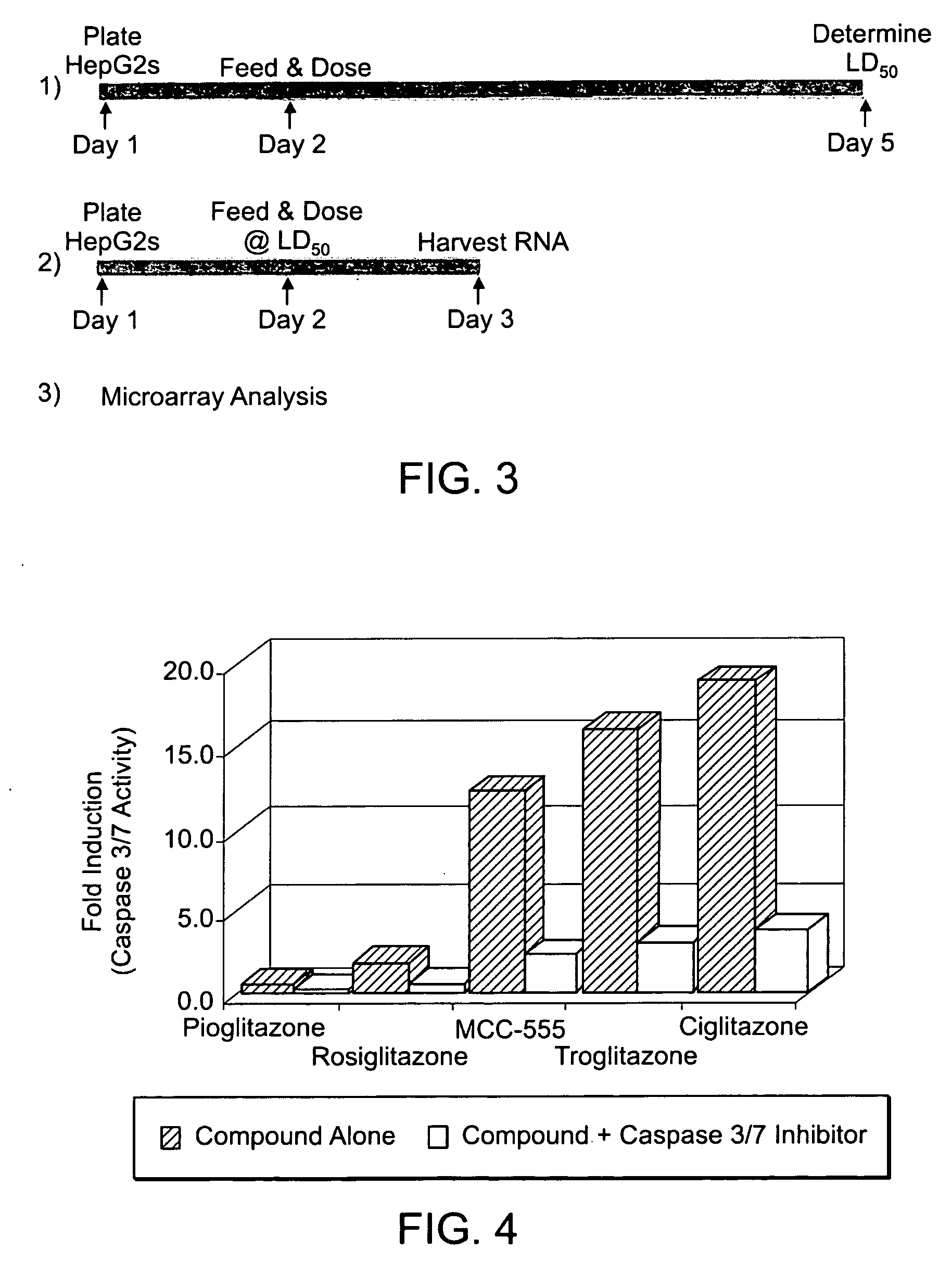
[0065] Apoptosis was assessed using the Apo-OneR Homogeneous Caspase-3 / 7 Assay (Promega) to determine the activity of an early apoptotic event: Caspase 3 / 7 activation. Cells (˜40,000 cells / well) were plated in 96 well plates (Corning, Acton, Mass., cat. no. 3595) using plating media (MEM, 1× Sodium Pyruvate, 1× NEAA, 10% FBS). Cells were then allowed to grow for 24 hours at 37° C., 5% CO2, and then serum starved by changing to serum free media (MEM, 1× Sodium Pyruvate, 1× NEAA, 0.1% BSA). Cells were allowed to remain in the serum free media for a further 24 hours. At 48 hours post-plating the media was removed and replaced with a test compound diluted in serum free media. A dilution series was created for each compound through serial dilutions performed in a separate plate and later transferred to the cells. Initially, a broad dilution series was conducted from ˜300 μM to ˜1 μM to determine approximate maximum tolerat...

Example

Example 3

Preparation of RNA

[0066] RNA for microarray analysis was obtained from cells treated for 24 hours at the determined LD50. Typically, ˜1.5×10⁶ cells were plated in a p100 dish and allowed to settle for 24 hours by incubation at 37° C., 5% CO2 in MEM+10% FBS without antibiotics. Old media was removed and fresh MEM+0.1% BSA without antibiotics containing a test compound at LD50 concentration and 0.25% DMSO was added to the flask. A vehicle-only treatment was also performed. Duplicate treatments were performed for each compound as well as for vehicle-only controls. The cells were incubated with compound for 24 hours at 37° C., 5% CO2 and were harvested by scraping (without trypsinization) and centrifugation. The cell pellets were flash frozen and stored at −80° C. until ready for RNA extraction.

[0067] Total RNA was isolated using RNeasy Midi or Maxi kits (Qiagen) according to methods described by the manufacturer. Total RNA (100 μg) was routinely treated with 40 Units DNaseI...


PUM

PropertyMeasurementUnit
Timeaaaaaaaaaa
Timeaaaaaaaaaa
Molar densityaaaaaaaaaa

Abstract

The invention combines a microarray and cell-based screening strategy that enables rapid identification of possible mechanisms underpinning the pharmacology and toxicology of drug candidates. The methods of the invention identified unique properties relating to apoptosis and the anti-inflammatory response elicited by several peroxisome proliferator activated receptor gamma (PPARγ) ligands. The methods illustrate, for example, that PPARγ ligands that are safe and effective drugs (e.g., Actos, Avandia) either do not induce apoptosis or only modestly induce apoptosis. Conversely, PPARγ ligands that have failed clinical development (e.g., Ciglitazone; Day, C., Diabet. Med., 16: 179-192 (1999)) or that have been withdrawn from the market (e.g., Troglitazone (Rezulin)) due to hepatotoxicity are potent inducers of apoptosis. The methods of the invention also illustrate that suppression of gene expression and protein expression for several pro-inflammatory factors by some PPARγ ligands occurs as a consequence of apoptotic induction (i.e., apoptosis produces an anti-inflammatory response). The invention also provides biomarkers for cellular pathways and methods for stratifying patient groups according to their biomarker expression as well as biomarkers that discriminate safe and effective drugs from compounds that have acute toxicities. These biomarkers provide novel insights into the mechanism of action and toxicity for test compounds, including cell death, anti-inflammatory activity, hepatotoxicity, and carcinogenicity. The methods are highly scalable and have broad application from discovery to the clinic, including compound prioritization, predictive pharmacology and toxicology; mechanism of action studies; and prognostic and diagnostic biomarker discovery.

Description

CROSS-REFERENCE TO RELATED APPLICATION [0001] This application claims priority to U.S. Provisional Patent Application No. 60 / 687,966, filed on Jun. 7, 2005 the entire contents of which is incorporated by reference.GOVERNMENT LICENSE RIGHTS [0002] The U.S. Government has a paid-up license in this invention and the right in limited circumstances to require the patent owner to license others on reasonable terms as provided for by the terms of DAMD17-03-1-0516 awarded by The Department of Defense(DOD) Breast Cancer Research Program.FIELD OF THE INVENTION [0003] The invention relates to ex vivo methods for identifying or predicting drug pharmacology and toxicology in vivo and for identifying biomarkers using a microarray and / or cell-based assay. BACKGROUND OF THE INVENTION [0004] Preclinical testing of drug pharmacology and toxicology is generally based on the results from a series of biochemical, cellular and animal studies that together are used to select the most promising drug candid...

Claims


Application Information

Patent Timeline
IPC IPC(8): C12Q1/68
CPCC12Q1/6809C12Q1/6837C12Q2600/136C12Q2600/142C12Q2600/158C12Q1/6883
Inventor HENDERSON, BARRYCHEATHAM, RICHARD
Owner RIBONOMICS
Why Eureka
Try Eureka