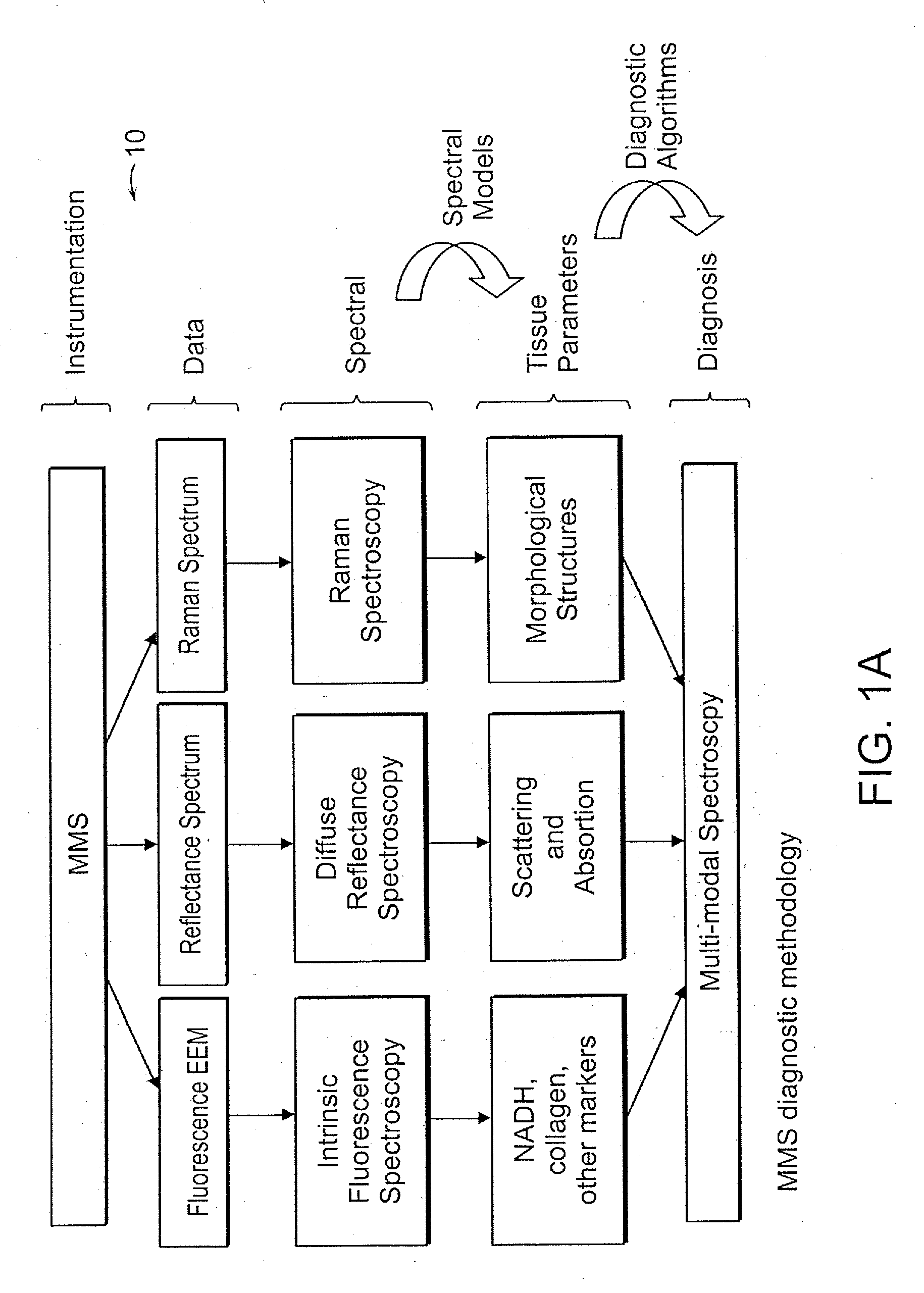
[0009]The combination of modalities in the
modal spectroscopy (TMS) has several advantages over the single modalities alone. First,
fluorescence spectroscopy provides information about tissue metabolites, such and NADH, that are not provided by
Raman spectroscopy. Second, TMS uses diffuse
reflectance spectroscopy (DRS) to overcome
distortion of
fluorescence signatures by the effects of tissue absorption and scattering, and extract the
intrinsic fluorescence signature (IFS). Third, in addition to its value in extracting IFS, DRS provides critical information about the tissue absorbers and scatterers themselves. Finally, while DRS provides information about tissue components responsible for diffusive scattering,
light scattering spectroscopy (LSS) provides information about tissue components responsible for single backscattering. The combination of techniques into TMS, therefore, provides a wealth of information about tissue fluorophores, absorbers and scatterers, which creates a much more complete biochemical, morphologic and metabolic tissue profile.
[0013]The combination of TMS and
Raman spectroscopy in MMS provides a more complete and complementary biochemical, morphologic and metabolic tissue profiles than either TMS or Raman spectroscopy alone resulting in better
diagnostic accuracy. Another key
advantage in combining both techniques is the potential for depth sensing. TMS and Raman spectroscopy can use different excitation wavelengths, and therefore sample different tissue volumes because of
wavelength-dependent differences in absorption and scattering. A Raman source preferably emits in a range of 750 nm to 1000 nm while the fluorescence source can employ one or more
laser sources or a filtered
white light source. Reflectance measurements preferably use a
broadband source such as
xenon flash lamp.
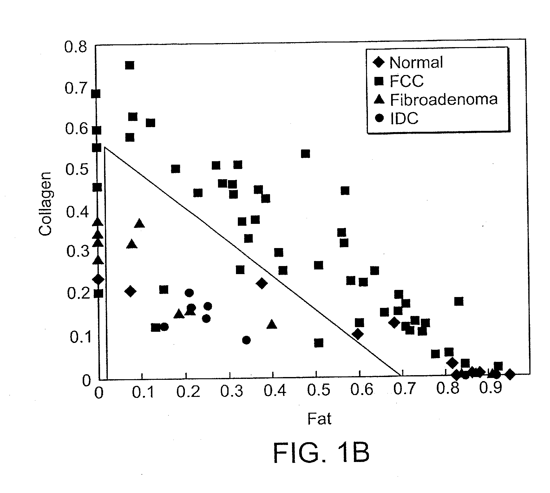
[0014]This difference in sampling volume can be exploited to provide information about the depth (or thickness) of certain tissue structures or
layers. For example, the thickness of the fibrous cap is used for the diagnosis of vulnerable atherosclerotic plaque. The fibrous cap is composed largely of collagen. IFS and Raman spectroscopy both provide information about the contribution of collagen to tissue spectra. Comparison of the results from these two techniques, which use different excitation wavelengths and sample different tissue volumes, may provide information about the thickness of the fibrous cap. DRS and Raman spectroscopy both provide information about the contribution of deoxy-
hemoglobin to the tissue spectra. Comparison of the results of these two techniques, which again use different excitation wavelengths and sample different tissue volumes, can provide depth-sensitive information useful in mapping cancers and pre-cancers of
breast tissue.
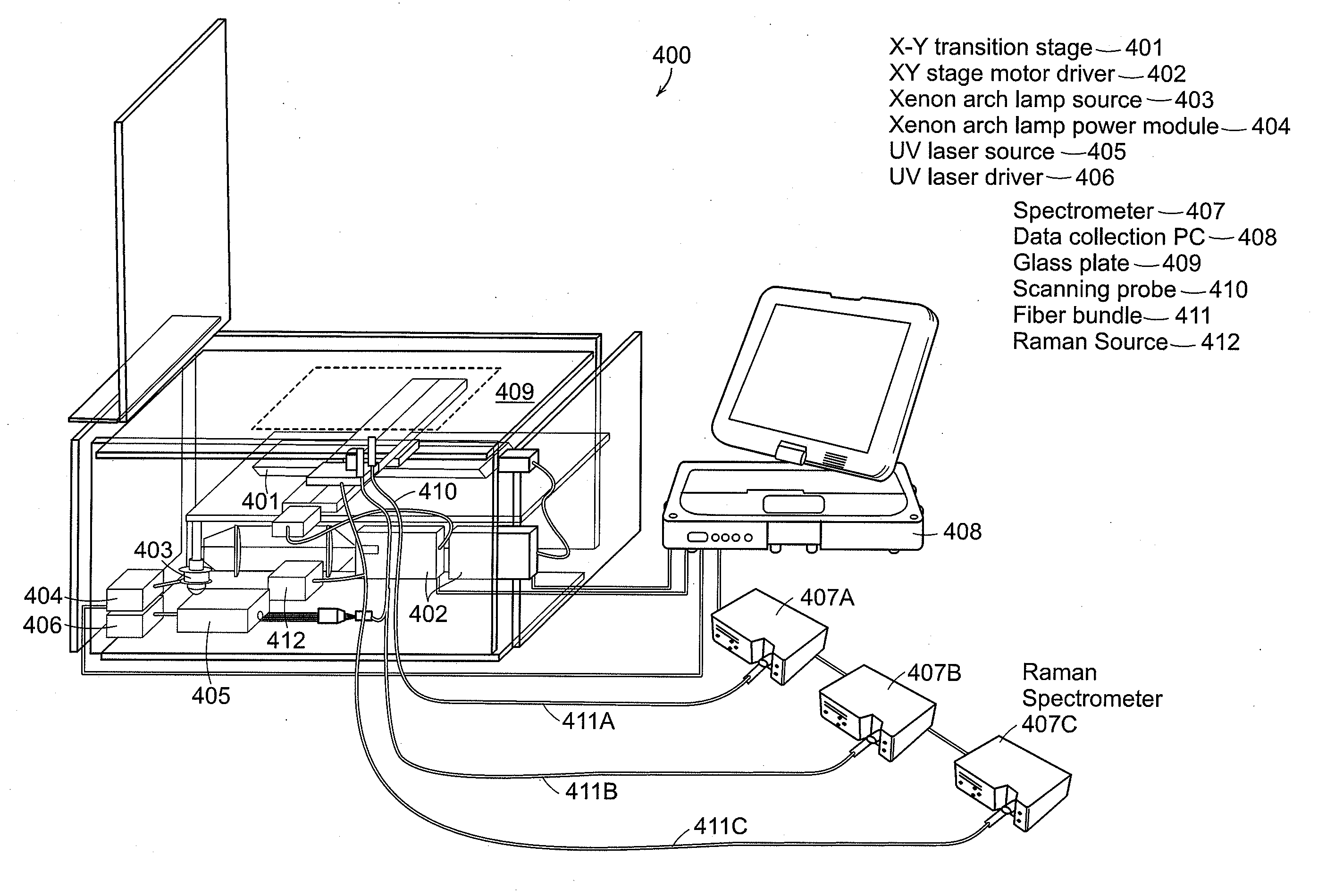
[0018]Preferred embodiments of the invention relate to a portable, quantitative,
optical fiber probe-based, spectroscopic tissue
scanner designed for intraoperative diagnostic imaging of surgical margins. The tissue
scanner combines diffuse
reflectance spectroscopy (DRS) and
intrinsic fluorescence spectroscopy (IFS), and has
hyperspectral imaging capability, acquiring full DRS and IFS spectra for each scanned image pixel. A preferred embodiment can incorporate Raman detection into the probe used for scanning the
region of interest. Modeling of the DRS and IFS spectra yields quantitative parameters that reflect the metabolic, biochemical and morphological state of tissue, which are translated into
disease diagnosis. The tissue
scanner has
high spatial resolution (0.25 mm) over a
wide field of view (10×10 cm), for example, and both high
spectral resolution (2 nm) and high spectral contrast, readily distinguishing tissues with widely varying optical properties (bone,
skeletal muscle, fat and
connective tissue). Tissue-simulating phantom measurements confirm that the tissue scanner can quantitatively measure spectral parameters, such as
hemoglobin concentration, in a physiologically relevant range with a high degree of accuracy (<5% error). Measurements using
human breast tissues showed that the tissue scanner can detect small foci of
breast cancer in a background of
normal breast tissue. This tissue scanner is simpler in design, images a larger
field of view at higher resolution and provides a more physically meaningful tissue diagnosis than existing spectroscopic imaging systems. This spectroscopic tissue scanner can provide real-time, comprehensive diagnostic imaging of surgical margins in excised tissues, overcoming the sampling limitation in current
histopathology margin assessment. Preferred embodiments can use a
fiber optic probe for manual scanning of surgical sites.
[0022]A preferred embodiment of the present invention includes a portable, quantitative,
optical fiber probe-based, spectroscopic tissue scanner that can provide real-time comprehensive assessment of surgical margins in excised tissue specimens. The scanner significantly extends existing
optical fiber probe-based spectroscopy instruments, which can be employed in the diagnosis of oral, esophageal, cervical,
skin and
breast cancer but previously unavailable in a
wide field,
high resolution imaging
system. This tissue scanner is simpler in design, images a larger
field of view at higher resolution and provides a more physically meaningful tissue diagnosis. The tissue scanner can provide fast, accurate, diagnostic images of the entire margin of excised surgical specimens, overcoming the sampling limitation in current
pathology margin assessment.
[0023]A preferred embodiment can employ an inverted geometry with the sample placed on a optically transparent support and scanned by providing relative movement between a
fiber optic
light delivery and
collection system and the
tissue sample. Either the
tissue sample or the
fiber optic probe can be scanned to achieve the desired scan area, resolution and
scan time. These scan parameters can be selected based on the size and geometry of the
tissue sample. Note that pressure can be applied uniformly across the
tissue surface to provide contact with the scanned surface. This provides precise spot size and distance to the probe to achieve this required pixel by pixel registration of images.

