Dyes for analysis of protein aggregation

a protein aggregation and dye technology, applied in the field of dyes, can solve the problems of irreversible aggregation, protein aggregation mechanism, and eventual precipitation, and achieve the effects of reducing the number of dyes, and improving the quality of dyes

Inactive Publication Date: 2013-05-30
ENZO LIFE SCI INC
View PDF2 Cites 8 Cited by
  • Summary
  • Abstract
  • Description
  • Claims
  • Application Information

AI Technical Summary

Benefits of technology

[0143]Homogeneous probe dependent assays are also well known in the art and may take advantage of the present invention. Examples of such methods are energy transfer between adjacent probes (U.S. Pat. No. 4,868,103), the Taqman exonuclease assay (U.S. Pat. No. 5,538,848 and U.S. Pat. No. 5,210,015), Molecular Beacons (U.S. Pat. No. 5,118,801 and U.S. Pat. No. 5,925,517) and various real time assays (US Patent Application Publication 2005 / 0137388), all of which are incorporated by reference.
[0144]Antibodies labeled with dyes of the present invention may be used in various formats. For example, an antibody with one of the dyes of the present invention may be used in an immunofluorescent plate assay or in situ analysis of the cellular location and quantity of various antigenic targets. Antibodies labeled with dyes may also be used free in solution in cell counting or cell sorting methods that use a flow cytometer or for in-vitro and in-vivo imaging of animal models.
[0145]The presence or absence of a signal may then be used to indicate the presence or absence of the target itself. An example of this is a test where it is sufficient to know whether a particular pathogen is present in a clinical specimen. On the other hand, quantitative assays may also be carried out where it is not so much the intention of evaluating if a target is present but rather the particular amount of target that is present. An example of this is the previously cited microarray assay where the particular rise or fall in the amount of particular mRNA species may be of interest.
[0146]In another embodiment of the present invention, dyes that have been disclosed above as well as dyes described previously in the literature may be attached to a carrier with a more general affinity. Dyes may be attached to intercalators that in themselves do not provide signal generation but by virtue of their binding may bring a dye in proximity to a nucleic acid. A further example is attachment of dyes to SDS molecules thereby allowing dyes to be brought into proximity to proteins. Thus this embodiment describes the adaptation of a dye or dyes that lack affinity to a general class of molecules may be adapted by linking them to non-dye molecules or macromolecules that can convey such properties.
[0147]Various applications may enjoy the benefits of binding the dyes of the present invention to appropriate targets. As described above, staining of macromolecules in a gel is a methodology that has a long history of use. More recent applications that also may find use are real time detection of amplification (U.S. Pat. No. 5,994,056, U.S. Pat. No. 6,174,670 and US Patent Application Publication 2005 / 0137388, all of which are hereby incorporated by reference), and binding of nucleic acids to microarrays. In situ assays may also find use where the binding of dyes of the present invention is used to identify the location or quantity of appropriate targets.
[0148]In other aspects, this invention provides a composition comprising a solid support to which is attached any of the above-described reactive compounds. In some embodiments, the solid support comprises glass particle, glass surface, natural polymers, synthetic polymers, plastic particle, plastic surface, silicaceous particle, silicaceous surface, glass, plastic or latex beads, controlled pore glass, metal particle, metal oxide particle, microplate or microarray, or any combination thereof. The aforementioned reactive group for attachment comprises or may have comprised an electrophilic reactive group comprising isocyanate, isothiocyanate, monochlorotriazine, dichlorotriazine, 4,6,-dichloro-1,3,5-triazines, mono- or di-halogen substituted pyridine, mono- or di-halogen substituted diazine, maleimide, haloacetamide, aziridine, sulfonyl halide, acid halide, hydroxysuccinimide ester, hydroxysulfosuccinimide ester, imido ester, hydrazine, azidonitrophenol, azide, 3-(2-pyridyl dithio)-propionamide, glyoxal or aldehyde groups, a nucleophilic reactive group comprising reactive thiol, amine or hydroxyl, a nickel coordinate group, a platinum coordinate group, a terminal alkene or a terminal alkyne, and any combination of the foregoing. As in the case of other embodiments previously described above, a linker arm can be usefully positioned between the compound and the reactive group, or between the solid support and the reactive group.Reagent Kits

Problems solved by technology

Structurally altered proteins have an especially strong tendency to aggregate, often leading to their eventual precipitation.
Irreversible aggregation is a major problem for the long-term storage and stability of therapeutic proteins and for their shipment and handling.
In the bioprocessing arena, the mechanisms of protein aggregation are still not fully understood, despite the fact that aggregation is a major problem in therapeutic protein development (Arakawa et al., 2006).
If the contaminant is the damaged protein itself, then its aggregation may lead to soluble oligomers, which become larger aggregates, visible particulates, or insoluble precipitates.
Damaged forms of a protein product can also arise from chemical modification (such as oxidation or deamidation) and from conformationally damaged forms arising from thermal stress, shear, or surface-induced denaturation.
Further, reversible self-association of proteins can significantly alter overall pharmaceutical properties of product solutions, such as solution viscosity.
Detection of reversible aggregates can be an especially challenging task.
One consequence of the complexities of monitoring aggregate formation processes is the difficulty of linking the effect (presence of aggregates) to its underlying cause, particularly because the key damage may occur at a time or place quite separated from the observed consequence.
Since the earliest clinical applications of protein pharmaceuticals in medicine, aggregation problems have been implicated in adverse reactions in humans and other safety issues.
AUC-SV suffers from lower precision than SEC, however.
Despite its advantages, AUC-SV is not yet readily amenable for use as a routine release test in the biotechnology industry because of issues related to low throughput, the need for specialized equipment, performance problems at high protein concentrations, the need for skilled practitioners of the method, and difficulty in validating data analysis software.
This method is highly sensitive to large aggregates because the intensity of scattered light increases proportionally with molecular weight.
Although this method is ideal for detecting very low mass fractions of large aggregates, it cannot resolve species that are similar in size.
DLS is also not amenable to use as a control method because it is semi-quantitative and very sensitive to dust or other extraneous particles.
When compared with SEC, the precision and limit of detection of aFFF is inferior in the high-molecular-weight range, because of increased baseline noise.
Experimental conditions (e.g., cross-flow rate) for reasonable separations in one size range are also not generally applicable to other size ranges, making the technique cumbersome, especially when analyzing a broad range of masses.
Along with other limitations, such as the need for specialized equipment and a skilled operator, and the difficulty in validating the method prevents the use of aFFF in applications for release and stability monitoring.
SEC cannot handle a large range of sizes because the pore size or degree of polymerization of the resin must be adjusted to the size of the protein species.
If a protein sample contains widely different sizes, many techniques are unsuitable for analyzing all sizes simultaneously.
FFF and DLS can cover a very large range of sizes, but in the case of DLS, resolution is generally fairly poor, and FFF entails some trade-off between resolution and dynamic range.
The resolution of SV-AUC is generally not ideal for separating monomer from dimer, compared with the best SEC columns (especially for lower molecular weight proteins).
The quantification of species that elute from SEC or FFF is quite good, but aggregates can easily be lost during the separation process.
Thus, SEC and FFF may provide good precision but poor accuracy.
For SV-AUC, loss of protein aggregates to surfaces is usually not a problem, but accurate quantification of small oligomers (dimer-tetramer) at total levels of ˜2% or less is quite difficult.
DLS is for all practical purposes useless for detecting oligomers smaller than an octamer, because the technique cannot resolve such oligomers from monomeric species, and for those protein aggregate species that are resolved, the accuracy of the weight fractions is quite poor, typically plus or minus factors of two to ten.
Overall, no single analytical technique is ideal for every protein or is optimal for analyzing the wide range of aggregation problems that can arise with protein pharmaceutical formulation.
Amyloid fibril detection assays have suffered from several drawbacks, however, when using thioflavin T, Congo red and their derivatives.
Furthermore, the binding of dyes can influence the stability of amyloid aggregates, and the interplay with other components (for example, during testing of potential amyloid inhibitors) is unpredictable (Murakami et al., 2003).
Despite the widespread use of thioflavin T, its application to amyloid quantification often generates inconsistent and inaccurate results.
Variations in spectral properties caused by buffer conditions and protein-dye ratios result in poor reproducibility, complicating the use of thioflavin T for quantitative assessment of fibril formation.
When the delivery device is worn close to the body or implanted within the body, a patient's own body heat and body motion, plus turbulence generated in the delivery tubing and pump, impart a high level of thermo-mechanical stress to a protein formulation.
In addition, infusion delivery devices expose the protein to hydrophobic interfaces in the delivery syringes and catheters.
These interfacial interactions tend to destabilize the protein formulation by inducing denaturation of the native structure of the protein at these hydrophobic interfaces.
The formulation of protein drugs is a difficult and time-consuming process, mainly due to the structural complexity of proteins and the very specific physical and chemical properties they possess.
The conventional analytical methods usually require a long period of time to perform, typically twenty or more days, as well as manual intervention during this period.
The development of new formulations is costly in terms of time and resources.
Moreover, even for a known protein formulation, batch to batch quality control analysis is often less than optimal using the current state of the art methods.
This type of technology usually requires a high protein concentration, therefore, it is not cost-effective.
In addition, thermal shift technology cannot work effectively on formulations with low protein concentrations or finalize protein formulations which require a very low detection limit (typically ˜1-5% protein aggregates).
One of the major complications encountered in both in vitro and in vivo protein folding is aggregation resulting from the commonly encountered low solubility of the unfolded protein or different folding intermediates.
Upon heating, BLG aggregates, which can be accelerated by subjecting the protein to either an elevated pH or through the additional of DTT. α-crystallin prevents heat-induced BLG aggregation, acting as a chaperone in the absence of DTT; in the presence of DTT, however, this chaperone activity is less efficient due to faster aggregation of heated and reduced beta-lactoglobulin.

Method used


Image

  • Dyes for analysis of protein aggregation
  • Dyes for analysis of protein aggregation
  • Dyes for analysis of protein aggregation

Examples


example 1

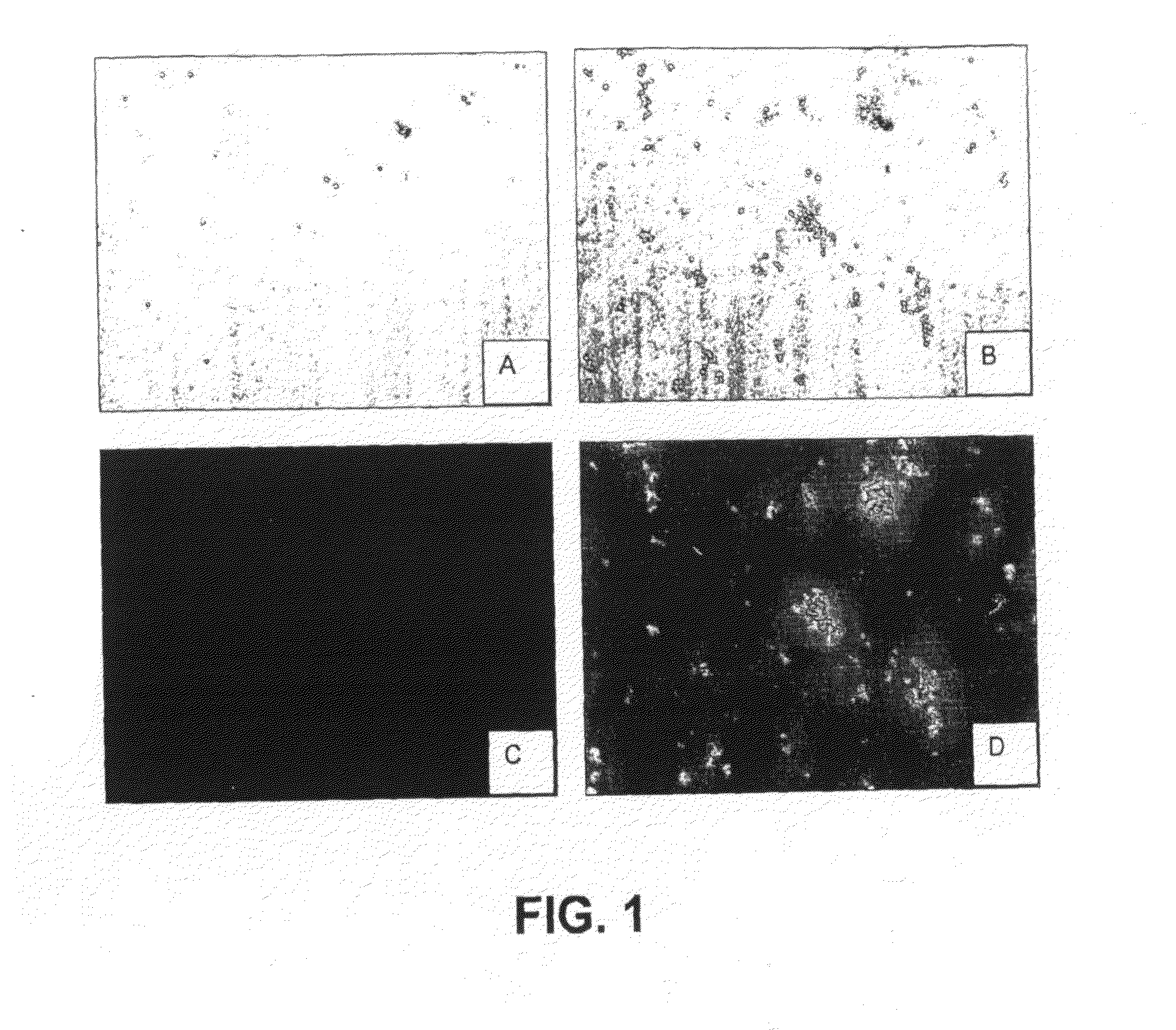
Testing Compounds for Ability to Sense Protein Aggregation

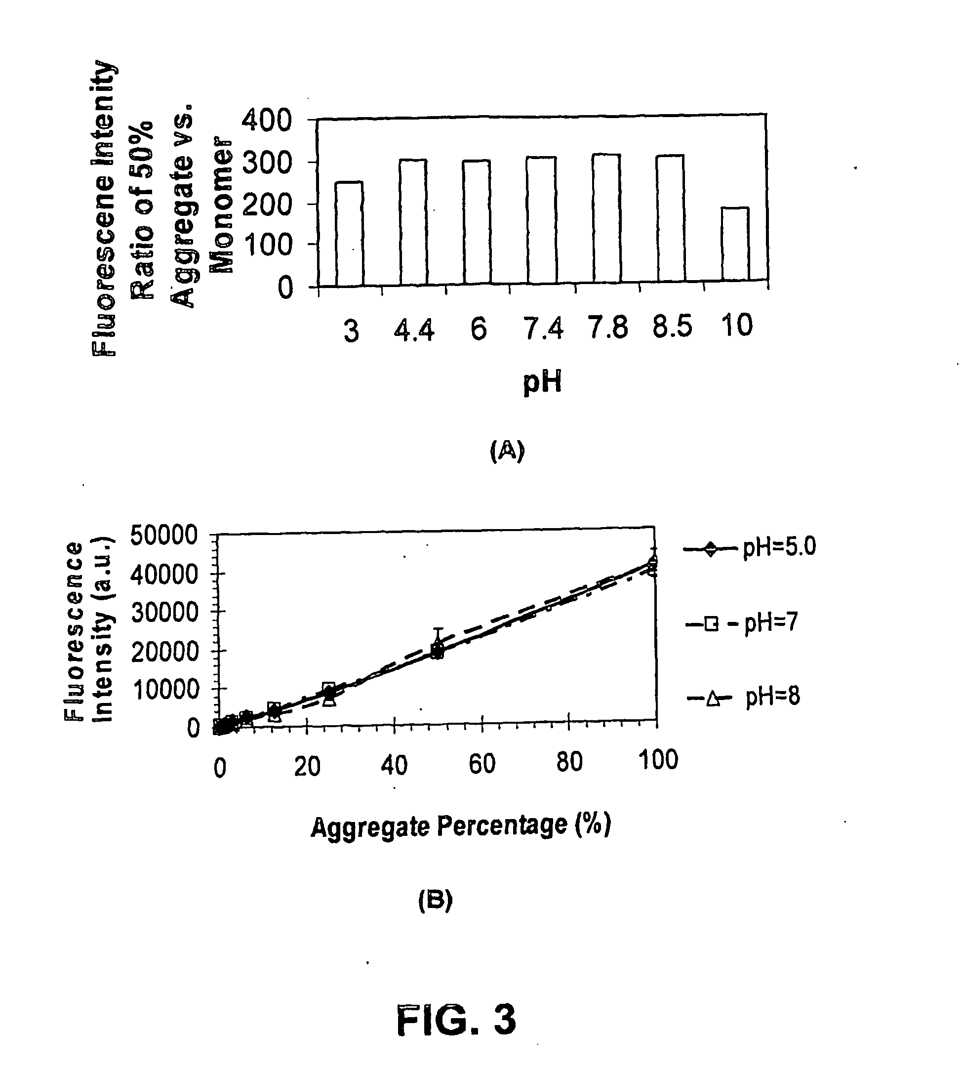
[0225]Fluorescent readings were carried out in 50 mM Tris-HCl, pH 7.8 using 10 μM dye. When present, 1 μM recombinant human α-synuclein (ASN, Sigma-Aldrich, St. Louis, Mo.) as monomers, or aggregated as described in van Raaij et al. (2006) was included. Fluorescence excitation and emission spectra were collected on a Cary Eclipse fluorescence spectrophotometer (Varian, Australia). Fluorescence spectra were measured with excitation and emission slit widths set to 5 nm, and at a constant PMT voltage. Spectroscopic measurements were performed in standard quartz cells. All measurements were made at the respective excitation maxima of each dye. All measurements were carried out at room temperature. Results are summarized in Tables 1 and 2.

example 2

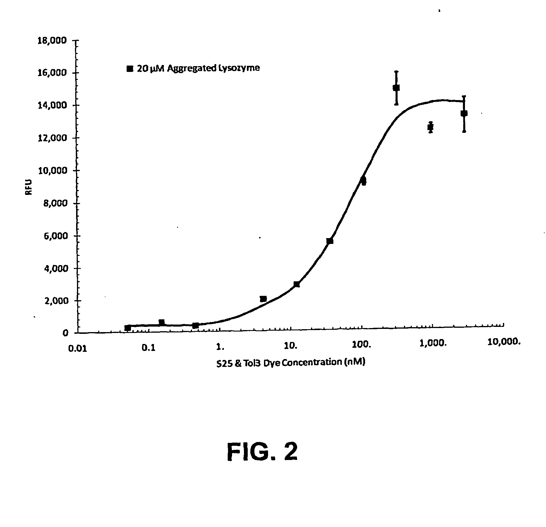
Fluorescence Sensitivity of Different Protein Aggregate-Sensing Dyes in the Presence of Excipients

[0226]IgG aggregate was prepared by adjusting 5.83 mg / ml of purified goat-anti-mouse IgG (H&L, Pel Freez, Rogers, Ark.) to pH 2.7 using HCl and incubating at 22° C. for 24 hours. The assay was performed using 2.8 μM IgG, either native or aggregated, and a dye concentration of 0.625 μM. The protein and dye were mixed together for 15 minutes at 22° C., then further incubated in the presence of the excipients shown in Table 5. The fluorescence intensity of S-25, Tol3 and Y2150 were determined with a FLUOstar OPTIMA plate reader (BMG LABTECH) at excitation wavelength of 550 nm and emission wavelength of 610 nm; while the fluorescence intensity for thioflavin-T was determined using a SpectraMAX GeminiXS (Molecular device, with Softmax Pro 7.0) using an excitation wavelength of 435 nm and emission wavelength of 495 nm. The fluorescence enhancement (aggregate / native IgG) is shown.

TABLE 5Effect...

example 3

Synthesis of S25

(a) Preparation of 6-methylsulfonyloxyhexyl methylsulfonate (Compound 1)

[0227]A solution of 1,6-hexanediol (13.15 g, 111.3 mmol) in 70 mL of anhydrous pyridine was cooled to 0° C. using ice bath. To this methanesulfonyl chloride (27 g, 235.7 mmol) was slowly added under mixing such that the temperature was maintained at 5-6° C. The combined mixture was stirred overnight at the temperature below 10° C. and the precipitate formed was filtered off, washed with 20% HCl (3×), water (3×), 5% solution of sodium bicarbonate (3×), and then again with water (3×). Product was dried under vacuum to obtain Compound 1 as a white solid (yield 32.8%). The structure of Compound 1 is given below:

(b) Preparation of Compound 2

[0228]A mixture of 4-methylpyridine (3.06 g, 32.9 mmol) and Compound 1 (4.11 g, 15 mmol) was heated at 120° C. for 3 hours. The reaction mixture was cooled and then 4 mL of isopropyl alcohol was added and the combined mixture was refluxed for an hour. After cooling...


PUM

PropertyMeasurementUnit
temperaturesaaaaaaaaaa
time intervalsaaaaaaaaaa
temperatureaaaaaaaaaa

Abstract

Provided are dyes and compositions which are useful in a number of applications, such as the detection and monitoring protein aggregation, kinetic studies of protein aggregation, neurofibrillary plaques analysis, evaluation of protein formulation stability, and analysis of molecular chaperone activity.

Description

CROSS-REFERENCE TO RELATED APPLICATIONS[0001]This is a continuation-in-part application of U.S. application Ser. No. 12 / 592,639, filed Nov. 30, 2009.BACKGROUND OF THE INVENTION[0002](1) Field of the Invention[0003]The present application generally relates to dyes and compositions comprising dyes. More particularly, provided are dyes and compositions for identifying and quantifying protein aggregation.[0004](2) Description of the Related Art[0005]The deposition of insoluble protein aggregates, known as amyloid fibrils, in various tissues and organs is associated with a number of neurodegenerative diseases, including Alzheimer's, Huntington's and Parkinson's diseases, senile systemic amyloidosis and spongiform encephalopathies (Volkova et al., 2007; Stefani & Dobson, 2003). Fibrillar deposits with characteristics of amyloid are also formed by several other proteins unrelated to disease, including the whey protein beta-lactoglobulin (BLG). All amyloid fibers, independent of the protein...

Claims


Application Information

Patent Timeline
Patent Type & Authority Applications(United States)
IPC IPC(8): C07D401/12
CPCC07D401/12C09B23/141G01N2800/2821G01N2800/2835C09B23/0008C09B23/145C09B23/0058C09B23/0066C09B23/04C09B23/06C09B23/102C09B23/0025G01N33/582G01N33/6845G01N2458/30
Inventor PATTON, WAYNE FORRESTYARMOLUK, SERGIY M.PANDE, PRAVEENKOVALSKA, VLADYSLAVADAI, LIJUNVOLKOVA, KATERYNACOLEMAN, JACKLOSYTSKYY, MYKHAYLOLUDLAM, ANTHONYBALANDA, ANATOLIYSHEN, DEE
Owner ENZO LIFE SCI INC
Why Eureka
Try Eureka