
Bacteriophages expressing amyloid peptides and uses thereof

a technology of amyloid peptides and bacteria, applied in the direction of peptides, drug compositions, peptides, etc., can solve the problems of difficult disinfection of other industrial surfaces, difficult elimination of bacteria biofilms, and contaminating surfaces of biofilms, so as to prevent the self- inhibit amyloid formation, and prevent the aggregation of amyloid peptides

Inactive Publication Date: 2012-11-29
TRUSTEES OF BOSTON UNIV
View PDF1 Cites 16 Cited by
  • Summary
  • Abstract
  • Description
  • Claims
  • Application Information

AI Technical Summary

Benefits of technology

[0225]One particular benefit of an anti-amyloid peptide engineered bacteriophage expressing an anti-amyloid peptide, and a method of using it according to methods disclosed herein is the presence of the anti-amyloid in the immediate locality of the bacteriophage, thus the anti-amyloid peptide is released from bacterial host cells infected with the bacteriophage, via either lysis or being secreted, allowing the anti-amyloid peptide to inhibit the formation of, or maintainance of amyloid. Additionally, another advantage of delivering the anti-amyloid peptides by being expressed by a bacteriophage is that it enables the anti-amyloid peptides to come into contact with amyloids which may not be accessible using conventional methods, for example it allows the anti-amyloid peptides to be within the locality of biofilms in difficult to reach places due to the bacteria being located in a difficult to access location, such as a small space or between two pieces of material. As such, another advantage of the present invention is an improved genetically engineered bacteriophage which express anti-amyloid peptides within the near vicinity of amyloids, such as curli amyloid in biofilms produced by bacterial cells, which may not be accessible to anti-amyloid peptides delivered by other means.
[0226]Without wishing to be bound to theory, when proteins are expressed by a cell, including a bacterial cell, the proteins are targeted to a particular part in the cell or secreted from the cell. Thus, protein targeting or protein sorting is the mechanism by which a cell transports proteins to the appropriate positions in the cell or outside of it. Sorting targets can be the inner space of an organelle, any of several interior membranes, the cell's outer membrane, or its exterior via secretion. This delivery process is carried out based on information contained in the protein itself. Correct sorting is crucial for the cell; errors can lead to diseases.
[0227]With some exceptions, bacteria lack membrane-bound organelles as found in eukaryotes, but they may assemble proteins onto various types of inclusions such as gas vesicles and storage granules. Also, depending on the species of bacteria, bacteria may have a single plasma membrane (Gram-positive bacteria), or both an inner (plasma) membrane and an outer cell wall membrane, with an aqueous space between the two called the periplasm (Gram-negative bacteria). Proteins can be secreted into the environment, according to whether or not there is an outer membrane. The basic mechanism at the plasma membrane is similar to the eukaryotic one. In addition, bacteria may target proteins into or across the outer membrane. Systems for secreting proteins across the bacterial outer membrane may be quite complex and play key roles in pathogenesis. These systems may be described as type I secretion, type II secretion, etc.
[0228]In most Gram-positive bacteria, certain proteins are targeted for export across the plasma membrane and subsequent covalent attachment to the bacterial cell wall. A specialized enzyme, sortase, cleaves the target protein at a characteristic recognition site near the protein C-terminus, such as an LPXTG (SEQ ID NO: 197) motif (where X can be any amino acid), then transfers the protein onto the cell wall. An system analogous to sortase / LPXTG, termed exosortase / PEP-CTERM, is proposed to exist in a broad range of Gram-negative bacteria.
[0230]By way of background but not wishing to be bound by theory, secretion is present in bacteria and archaea as well. ATP binding cassette (ABC) type transporters are common to all the three domains of life. The secretory system in bacteria, also referred to in the art as the “Sec system” is a conserved secretion system which generally requires the presence of an N-terminal signal peptide on the secreted protein. Gram negative bacteria have two membranes, thus making secretion topologically more complex. There are at least six specialized secretion systems (Type I-VI) in Gram negative bacteria.
[0232]It is similar to the ABC transporter, however it has additional proteins that, together with the ABC protein, form a contiguous channel traversing the inner and outer membranes of Gram-negative bacteria. It is a simple system, which consists of only three protein subunits: the ABC protein, membrane fusion protein (MFP), and outer membrane protein (OMP). Type I secretion system transports various molecules, from ions, drugs, to proteins of various sizes (20-900 kDa). The molecules secreted vary in size from the small Escherichia coli peptide colicin V, (10 kDa) to the Pseudomonas fluorescens cell adhesion protein LapA of 900 kDa. The best characterized are the RTX toxins and the lipases. Type I secretion is also involved in export of non-proteinaceous substrates like cyclic β-glucans and polysaccharides. Many secreted proteins are particularly important in bacterial pathogenesis. [Wooldridge K (2009). Bacterial Secreted Proteins: Secretory Mechanisms and Role in Pathogenesis. Caister Academic Press]

Problems solved by technology

Bacterial biofilms are sources of contamination that are difficult to eliminate in a variety of industrial, environmental and clinical settings.
Bacterial biofilms are associated with many human and animal health and environmental problems.
Biofilms also contaminate surfaces such as water pipes and the like, and render also other industrial surfaces hard to disinfect.
Their use is often severely compromised as a result of bacterial biofilm infection which is associated with significant mortality and increased costs.
Treatment of CVC-associated infections with conventional antimicrobial agents alone is frequently unsuccessful due to the extremely high tolerance of biofilms to these agents.
This is a costly and risky procedure and re-infection can quickly occur upon replacement of the catheter.
Naturally occurring bacteriophages are incapable of infecting anything other than specific strains of the target bacteria, undermining their potential for use as control agents.
Such multiple administration is not always possible or practical.
Moreover, bacterial infections can persist and propagate if surrounded by a biofilm, and use of bacteriophage to effectively reduce bacterial infections can be limited by the requirement for the bacteriophage to find and infect bacteria before it can destroy the surrounding biofilm, providing a formidable obstacle when the bacterial concentration in the biofilm is low or when most of the bacteria have been destroyed and some bacterial isolates are still protected by a large mass of biofilm.
There is a lack of effective treatments for diseases which involve amyloidosis.
Small-molecule inhibition of amyloids is hard to achieve since protein-protein interfaces need to be disrupted (Arkin et al., Small-molecule inhibitors of protein-protein interactions: progressing towards the dream.
Peptide-based inhibitors of amyloids are difficult to deliver to sites of disease or to bacterially infected surfaces which are difficult to access by conventional routes of administration.

Method used


Image

  • Bacteriophages expressing amyloid peptides and uses thereof
  • Bacteriophages expressing amyloid peptides and uses thereof
  • Bacteriophages expressing amyloid peptides and uses thereof

Examples


example 1

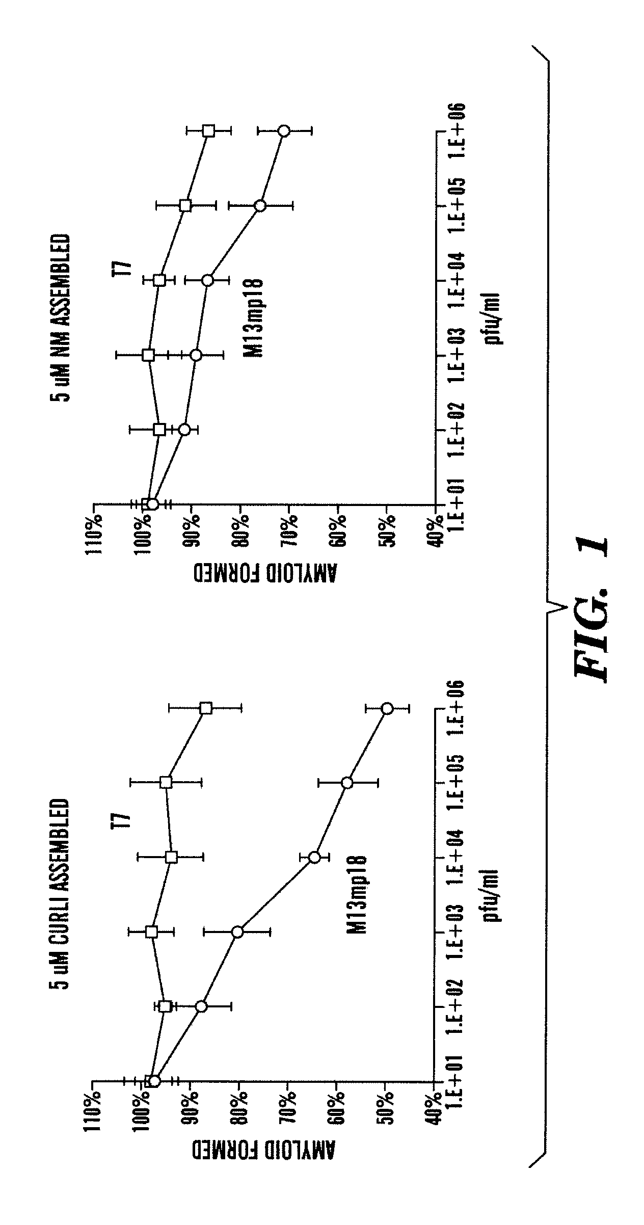
[0472]There are amyloids found in humans, yeast, and bacteria. Curli protein in E. coli constitute amyloids (Chapman, M. R. et al. Science 295 (2002)). The inventors first tested whether unmodified phage could block amyloid formation in the absence of capsid-bound peptide-based curli modulators. The inventors monitored in vitro CsgA fiber assembly using ThT fluorescence. As shown in FIG. 1, wild-type T7 phage (T7-wt) exhibited minimal inhibition of curli and Sup35-NM amyloid fiber formation (<15%) while unmodified M13mp18 phage was effective at inhibiting curli amyloids (˜50%) and Sup35-NM amyloids (˜25%). The inventors demonstrated that bacteriophage alone (i.e. non-engineered bacteriophage) was able to block curli formation in vitro. The inventors determined that M13mp18, a filamentous and lysogenic bacteriophage, was more effective than T7, a lytic bacteriophages, at preventing amyolid formation by curli.

example 2

[0473]CsgA is the major curli subunit and is nucleated by CsgB and CsgF (Chapman, M. R. et al. Science 295 (2002); Loferer, H. et al. Mol Microbiol 26 (1997); Hammar, et al. Mol Microbiol 18 (1995)). The inventors designed potential peptide-inhibitors for curli based off of the native amino acid sequences of CsgA (SEQ ID NO: 1) and CsgB (SEQ ID NO:2).

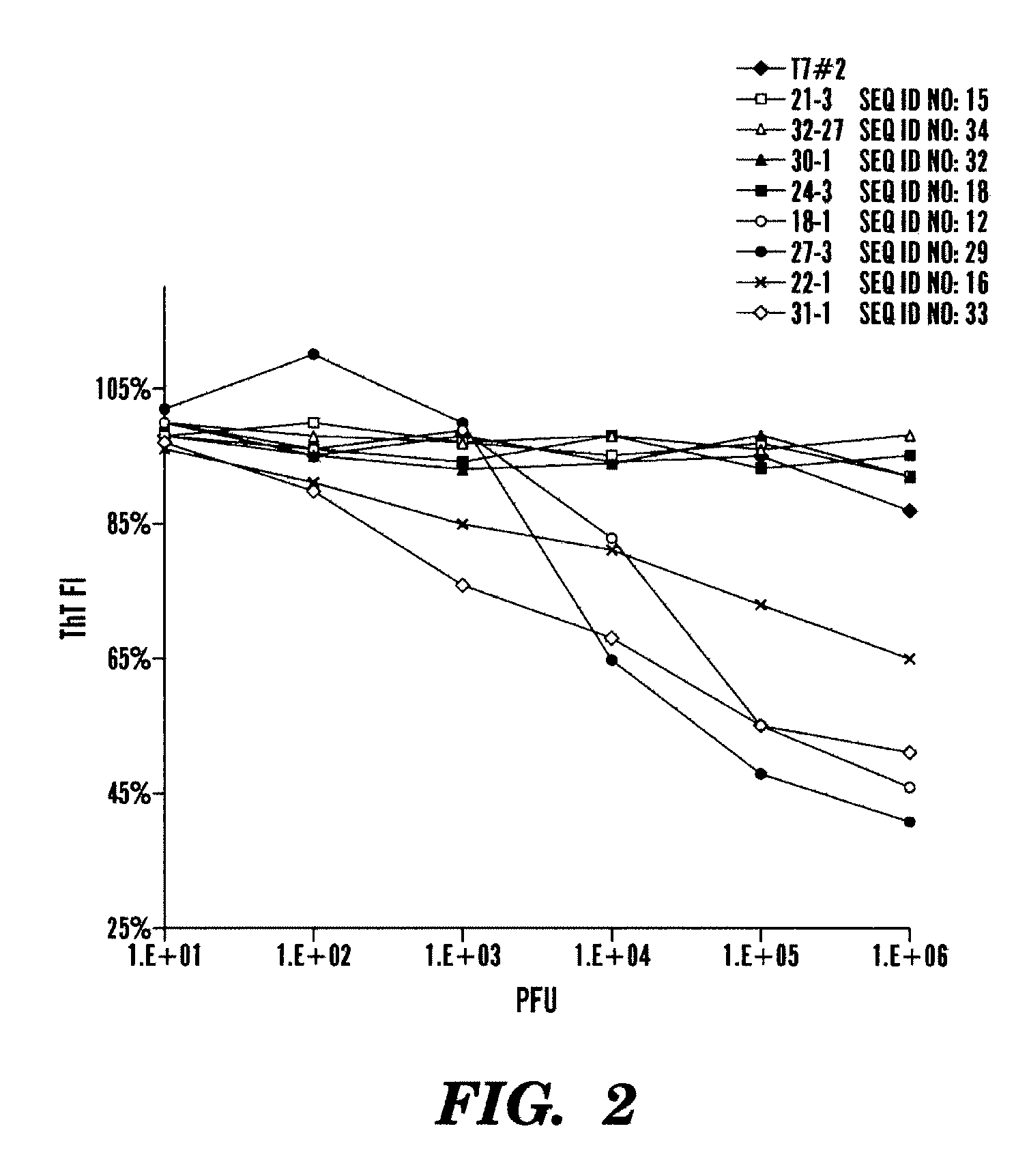
[0474]The inventors designed and expressed specific peptide sequences derived from CsgA polypeptide sequence (SEQ ID NO: 1), as shown in Table 3 and cloned them into the EcoRI and HindIII sites of T7select-415 plasmid from Novagen.

TABLE 3Sequences derived from CsgA that were cloned into T7select-415 plasmid between EcoRI andHindIII restriction sites, the CsgA sequence is highlighted in bold between the EcoRIand HindIII restriction sites (not bold). The nucleic acid sequences(SEQ ID NO: 3-10) encode polypeptides SEQ ID NOs 11-18 respectively.Relevant PeptideNUMBERDNA SequenceSequence17GGGGATCCGAATTCGTCTGAGCTGAACATTTACCAGTACGGTGGCAASELNIY...

example 3

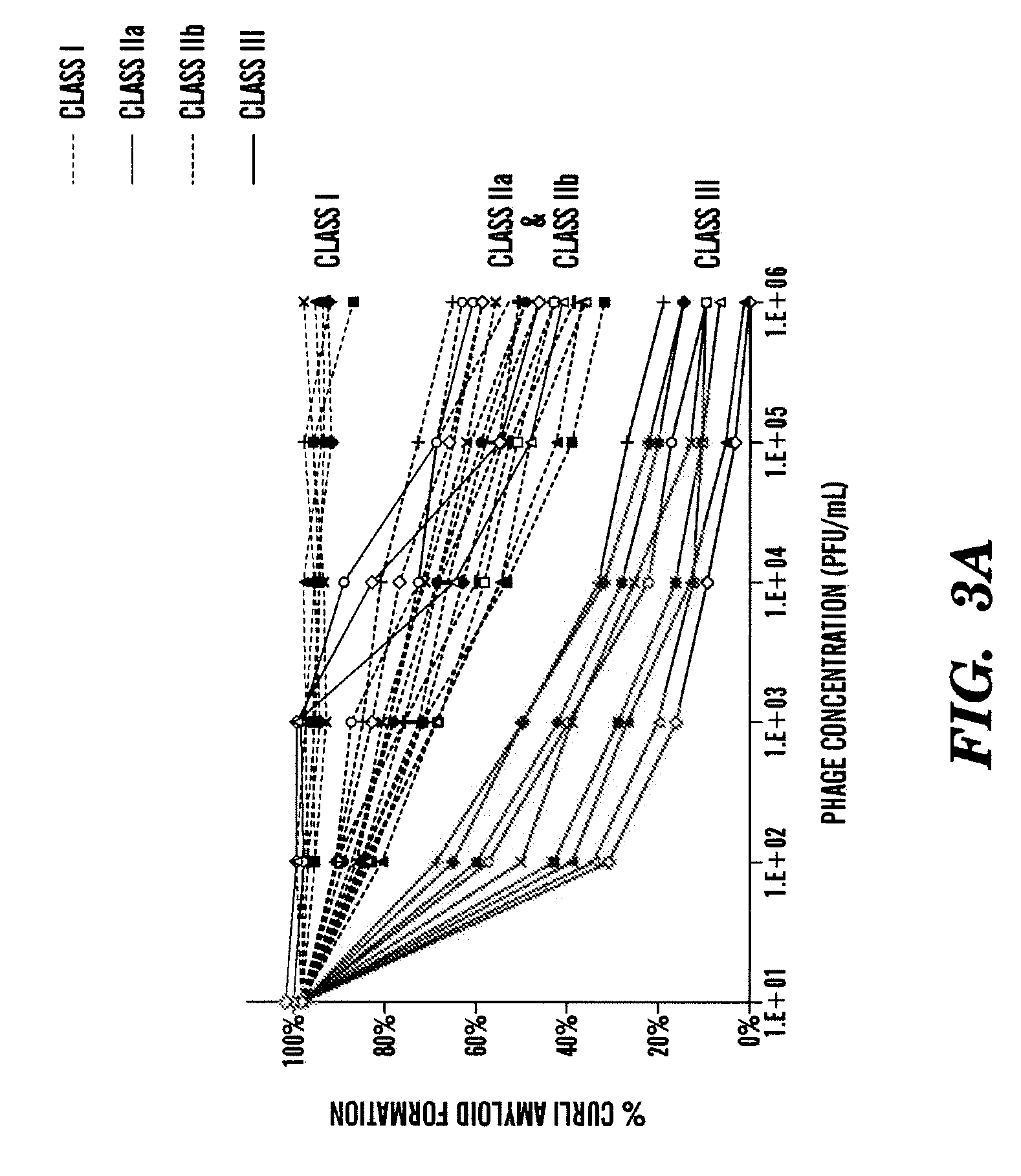
[0479]Next the inventors generated a new set of CsgA and CsgB peptide sequences expressed by bacteriophages for enhanced anti-amyloid activity. The new peptide sequences, shown in Table 5, are variant sequences (i.e. one or more changes amino acid) from the peptides shown in Tables 3 and 4. In particular, the inventors modified (i.e. added, deleted or substituted) one or more amino acid of the CsgA peptides (SEQ ID NOs: 11 or 12), or modified (i.e. added, deleted or substituted) one or more amino acid of the CsgB peptides (SEQ ID NO: 29).

[0480]Further, to see if charged mutations could enhance blocking of CsgA fiber assembly, the inventors mutated key residues within CsgA43-52 (SEQ ID NO: 11), CsgA55-64 (SEQ ID NO: 12), and CsgB133-142 (SEQ ID NO: 29) to lysines (Table 5). The inventors also constructed charged mutations in CsgB142-151 (SEQ ID NO: 30) since the peptide arrays showed that the peptides CsgB130-149 was important for nucleation (Table 5). In addition, the inventors cons...


PUM

PropertyMeasurementUnit
Fractionaaaaaaaaaa
Volumeaaaaaaaaaa
Volumeaaaaaaaaaa

Abstract

The present invention generally relates to engineered bacteriophages which express amyloid peptides for the modulation (e.g. increase or decrease) of protein aggregates and amyloid formation. In some embodiments, the engineered bacteriophages express anti-amyloid peptides for inhibiting protein aggregation and amyloid formation, which can be useful in the treatment and prevention of and bacterial infections and biofilms. In some embodiments, the engineered bacteriophages express amyloid peptides for promoting amyloid formation, which are useful for increasing amyloid formation such as promoting bacterial biofilms. Other aspects relate to methods to inhibit bacteria biofilms, and methods for the treatment of amyloid related disorders, e.g., Alzheimer's disease using an anti-amyloid peptide engineered bacteriophages. Other aspects of the invention relate to engineered bacteriophages to express the amyloid peptides on the bacteriophage surface and / or secrete the amyloid peptides, e.g., anti-amyloid peptides and pro-amyloid peptides, and uses thereof for modulation protein aggregates and amyloid formation.

Description

CROSS REFERENCE TO RELATED APPLICATIONS[0001]This application claims benefit under 35 U.S.C. 119(e) of U.S. Provisional Patent Application Ser. No. 61 / 229,703 filed Jul. 29, 2009, and U.S. Provisional Patent Application Ser. No. 61 / 233,697 filed Aug. 13, 2009, the contents of each are incorporated herein in their entirety by reference.GOVERNMENT SUPPORT[0002]This invention was made with government support under R01 GM 025874-29 and OD003644 awarded by the National Institites of Health (NIH). The Government has certain rights to the invention.FIELD OF THE INVENTION[0003]The present invention relates to the field of treatment and prevention of bacteria and bacterial infections. In particular, the present invention relates to engineered bacteriophages that have been engineered to express and secrete amyloid peptides, including anti-amyloid peptides and pro-amyloid peptides.BACKGROUND OF THE INVENTION[0004]Bacterial biofilms are sources of contamination that are difficult to eliminate i...

Claims


Application Information

Patent Timeline
IPC IPC(8): C12N7/01C12S9/00A61K35/76
CPCA61K35/76C07K14/4711C07K2319/735C12N2795/10232C12N2795/14143A61K38/00C12N2795/14132C12N2795/10243A61K2300/00A61P25/28Y02A50/30
Inventor LU, TIMOTHY KUAN-TALINDQUIST, SUSANKRISHNAN, RAJARAMANCOLLINS, JAMESO'DONNELL, CHARLES W.LEIGHTON, BONNIE BERGERDEVADAS, SRINIVAS
Owner TRUSTEES OF BOSTON UNIV
Learn More