Electrochemical detection of proteases using ac voltammetry on nanoelectrode arrays

a technology of ac voltammetry and proteases, which is applied in the field of electrochemical detection of proteases using ac voltammetry on nanoelectrode arrays, can solve the problems of large amount of sample and reagents, and workstation in a centralized lab is required

Active Publication Date: 2015-01-08
KANSAS STATE UNIV RES FOUND
View PDF12 Cites 10 Cited by
  • Summary
  • Abstract
  • Description
  • Claims
  • Application Information

AI Technical Summary

Benefits of technology

[0010]According to one embodiment of the present invention there is provided a nanoelectrode array comprising a substrate having a plurality of carbon nanofibers extending vertically therefrom. The array is capable of detecting the presence of one or more enzymes present within a biological sample. In particular embodiments, the nanoelectrode arrays comprise one or more peptides or peptide residues covalently attached to the exposed distal tips of the carbon nanofibers. When contacted with a sample comprising one or more target enzymes, the enzymes cleave at least some of the peptides or peptide residues, which cause a change in the electrical signal of the array as measured by AC volatammetry.
[0011]According to another embodiment of the present invention there is provided an electronic chip comprising a plurality of the nanoelectrode arrays as described herein. In particular embodiments, the electronic chip is useful for detecting specific enzymes, the presence of which are biomarkers of various cancers such as breast, lung and liver cancer, and other diseases such as Alzheimer's disease and various viral infections. Thus, the nanoelectrode arrays and electronic chips comprised of the arrays can be applied for rapid screening tests and monitoring of treatment response. In addition, the electronic chips may comprise a nanoelectrode arrays configured to detect a plurality of different enzymes within a biological sample.
[0012]According to still another embodiment of the present invention there is provided a method of detecting an enzyme within a biological sample comprising contacting said biological sample with one or more nanoelectrode arrays as described he...

Problems solved by technology

There are three main limitations: 1) only one protease can be analyzed in each measurement, 2) a ...

Method used


Image

  • Electrochemical detection of proteases using ac voltammetry on nanoelectrode arrays
  • Electrochemical detection of proteases using ac voltammetry on nanoelectrode arrays
  • Electrochemical detection of proteases using ac voltammetry on nanoelectrode arrays

Examples


example 1

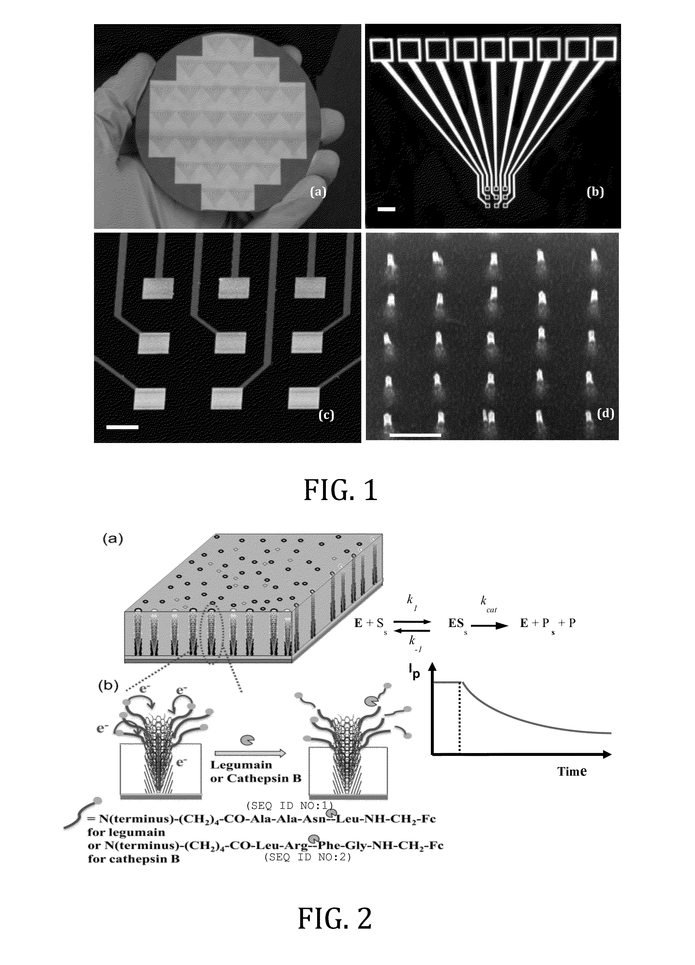
[0049]In this Example, two types of enzymatic biosensors were prepared and tested: (1) a redox labeled ACV biosensor for protease (legumain) detection, and (2) a label-free REIS biosensor for kinase / phosphatase detection. These enzymes are critical in regulating cell behaviors, whose overexpression in human body is known to associate with various cancers. These electrochemical methods, particularly with the enhancement by NEAs, have the potential as multiplex portable techniques for rapid diagnosis or treatment monitoring of cancer patients. These two enzymatic sensors share similar fabrication techniques, but differ in the detection mechanism due to the nature of the reactions.

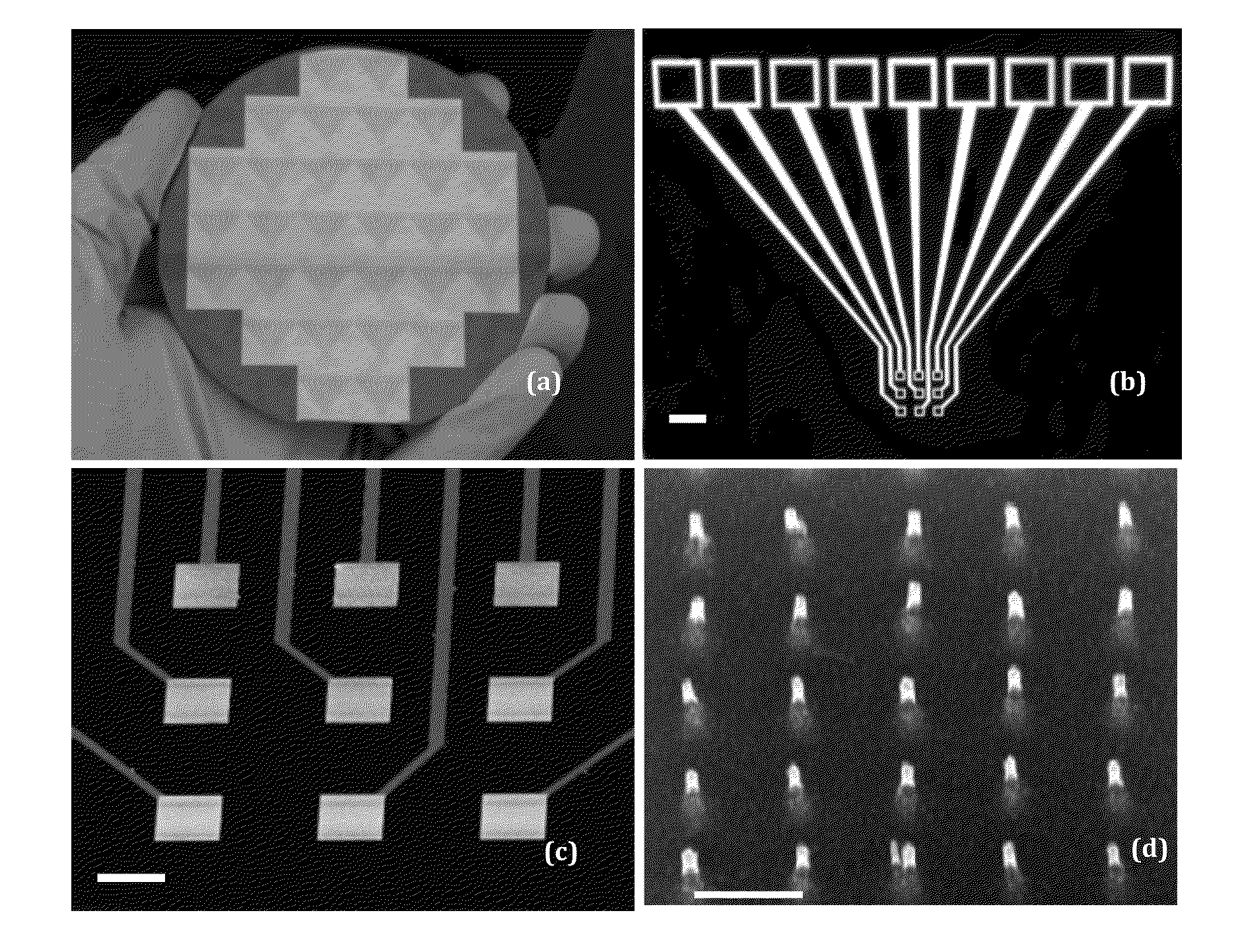
[0050]In the protease study, a tetrapeptide H2N—(CH2)4—CO-(SEQ ID NO:1)-NH—CH2-Fc is covalently attached to the exposed tip of a VACNF NEA as shown in FIG. 2. Fc provides reliable redox signals at 0.25 V vs. Ag / AgCl (3M KCl) in ACV measurements. As shown in later sections, the unique structure of VACNF NE...

example 2

[0077]In this example, the proteolytic activity of a cancer-related enzyme cathep sin B is measured with alternating current voltammetry (ACV) using ferrocene (Fc) labeled tetrapeptides attached to nanoelectrode arrays (NEAs) fabricated with vertically aligned carbon nanofibers (VACNFs).

Materials and Methods

[0078]The electrochemical measurements were conducted with an electrochemical analyzer (Model 440A, CH Instruments, Austin, Tex.). Fluorescence assay was performed on GloMax-Multi+Microplate Multimode Reader (Promega, Madison, Wis.) with black polystyrene 96 well plates obtained from Whatman (Picataway, N.J.). 3-Aminopropyl-triethoxysilane (APTES), 2-(2-methoxyethoxy)ethoxyacetic acid, 1-ethyl-3-(3-dimethylaminopropyl)carbodiimide hydrochloride (EDC), 1-hydroxy-2,5-dioxopyrrolidine-3-sulfonic acid sodium salt (sulfo-NHS), sodium hydroxide, and 6-amino-1-hexanol were purchased from Sigma-Aldrich (Saint Louis, Mo.). 2-(4-Morpholino)ethane sulfonic acid (MES) and dithiothreitol (DTT...

example 3

[0110]In this example, ACV techniques on VACNF NEAs were applied to demonstrate the possible values of cathepsin B concentrations and activities in breast cancer cell lysates and their correlation with stages of the breast cancer for the first time. The results showed that this electrochemical method has great potential as a portable multiplex system for simple and rapid protease profiling of lysate, serum or urine samples for cancer early diagnosis and treatment monitoring.

Materials

[0111]The electrochemical measurements were carried out with an electrochemical analyzer (Model 440A, CH Instruments, Austin, Tex.). Fluorescence assay was performed on GloMax-Multi+Microplate Multimode Reader (Promega, Madison, Wis.) with black polystyrene 96 well plates obtained from Whatman (Picataway, N.J.). 3-Aminopropyl-triethoxysilane (APTES), 2-(2-methoxyethoxy)ethoxyacetic acid, 1-ethyl-3-(3-dimethylaminopropyl)carbodiimide hydrochloride (EDC), 1-hydroxy-2,5-dioxopyrrolidine-3-sulfonic acid sodi...


PUM


Abstract

An electrochemical method for measuring the activity of enzymes using nanoelectrode arrays fabricated with vertically aligned carbon nanofibers. Short peptide substrates specific to disease-related enzymes are covalently attached to the exposed nanofiber tips. A redox moiety, such as ferrocene, can be linked at the distal end of the nanofibers. Contact of the arrays with a biological sample containing one or more target enzymes results in cleavage of the peptides and changes the redox signal of the redox moiety indicating the presence of the target enzymes.

Description

CROSS-REFERENCE TO RELATED APPLICATION[0001]This application claims the benefit of U.S. Provisional Patent Application No. 61 / 842,457, filed Jul. 3, 2013, which is incorporated by reference herein in its entirety.STATEMENT REGARDING FEDERALLY SPONSORED RESEARCH OR DEVELOPMENT[0002]This invention was made with U.S. Government support under grant number R15CA159250 awarded by the National Institutes of Health. The government has certain rights in the invention.SEQUENCE LISTING[0003]The following application contains a sequence listing in computer readable format (CRF), submitted as a text file in ASCII format entitled “SequenceListing,” created on Jun. 30, 2014 as 2 KB. The content of the CRF is hereby incorporated by reference.BACKGROUND OF THE INVENTION[0004]1. Field of the Invention[0005]The present invention is generally directed toward an electrochemical method for measuring the activity of proteases using nanoelectrode arrays (NEAs) fabricated with vertically aligned carbon nano...

Claims


Application Information

Patent Timeline
IPC IPC(8): C12Q1/37G01N27/414B01J19/00
CPCC12Q1/37B01J19/0046G01N27/4146G01N2333/96466B01J2219/00653G01N2800/7028G01N27/4145B01J2219/00659B01J2219/00725B82Y15/00G01N27/27G01N27/308
Inventor LI, JUNHUA, DUYSYED, LATEEF UDDINPRIOR, ALLANSWISHER, LUXI
Owner KANSAS STATE UNIV RES FOUND
Why Eureka
Try Eureka