Passive immunization for staphylococcus infections

a staphylococcus and immunization technology, applied in the field of passive immunization against staphylococcus infection, can solve the problems of infection rate, osteomyelitis (om) remains a serious problem, amputation or death, etc., to promote phagocytosis, inhibit one or both cell division and biofilm formation, and promote adhesion and immune evasion.

Active Publication Date: 2016-10-27
UNIVERSITY OF ROCHESTER
View PDF0 Cites 2 Cited by
  • Summary
  • Abstract
  • Description
  • Claims
  • Application Information

AI Technical Summary

Benefits of technology

[0019]The monoclonal antibodies and binding portions thereof, as well as pharmaceutical compositions containing the same, are therapeutic agents suitable for immunotherapy in patients with or at risk for infection by Staphylococcus strains. The power of these monoclonal antibodies is derived from their multiple activities that will hinder growth, adhesion, and immune evasion by Staphylococcus strains. First, as antibodies, they will promote phagocytosis by neutrophils at the site of incipient Staphylococcus infections. Second, as inhibitors of the Staphylococcus amidase, an enzyme with multiple roles in Staphylococcus survival and surface colonization, these antibodies potentially hinder one or both of cell division and biofilm formation. Finally, as demonstrated herein, the disclosed antibodies reduce Staphylococcus spread, as evidenced by the formation of fewer abscesses, and afford macrophage invasion of abscesses, which promotes the formation of sterile abscesses and accelerates bone healing.

Problems solved by technology

Although improvements in surgical technique and aggressive antibiotic prophylaxis have decreased the infection rate following orthopedic implant surgery to 1-5%, osteomyelitis (OM) remains a serious problem and appears to be on the rise from minimally invasive surgery (Mahomed et al., “Rates and Outcomes of Primary and Revision Total Hip Replacement in the United States Medicare Population,”J.
Bone Joint Surg. Br. 88(2):149-55 (2006)), this result may lead to amputation or death.
However, the greatest concern is the emergence of drug-resistant staphylococcal strains, most notably MRSA, which has surpassed HIV as the most deadly pathogen in North America, and continues to make the management of chronic OM more difficult and expensive, resulting in a great demand for novel therapeutic interventions to treat patients with these infections.
Presently, there are no prophylactic treatments that can protect high-risk patients from MRSA, most notably the aging “baby boomers” who account for most of the 1.5 million TJR performed annually in the United States.
First, although the total number of osteomyelitis cases is high, its incidence of 1-5% is too low for rigorous prospective clinical studies, with the possible exception of revision arthroplasty.
Second, it is well known that in vitro cultures rapidly select for growth of organisms that do not elaborate an extracellular capsule, such that biofilm biology can only be studied with in vivo models (Costerton et al., “Bacterial Biofilms: A Common Cause of Persistent Infections,”Science 284(5418):1318-22 (1999)).
This leads to the “greatest obstacle” in this field, which is the absence of a quantitative animal model that can assess the initial planktonic growth phase of the bacteria prior to biofilm formation.
Thus, they cannot be efficiently used to assess drug effects, bacterial mutants, and the role of host factors with transgenic mice.
However, at this point the infection is now chronic and cannot be eradicated by drugs or host immunity.
Unfortunately, S. aureus has many adhesins, such that inhibition of one or more may not be sufficient to prevent bacterial attachment.
Furthermore, bacterial clearance by phagocytic cells may be limited in avascular tissue such as bone such that an antibody alone may need additional anti-microbial mechanisms of action to significantly reduce the in vivo planktonic growth of S. aureus and prevent the establishment of chronic OM or reinfection during revision total joint replacement surgery.

Method used


Image

  • Passive immunization for staphylococcus infections
  • Passive immunization for staphylococcus infections
  • Passive immunization for staphylococcus infections

Examples


example 1

Preparation of Antigen

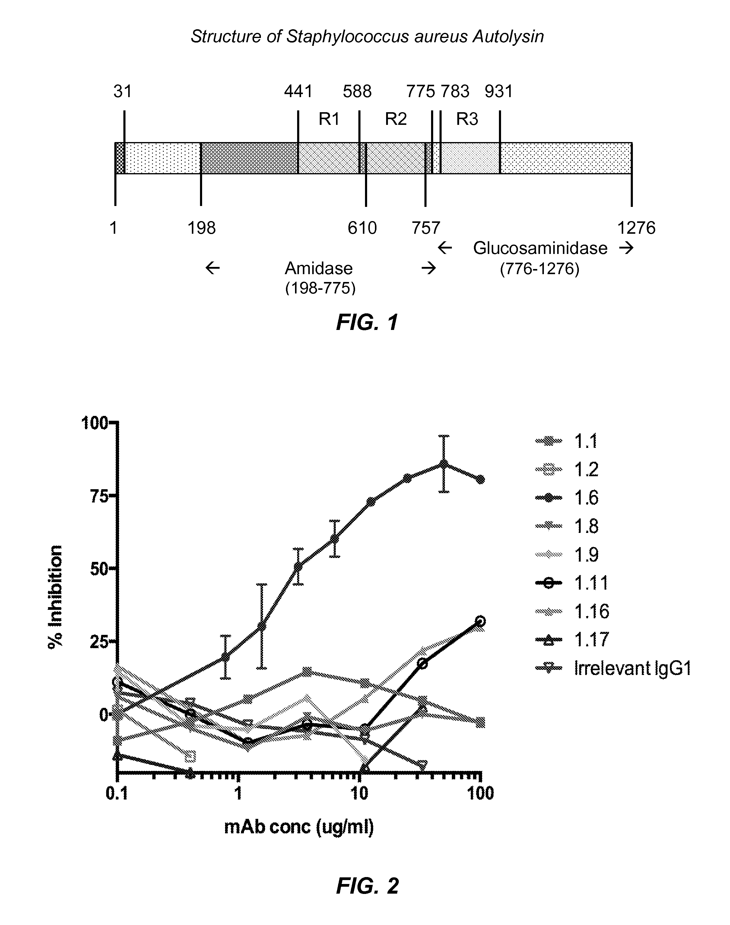
[0122]A recombinant form of the entire amidase domain of S. aureus autolysin that includes a hexa-histidine sequence near its N-terminus (His-Amd) was prepared. The open reading frame for His-Amd was designed by collecting known sequences of S. aureus autolysin, determining the consensus protein sequence using Geneious™ software, and then optimizing codon usage for expression in E. coli. The encoded consensus protein and encoding open reading frame sequences for His-Amd are identified as SEQ ID NOS: 1 and 2 below.

SEQ ID NO: 1 (Hex-histidine leader sequence plus Autolysin aa 198-775)MHHHHHHSASAQPRSVAATPKTSLPKYKPQVNSSINDYIRKNNLKAPKIEEDYTSYFPKYAyRNGVGRPEGIVVHDTANDRSTINGEISYMKNNYQNAFVHAFVDGDRIIETAPTDYLSWGVGAVGNPRFINVEIVHTHDYASFARSMNNYADYAATQLQYYGLKPDSAEYDGNGTVWTHYAVSKYLGGTDHADPHGYLRSHNYSYDQLYDLINEKYLIKMGKVAPWGTQSTTTPTTPSKPTTPSKPSTGKLTVAANNGVAQIKPTNSGLYTTVYDKTGKATNEVQKTFAVSKTATLGNQKFYLVQDYNSGNKFGWVKEGDWYNTAKSPVNVNQSYSIKPGTKLYTVPWGTSKQVAGSVSGSGNQTFKASKQQQIDKSIYLYGSVN...

example 2

Inoculation of Mice and Preparation of Hybridomas

[0126]For the initial hybridoma fusion (Fusion #1), six female Balb / c mice were immunized two times with 75 μg of His-AmdR1R2, in the Sigma Adjuvant System (Sigma, Cat. No. S6322) by intraperitoneal injection at seven-week intervals. Two of the mice with the highest titers in ELISA on immobilized His-AmdR1R2 were selected for hybridoma fusion. Each mouse received a final immunization of 350 μg of His-AmdR1R2, i.p., four days prior to sacrifice and hybridoma fusion.

[0127]For the second hybridoma fusion (Fusion #2), Balb / c mice were immunized two times: first dose with 120 μg of His-AmdR1R2-B from GenScript (Lot Number 222933505 / P20011303) in Sigma Adjuvant System (Sigma, Cat. No. S6322), and a second immunization with 100 μg of His-AmdR1R2-B conjugated with Keyhole limpet hemocyanin (KLH) (Imject EDC mcKLH Spin Kit; Thermo Scientific; Cat #77671) at twelve-week intervals. Two of the mice with the highest titers in ELISA on immobilized ...

example 3

Characterization of Monoclonal Antibodies

[0129]New monoclonal antibodies were screened on multiple related proteins to determine that they recognized native Amd (and not just the recombinant form) and whether their epitope was present on the catalytic (C) or cell wall binding domain (R1, R2 or R3). The proteins used for screening the monoclonal antibodies are identified in Table 1 below.

TABLE 1Proteins Used for Screening the Monoclonal AntibodiesProtein / Antigen NameRegion of Autolysin / Sequence DescriptionHis-AmdR1R2MGHHHHHH - Autolysin aa 198 to 775His-AmdcatMGHHHHHH - Autolysin aa 198 to 441Native AmdMixture of S. aureus UAMS-1 Δspa proteins includingfull length autolysin (aa 198-1276), Amd (aa 198 to775), and Gmd (aa 776-1276)His-AmdR1R2-BMGHHHHHH - Autolysin aa 198 to 775 -BirA biotinylation siteHis-R3Gmd-BMGHHHHHH - Autolysin aa 776 to 1276 -BirA biotinylation site

[0130]Screening assays were carried out by ELISA using the proteins identified in Table 1 as capture antigen. ELISA ...


PUM

PropertyMeasurementUnit
ODaaaaaaaaaa
concentrationaaaaaaaaaa

Abstract

Disclosed herein are monoclonal antibodies or binding portion thereof that bind specifically to a Staphylococcus spp. autolysin N-acetylmuramoyl-L-alanine amidase catalytic domain and/or cell wall binding domain, as well as pharmaceutical compositions containing the same. Cell lines expressing the monoclonal antibodies, including hybridomas, are also disclosed. Methods of using the monoclonal antibodies for installation of orthopedic implants, grafts or medical devices, treating or preventing a Staphylococcus infection, and treating osteomyelitis are described, as are diagnostic methods for the detection of Staphylococcus in a sample.

Description

[0001]This application claims the priority benefit of U.S. Provisional Patent Application Ser. No. 61 / 915,953, filed Dec. 13, 2013, which is hereby incorporated by reference in its entirety.FIELD OF USE[0002]Disclosed herein are methods and compositions for the passive immunization against Staphylococcus infection, particularly for the prevention or treatment of osteomyelitis and for infections arising from implantation of a medical device, or an orthopedic implant or graft. Antibodies that bind specifically to a Staphylococcus spp. autolysin N-acetylmuramoyl-L-alanine amidase catalytic domain and / or cell wall binding domain, and pharmaceutical compositions containing the same can be used for these purposes.BACKGROUND[0003]There is a great need for novel interventions of chronic osteomyelitis (OM) as approximately 112,000 orthopedic device-related infections occur per year in the US, at an approximate hospital cost of $15,000-70,000 per incident (Darouiche, “Treatment of Infections ...

Claims


Application Information

Patent Timeline
Patent Type & Authority Applications(United States)
IPC IPC(8): C07K16/40A61K45/06G01N33/569A61K39/40
CPCC07K16/40A61K39/40A61K45/06G01N33/56938C07K2317/14C07K2317/24A61K2039/505A61K2039/507C07K2317/21D21H27/30D21H27/38D21H11/02D21H11/08C07K16/1271C07K2317/92C07K2317/76A61P31/04A61P19/08A61P19/02A61P29/00A61P31/00A61P37/02A61P7/00A61F2/30A61K39/3955C12Y305/01028G01N33/532C07K2317/56B32B29/005B32B2250/26B32B2262/0261B32B2262/062B32B2262/067B32B2307/54B32B2307/72B32B2439/00B32B2439/70
Inventor DAISS, JOHN L.SCHWARZ, EDWARDVARRONE, JOHN J.BRODELL, JR., JAMESBELLO-IRIZARRY, SHEILA N.
Owner UNIVERSITY OF ROCHESTER
Why Eureka
Try Eureka