Selective sampling for assessing structural spatial frequencies with specific contrast mechanisms

a structural spatial frequency and contrast mechanism technology, applied in the field of diagnostic assessment of fine textures, can solve the problems of inability to accurately assess fine texture clinically, the robustness of the ct measurement of early stage disease remains limited, and the in-vivo assessment and measurement of fine texture remain outside the capabilities of current imaging technologies, so as to enhance the contrast between component tissue types

Inactive Publication Date: 2017-09-14
BIOPROTONICS
View PDF5 Cites 63 Cited by
  • Summary
  • Abstract
  • Description
  • Claims
  • Application Information

AI Technical Summary

Benefits of technology

[0021]The embodiments disclosed herein provide a method for pathology assessment employing tissue texture using magnetic resonance (MR) which may be used integrally with an MR imaging technique. A contrast mechanism is selected for enhancing the contrast between component tissue types in a multiphase biologic sample for measurement with a MR imaging process. The selected contrast mechanism is then applied and a volume of interest (VOI) is selectively excited employing a plurality of time varying radio frequency signals and applied gradients. An encoding gradient pulse is applied to induce phase wrap to create a spatial encode for a specific k-value and orientation, the specific k-value determined based on texture within the VOI. A series of gradients is initiated to produce k-value encodes, a resulting k-value set being a subset of that required to produce an image of the VOI. Multiple sequential samples of the NMR RF signal encoded with the k-value set are recorded. Post processing the recorded NMR signal samples is accomplished to produce a data set of signal vs k-values for k-values in the k-value set, to characterize textural features of tissue in the VOI. The MR imaging process is then performed as an integral or hybrid pulse sequence with the texture measurement.

Problems solved by technology

Though fine textural changes in tissue have long been recognized as the earliest markers in a wide range of diseases, robust clinical assessment of fine texture remains elusive, the main difficulty arising from blurring caused by subject motion over the time required for data acquisition.
Changes in fine tissue texture have been recognized for many years by diagnosticians, including radiologists and pathologists as the earliest harbinger of a large range of diseases, but in vivo assessment and measurement of fine texture has remained outside the capabilities of current imaging technologies.
For instance, differential diagnosis of obstructive lung disease relies on a textural presentation in the lung parenchyma, but the robustness of the Computed Tomography (CT) measure of early stage disease is limited.
Trabecular bone microarchitecture, the determinant of fracture risk in aging bone, has also remained elusive due to image blurring from patient motion during Magnetic Resonance (MR) imaging scans.
But post processing analysis is limited in effect as it doesn't deal with the underlying problem that prevents high resolution acquisition of textural information, i.e. subject motion.
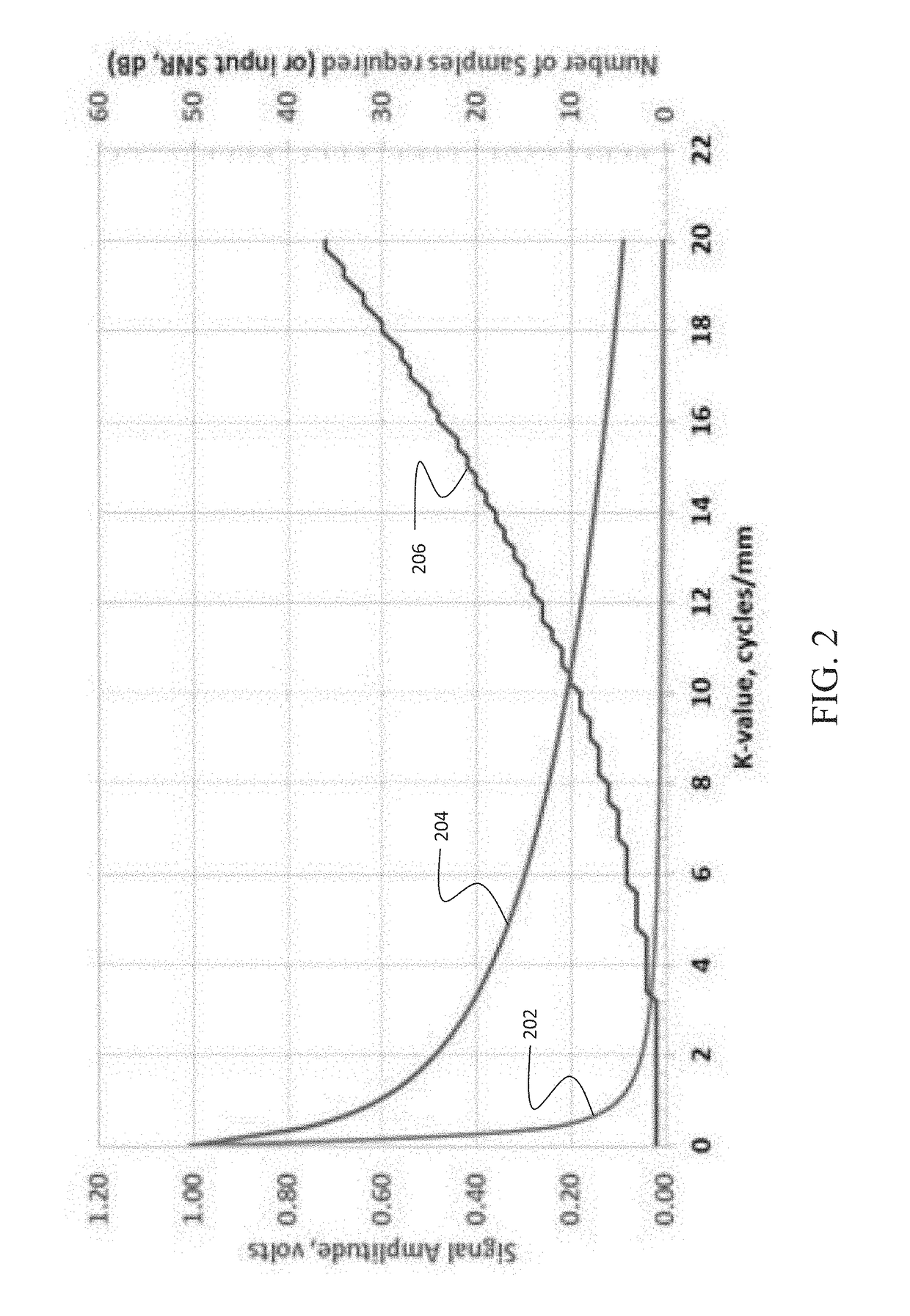
However, even with gating, there is sufficient motion between acquisitions to cause loss of spatial phase coherence at the high k-values of interest for texture measurements.
This problem is exacerbated by the fact that motion may not be perfectly cyclic, and often originates from combined sources.
Twitching is rapid, inducing random displacements, and hence it is not possible to maintain coherence at the high k-values of interest when measuring texture.
While Positron Emission Tomography (PET) provides valuable diagnostic information, it is not capable of resolution below about 5 mm and relies on the use of radioactive tracers for imaging as well as x-ray beams for positioning, raising dose concerns, especially if repeat scanning is needed.
Further, PET imaging is extremely costly, requiring a nearby cyclotron.
The non-negligible risk from the associated radiation dose makes CT problematic for longitudinal imaging and limits available resolution.
Along with serious dose concerns, digital x-ray resolution is limited because the 2-dimensional image obtained is a composite of the absorption through the entire thickness of tissue presented to the beam.
Current clinical diagnostics for the diseases that are the target of the embodiments disclosed herein are fraught with difficulties in obtaining sufficient in vivo resolution, or accuracy.
In some cases, no definitive diagnostic exists currently.
In other pathologies, particularly in breast and liver, diagnosis is dependent on biopsy, with its non-negligible risk of morbidity and even mortality, and which is prone to high read and sampling errors.
Bone health is compromised by ageing, by bone cancer, as a side effect of cancer treatments, diabetes, rheumatoid arthritis, and as a result of inadequate nutrition, among other causes.
Bone disease affects over ten million people annually in the US alone, adversely affecting their quality of life and reducing life expectancy.
But, though TBS does not yield a detailed assessment of trabecular architecture.
In MRI, though high contrast between bone and marrow is readily obtained, resolution is limited by patient motion over the long time needed to acquire an image with sufficient resolution to characterize the trabecular network.
Further, a large data matrix, hence long acquisition time, is still required to obtain sufficient image information to determine trabecular spacing and element thickness.
This long acquisition time results in varying levels of motion-induced blurring, depending on patient compliance—twitching is still a serious problem even when measuring extremities.
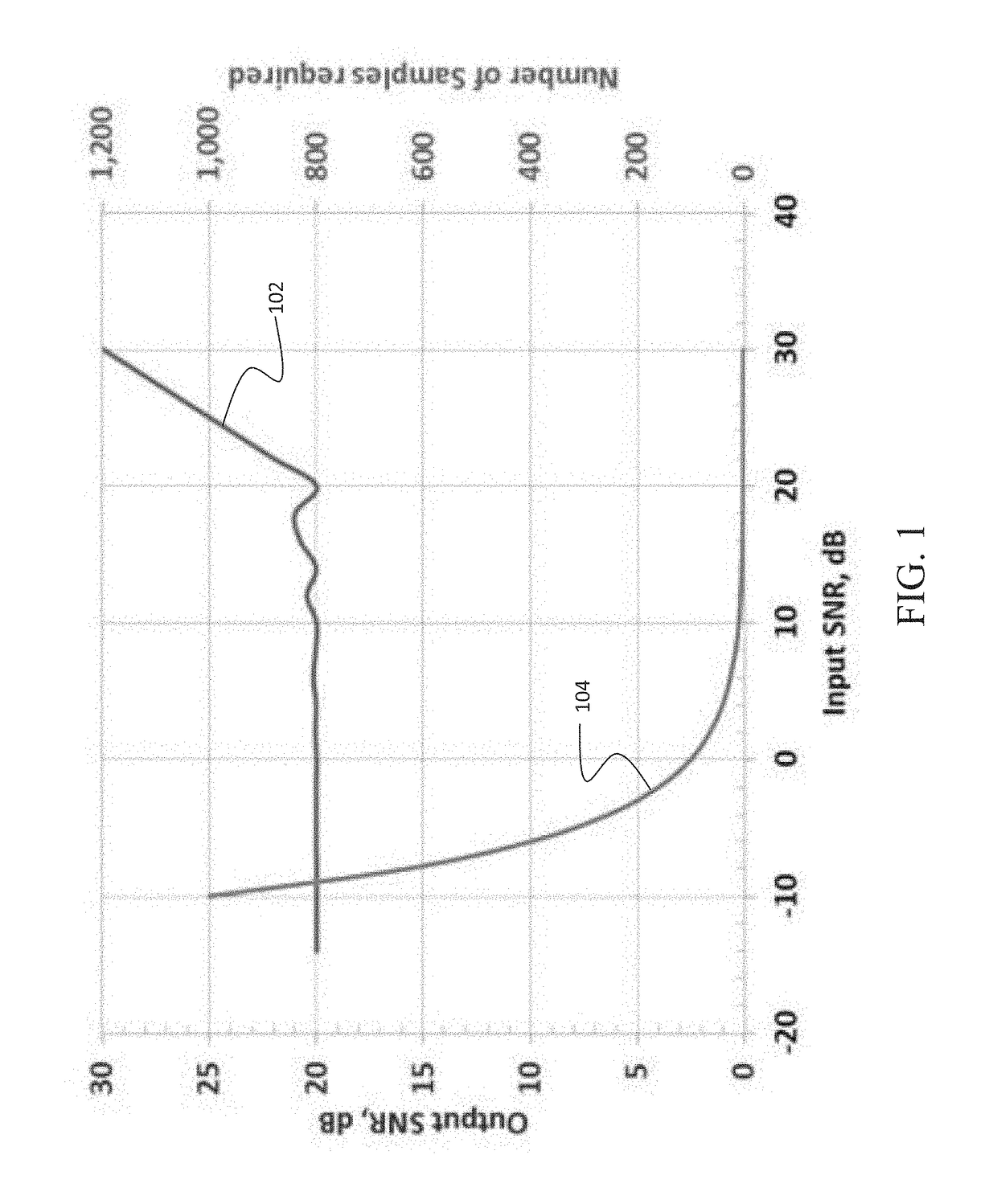
However, as the gradient encoded echoes, are very low Signal to Noise (SNR), noise averaging is required.
Thus, resolution improvement obtainable by the technique is limited.
Until now, no clinical technique has been able to provide this capability.
Currently, the gold standard for pathology assessment is tissue biopsy—a highly invasive and often painful procedure with a non-negligible morbidity—and mortality—risk (patients need to stay at the hospital for post-biopsy observation for hours to overnight), and one that is prone to sampling errors and large reading variation.
Ultrasound, another modality often used to assess tissue damage in liver disease, is only able to provide adequate assessment in the later stages of the disease—it is used to diagnose cirrhosis.
Magnetic Resonance-based Elastography (MRE), which has been under development for some time for use in assessment of liver disease, is not capable of early-stage assessment—the read errors are too large prior to significant fibrotic invasion (advanced disease).
Further, this technique requires expensive additional hardware, the presence of a skilled technician, and takes as much as 20 minutes total set up and scanning time, making it a very costly procedure.
The ability to image fibrotic texture directly by MR imaging is compromised both by patient motion over the time necessary to acquire data and by lack of contrast between the fibers and the surrounding tissue.
Even acquisition during a single breath hold is severely compromised by cardiac pulsatile motion and noncompliance to breath hold, which results in significant motion at many organs, such as liver and lungs.
And SNR is low enough that motion correction by combining reregistered MR-intensity profiles obtained from successive echoes is extremely problematic.
Similarly, assessment of the amount of cardiac fibrosis in early stage disease using MRI is seriously hampered by cardiac pulsation over the time of the measurement.
However, ability to assess such changes in the brain is only available post mortem.
Various in vivo diagnostic techniques are available for AD and other dementias, but none of them are definitive.
As discussed previously, PET imaging is extremely expensive, cannot provide high resolution, and relies on use of radioisotopes and positioning x-ray beams, complicating approval for longitudinal use due to dose concerns.
Use of CSF biomarkers for dementia diagnosis is painful and highly invasive and cannot differentiate signal levels by anatomic position in the brain, as is possible with imaging biomarkers.
As various forms of dementia are found to have different spatial / temporal progression through the brain, this is a serious drawback to use of liquid biopsy.
This textural size, and the fact that the cortex is extremely thin, makes speed of acquisition paramount, as even tiny patient motion will make data collection impossible.
However, in order to circumvent the problem of signal degradation due to patient motion, data must be taken on a time scale not previously possible.

Method used


Image

  • Selective sampling for assessing structural spatial frequencies with specific contrast mechanisms
  • Selective sampling for assessing structural spatial frequencies with specific contrast mechanisms
  • Selective sampling for assessing structural spatial frequencies with specific contrast mechanisms

Examples


Embodiment Construction

[0055]The following definition of terms as used herein is provided:

180° inversion pulse RF pulse that inverts the spins in a tissue region to allow refocusing of the MR signal.

180° pulse An RF pulse that tips the net magnetic field vector antiparallel to B0

90° pulse An RF pulse that tips the net magnetic field vector into the transverse plane relative to B0

3 T 3 Tesla

[0056]A / D Analog to digital converter

AD Alzheimer's Disease

[0057]ADC Average diffusion coefficient measured in Diffusion Weighted Imaging

Adiabatic pulse excitation Adiabatic pulses are a class of amplitude- and frequency-modulated RF-pulses that are relatively insensitive to 6 inhomogeneity and frequency offset effects.

ASL Arterial Spin Labelling

[0058]AWGN Additive White Gaussian Noise Additive white Gaussian noise (AWGN) is a basic noise model used in Information theory to mimic the effect of many random processes that occur in nature.

BPH Benign Prostatic Hyperplasia

[0059]Biopsy A biopsy is a sample of tissue extract...


PUM


Abstract

The disclosed embodiments provide a method for acquiring MR data at resolutions down to tens of microns for application in in vivo diagnosis and monitoring of pathology for which changes in fine tissue textures can be used as markers of disease onset and progression. Bone diseases, tumors, neurologic diseases, and diseases involving fibrotic growth and/or destruction are all target pathologies. Further the technique can be used in any biologic or physical system for which very high-resolution characterization of fine scale morphology is needed. The method provides rapid acquisition of signal at selected values in k-space, with multiple successive acquisitions at individual k-values taken on a time scale on the order of microseconds, within a defined tissue volume, and subsequent combination of the multiple measurements in such a way as to maximize SNR. The reduced acquisition volume, and acquisition of only signal values at select places in k-space, along selected directions, enables much higher in vivo resolution than is obtainable with current MRI techniques.

Description

REFERENCE TO RELATED APPLICATIONS[0001]This application is a continuation in part of application Ser. No. 15 / 288,974 filed on Oct. 7, 2016 which is a continuation in part of Ser. No. 15 / 167,828 filed on May 27, 2016 which is a continuation in part of application Ser. No. 14 / 840,327 filed on Aug. 31, 2015 now U.S. Pat. No. 9,366,738. Application Ser. No. 14 / 840,327 relies on the priority of U.S. provisional application Ser. No. 62 / 044,321 filed on Sep. 1, 2014 entitled SELECTIVE SAMPLING MAGNETIC RESONANCE-BASED METHOD FOR ASSESSING STRUCTURAL SPATIAL FREQUENCIES, Ser. No. 62 / 064,206 filed on Oct. 15, 2014 having the same title and Ser. No. 62 / 107,465 filed on Jan. 25, 2015 entitled MICRO-TEXTURE CHARACTERIZATION BY MRI, the disclosures of which are incorporate herein by reference. Application Ser. No. 15 / 167,828 additionally relies on the priority of provisional application Ser. No. 62 / 302,577 filed on Mar. 2, 2016 entitled METHOD FOR ASSESSING STRUCTURAL SPATIAL FREQUENCIES USING H...

Claims


Application Information

Patent Timeline
Patent Type & Authority Applications(United States)
IPC IPC(8): G01R33/56G06T7/00G01R33/561
CPCG01R33/5601G06T2207/10088G06T7/0012G01R33/5619A61B5/055A61B5/7203A61B5/7207A61B2560/0238G01R33/4818G01R33/4833G01R33/4835G01R33/50G01R33/5602G01R33/5616G01R33/5617G01R33/56341
Inventor JAMES, KRISTINJAMES, TIMOTHY W.
Owner BIOPROTONICS
Why Eureka
Try Eureka