Natural Polymer-Derived Scaffold Material and Methods for Production Thereof

a scaffold material and natural polymer technology, applied in the field of tissue engineering, can solve the problems of difficult control of physical parameters such as degradation rate, affecting the release kinetics of growth factors, and inability to show beneficial effects for patients in trials, etc., and achieve the effect of increasing the polymer density and altering the in vitro degradation ra

Inactive Publication Date: 2019-02-28
ECOLE POLYTECHNIQUE FEDERALE DE LAUSANNE (EPFL)
View PDF0 Cites 0 Cited by
  • Summary
  • Abstract
  • Description
  • Claims
  • Application Information

AI Technical Summary

Benefits of technology

[0024]In one embodiment, the microbeads' in vitro degradation rate can be altered by crosslinking, using inhibitor m

Problems solved by technology

However, the larger phase II trials did not show the beneficial effect for patients (Simons M 2002).
However, it is hard to control some of their physical parameters such as degradation rate.
Furthermore, the alteration of the fibrinogen and thrombin concentration affected the growth factor release kinetics.
Due to its increased size, the heparin-growth factor complex diffuses more slowly through the fibrin matrix, delaying its release.
A major drawback to this delivery strategy is that the time course is not well controlled.
However, only a few publications reported pure fibrin micro-beads or fibrin micro-beads combined with other natural polymers.
(Goro

Method used


Image

  • Natural Polymer-Derived Scaffold Material and Methods for Production Thereof
  • Natural Polymer-Derived Scaffold Material and Methods for Production Thereof
  • Natural Polymer-Derived Scaffold Material and Methods for Production Thereof

Examples


example 1

on of Fibrin Beads

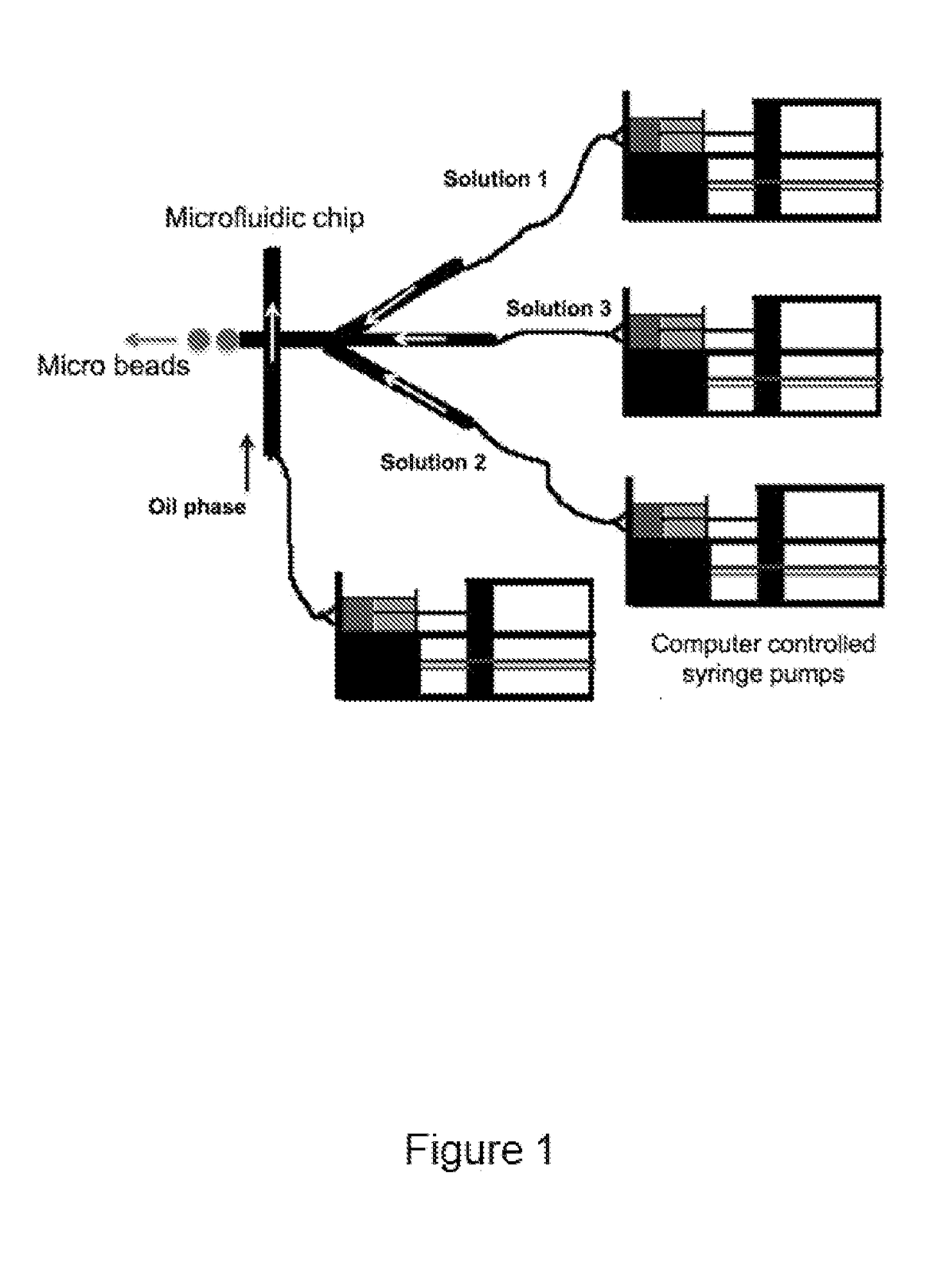
[0154]Fibrin beads were obtained by modifying a flow-focusing microfluidic system previously described in an article by Allazetta et al. (Allazetta S 2013), the content of which is incorporated herein by reference in its entirety. Computer-controlled syringe pumps (neMESYS from Cetoni, Germany) were used to adjust flow rates. Briefly, one microfluidic channel was loaded with 40 mg / mL of human fibrinogen (plasminogen, fibronectin-depleted; Enzyme Research Laboratories, South Bend, Ind., USA) at a flow rate of 1.5 μl / min and the second channel was loaded with an enzyme solution containing 200 U / mL human thrombin (Sigma Aldrich, Switzerland), 200 U / mL factor XIIIa (Fibrogammin, CSL Behring UK) and 10 mM Ca2+ in tris-buffered saline (TBS) at a flow rate of 1 μl / min. For the fabrication of bioactive fibrin beads, the enzyme solution was supplemented with a recombinant IGF-1 (α2PI1-8-MMP-IGF-1). The fibrinogen and enzyme solution were injected into an oil phase of 2% (w / ...

example 2

ical Characterization of Fibrin Beads

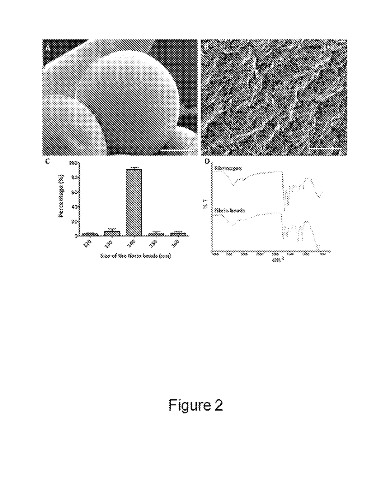
[0155]The surface morphology of fibrin beads was studied using Scanning Electron Microscopy (SEM) (FIG. 2A) as described below. The obtained fibrin beads were very homogeneous in terms of size and porosity, showing highly dense fiber bundling (FIG. 2B). The average diameter of the fibrin beads was around 140 μm as determined by bright-field microscopy and image analysis using the software ImageJ (FIG. 2C). Fibrin beads were analyzed for their functional groups via FT-IR analysis. The FT-IR spectrum of fibrinogen demonstrated its characteristic amide absorption bands at around 1643 cm-1, 1550 cm-1, and 1240 cm-1, which are indicative of amide I, II, and III groups, respectively (FIG. 2D). The same bands for fibrin were observed around 1687 cm-1, 1550 cm-1, and 1276 cm-1. The bands at around 1110 cm-1 can be attributed to C—N stretching (FIG. 2D). The absence of characteristic silicone bands around 800 cm-1, 1022 cm-1, and 1100 cm-1 showed the obta...

example 4

n of Fibrin Bead Degradation In Vitro

[0162]Fibrin beads were added to hSMCs seeded 12 well-plates and cultured for 7 days to determine their degradation behaviour in the presence of cells as described below. At different time points fibrin beads were visualized under a bright-file microscope and the number and the diameter of non-degraded beads was determined (FIGS. 5A and 5B). In vitro degradation results demonstrated that 93% of fibrin beads were degraded after 4 days of incubation with hSMCs (FIG. 5A). However, fibrin gels, having the same protein concentration as the fibrin bead samples, showed a considerably higher mass loss after 24 hours. Fibrin gels lost around 87% of their initial mass within the first day when incubated with cells (data not shown). In contrast, the average diameter of non-degraded fibrin beads did not change significantly at any time point (FIG. 5B).

[0163]Cell Culture

[0164]Human smooth muscle cells (hSMCs) were extracted from human ureter biopsies, and cul...


PUM

PropertyMeasurementUnit
Fractionaaaaaaaaaa
Fractionaaaaaaaaaa
Fractionaaaaaaaaaa

Abstract

The invention relates to a scaffold material comprising a carrier and embedded microbeads for use in tissue engineering applications such as soft tissues therapeutic treatment. The scaffold provides a short-term bulking effect coupled with a long-term functional activity. Both the carrier and the microbeads are substantially composed of natural or extracellular matrix-derived polymers, and the beads can comprise homogeneously distributed active agents, providing a regulated agent release along time. An aspect of the invention relates to a method for producing the microbeads of the invention by using an expressly designed microfluidic chip.

Description

TECHNICAL FIELD[0001]The present invention lies in the field of tissue engineering. More particularly, it relates to a novel kind of microbead-based biomaterial obtained from extracellular matrix (ECM) components, as well as to methods for producing thereof.BACKGROUND ART[0002]Collagen is the most abundant structural extracellular matrix protein in the human body, and therefore is widely used as a scaffold component for tissue engineering applications. Different methods have been developed to overcome its weak mechanical properties and to create scaffolds with appropriate strength. One option is the lyophilization (freeze-drying) of an aqueous collagen solution followed by chemical cross-linking (Macomber L 2005). Another possibility is freeze-drying of a collagen gel followed by thermal dehydration cross-linking, or the simple removal of excess water from a collagen gel, a process called plastic compression (Shimizu 1999) (Brown 2005). Furthermore, collagen hybrid structures have b...

Claims


Application Information

Patent Timeline
IPC IPC(8): A61L27/48A61L27/54A61L27/22A61L27/24A61L27/26
CPCA61L27/48A61L27/54A61L27/225A61L27/24A61L27/26A61L27/227A61L2400/06A61L2430/34C08L89/06C08L89/00
Inventor FREY, PETERHUBBELL, JEFFREY ALANLUTOLF, MATTHIASVARDAR, ELIFLARSSON, HANS MATTIASBALET, EVA-MARIAPINNAGODA, KALITHAALLAZETTA, SIMONE
Owner ECOLE POLYTECHNIQUE FEDERALE DE LAUSANNE (EPFL)
Why Eureka
Try Eureka