A degradable foldable biological amniotic membrane composite repair stent

a bio-amniotic membrane and composite technology, applied in the field of biomedical technology, to achieve the effects of promoting repair, reducing inflammation, and reducing scar formation

Active Publication Date: 2021-02-04
JIANGXI RUIJI BIOTECH CO LTD
View PDF0 Cites 0 Cited by
  • Summary
  • Abstract
  • Description
  • Claims
  • Application Information

AI Technical Summary

Benefits of technology

[0028]Compared with the prior art, the present invention has the following beneficial effects: the foldable reticulated polylactic acid stent coated with biological amniotic membrane has better fit to the lacrimal duct wall or intrauterine wall after stretching, so that the biological functions of amniotic membrane such as promoting repair, reducing scar formation, reducing inflammation, and preventing adhesion, can be fully exerted. The foldable reticulated polylactic acid stent has good strength and hardness. After being stretched, it can maintain its shape and can be stably supported in the lacrimal passage or uterine cavity, without blocking the lacrimal passage or uterine cavity, thereby maintaining the lacrimal passage or uterine cavity unblocked, with better drainage effect. The degradation time of foldable reticulated polylactic acid stent is selective, which can meet clinical needs. After the biological amniotic membrane coated on the outer surface of foldable reticulated polylactic acid stent is completely degraded, while the foldable reticulated polylactic acid stent is being degraded, the amniotic membrane powder filled in the micropore of the reticulated polylactic acid stent can continue to slowly play the repair role of amniotic membrane, and continue to maintain the biological activity of the amniotic membrane until the wound is completely healed.
[0029]The structure of amniotic membrane is mainly composed of basement membrane layer and amniotic membrane stem cells (the epithelial cell layer is attached to the surface of the basement membrane, and the mesenchymal stem cells are scattered in the reticular structure of the basement membrane). They have elasticity and have a thickness of 0.02-0.5 mm. Amniotic membrane cells can secrete a large amount of biologically active substances to promote and regulate tissue growth and development. More than 150 kinds of cell growth regulators and a variety of active proteases are detected in the amniotic membrane, and they form a complex network-like regulatory mechanism that works together to regulate cell growth, differentiation and activity, and can regulate the cytokine expression and secretion of local tissues in different time phases, and promote the repair and healing of injured tissues. The matrix layer collagen fibers (types I, III, IV, V, VII collagens) that make up the basement membrane of the amniotic membrane are interwoven into a network, with a mesh gap of about 0.5-1.5 μm; there are a large amount of fibronectins and laminin filled in the mesh gap that tightly combined with collagen fibers through hydrogen bonding, metal chelation, electrostatic attraction, etc., so that the collagen fiber network has better elasticity and toughness; and through these mechanisms, a large number of biologically active factors secreted by amniotic membrane stem cells are fixed in the basement membrane network structure to continue to play their biological roles. The special biomechanical structure is conducive to cell attachment, growth and prolongation, so that the amniotic membrane can act as a “transplanted basement membrane” and exert the functions of a new healthy and suitable matrix, to promote cell growth.
[0030]Through a large number of studies, the amniotic membrane has been found to have the effect of promoting tissue wound repair, inhibiting local inflammation, mechanical isolation and preventing adhesion of injured wounds, reducing and inhibiting scar formation, etc. because of the following: ① the complete structure of amniotic membrane basement membrane matrix is the key that determines its role in promoting tissue repair; ② A large number of cytoactive factors are an indispensable factor for amniotic membrane to exert its functions; ③ The time limit for maintaining a complete structure in the body is consistent with the tissue repair process (that is, to maintain a good separation effect during the period of inflammatory hyperplasia within 1 to 3 months (the tissue is prone to adhesion)); ④ The amniotic membrane stent body must be compatible with the implanted body cavity characteristics, and the amniotic membrane maintains a good attachment state to the tissue wound; {circle around (5)} The stent system has good semi-permeability, allowing gas and small molecular substances to pass through, to facilitate tissue respiration and discharge of metabolites.
[0031]Based on the above research results and the problems in the prior art, the tubular body of the present invention includes two parts: a foldable reticulated polylactic acid stent and a complete amniotic membrane wrapped in an outer layer. According to the treatment needs of different diseases, by adjusting the polymerization temperature, time, polymer molecular weight, glycolic acid addition and other key process factors during the polymerization process, the degradation time, compliance, support strength characteristics, and integrity of the polylactic acid material stent, the stent can be adjusted within 3 to 6 months, to meet the needs. Through laser engraving, etc., it is processed into a large number of nano-scale micropore fibers...

Problems solved by technology

Although non-surgical treatments such as punctum dilation, probing of lacrimal passage, and irrigation of lacrimal passage are simple, improper operation can easily cause iatrogenic injuries such as punctum dilation, lacrimal mucosa injury, and false duct formation.
However, the mucosal wall of the lacrimal passage after dredging may form an inner wall wound due to mechanical expansion or thermal burning, and the mucosal layer has not been completely epithelialized.
These methods have improved the long-term surgical effect of obstruction of lacrimal canaliculus, but there is still re-obstruction of lacrimal canaliculus after dredging, and various complications caused by catheter placement are still not resolved effectively (formation of wounds in the mucous membrane of the lacrimal canaliculus wall and the occurrence of traumatic inflammation and edema after dredging the lacrimal canaliculus part; the mucosal inner walls that are not completely repaired adhere to each other and the formation of scar contraction during the damage repair process causes re-obstruction after operation; adhesion of implanted canaliculus and cavity wall, and new trauma caused by extubation, re-obstruction of lumen of the lacrimal duct caused by wound re-adhesion), so re-obstruction or stenosis is prone to occur after the operation.
As foreign bodies, it can cause irritation to the skin, eye conjunctiva and lacrimal mucosa.
In severe cases, it may even cause rejection and reduce patient compliance.
During the operation of inserting the silicone tube, it is easy to form false channel or cause punctum splitting or tearing of the lacrimal canaliculus, resulting in unsmooth irrigation of lacrimal passage and failure of the operation.
The silicone tube is flexible and has poor conformability, and if it is not fixed properly after implanting lacrimal passage, it may cause spondylolisthesis; long-lasting expansion and compression of the tube wall is not only not conducive to the fading of inflammation and edema of the inner wall and the repair of the tube wall mucosa, but also can cause chronic inflammation of the lacrimal duct wall.
In severe cases, the silicone tube stimulates the lacrimal mucosa to form granulation tissues and wrap the silicone tube, which makes it difficult to extubate.
The long-term indwelling of the silicone tube can easily cause eyelid ectropion, which affects the appearance of the eyelids and the siphon and tear guiding functions of the lacrimal duct.
However, clinical practices have found that the silicone tube stent may cause injury to the lacrimal duct tissues due to friction during pulling.
When indwelling, adhesion with the lacrimal duct wall may occur, and when extubating, secondary injury occurs, causing re-obstruction of lacrimal passage, etc.
However, the product has no repair function, and the tube body needs to be pulled out, which is easy to cause secondary damage.
However, for patients with severe IUA, the basement membrane of the endometrium almost has no regenerative functions after severe damage.
It is difficult for intimal regeneration after electrosurgical separation in TCRA; in addition, with the presence of electrothermal effect during surgical resection and electrocoagulation to stop bleeding, th...

Method used


Image

  • A degradable foldable biological amniotic membrane composite repair stent

Examples


example 1

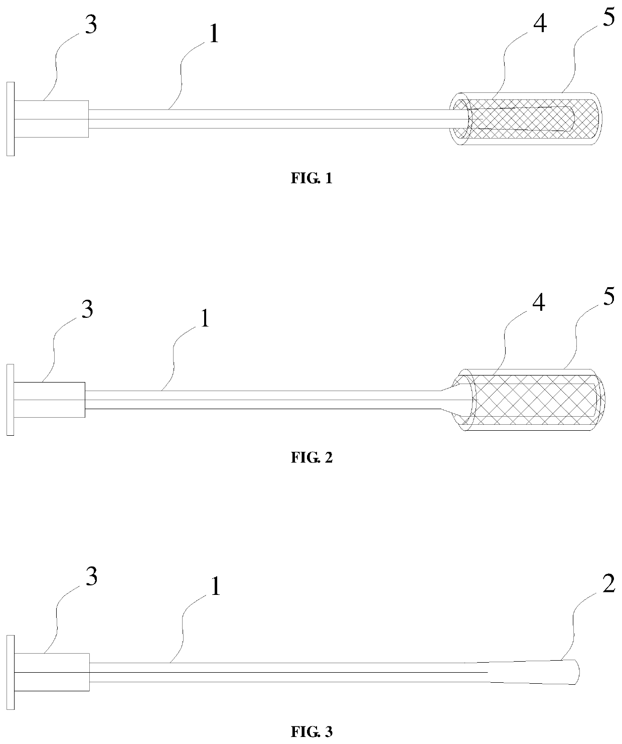
[0037]A degradable foldable biological amniotic membrane composite repair stent, comprising a tubular body 1 with an axially extending through hole, the front end of the tubular body 1 is provided with an elastic balloon 2, and the end of the tubular body 1 is connected to a one-way valve 3 which seals the through hole here, the elastic balloon 2 is arranged in the lumen of a foldable reticulated polylactic acid stent 4, the foldable reticulated polylactic acid stent 4 is made of polylactic acid material by laser engraving, the outer surface of the foldable reticulated polylactic acid stent 4 is coated with a biological amniotic membrane 5, and there are a plurality of micropores on meshes of the foldable reticulated polylactic acid stent 4, the plurality of micropores are filled with biological amniotic membrane powder; in the initial state, the elastic balloon 2, the foldable reticulated polylactic acid stent 4, and the biological amniotic membrane 5 are compressed into a tight st...

example 2

[0039]A degradable foldable biological amniotic membrane composite repair stent, comprising a tubular body 1 with an axially extending through hole, the front end of the tubular body 1 is provided with an elastic balloon 2, and the end of the tubular body 1 is connected to a one-way valve 3 which seals the through hole here, the elastic balloon 2 is arranged in the lumen of a foldable reticulated polylactic acid stent 4, the foldable reticulated polylactic acid stent 4 is woven from filamentous polylactic acid, the outer surface of the foldable reticulated polylactic acid stent 4 is coated with a biological amniotic membrane 5, and there are a plurality of micropores on meshes of the foldable reticulated polylactic acid stent 4, the plurality of micropores are filled with compound amniotic membrane gel; in the initial state, the elastic balloon 2, the foldable reticulated polylactic acid stent 4, and the biological amniotic membrane 5 are compressed into a tight state; in the use st...

example 3

[0041]A degradable foldable biological amniotic membrane composite repair stent, comprising a tubular body 1 with an axially extending through hole, the front end of the tubular body 1 is provided with an elastic balloon 2, and the end of the tubular body 1 is connected to a one-way valve 3 which seals the through hole here, the elastic balloon 2 is arranged in the lumen of a foldable reticulated polylactic acid stent 4, sheet-like polylactic acid and sheet-like amniotic membrane are laminated and wound on the outer surface of the elastic balloon 2, there are a plurality of micropores on the sheet-like polylactic acid and the plurality of micropores are filled with biological amniotic membranes; in the initial state, the elastic balloon 2, the sheet-like polylactic acid, and the sheet-like amniotic membrane are compressed into a tight state; in the use state, the elastic balloon 2 is injected with sterile gas or liquid to expand to adapt to the shape of the affected area, and stretc...


PUM

PropertyMeasurementUnit
Biodegradabilityaaaaaaaaaa

Abstract

The present invention discloses a degradable foldable biological amniotic membrane composite repair stent, comprising a tubular body with an axially extending through hole, the front end of the tubular body is provided with an elastic balloon, and the end of the tubular body is connected to a one-way valve which seals the through hole here, the outer face of the elastic balloon is coated with a foldable reticulated polylactic acid stent, the outer surface of the foldable reticulated polylactic acid stent is coated with a biological amniotic membrane, and there are a plurality of micropores on meshes of the foldable reticulated polylactic acid stent, the plurality of micropores are filled with biological amniotic membrane powder; in the initial state, the elastic balloon, the foldable reticulated polylactic acid stent, and the biological amniotic membrane are compressed into a tight state; in the use state, after being implanted in the body and expanded under pressure, it can conform to the lacrimal duct/uterine cavity to form a tubular or drop-like shape or other spatial shape that adapts to the body cavity.

Description

FIELD OF THE INVENTION[0001]The present invention generally relates to the biomedical technology, and more particularly to a multi-purpose degradable foldable biological amniotic membrane composite repair stent.BACKGROUND OF THE INVENTION[0002]Lacrimal duct obstruction diseases (LDOD) are a class of diseases with lacrimal duct obstruction (striction or / and occlusion of the lacrimal duct) as pathological features and epiphora as main clinical manifestation. According to incomplete statistics, LDOD accounts for about 3% of outpatients in ophthalmology clinics, and it is a common and frequently-occurring disease in ophthalmology. Lacrimal duct obstruction may occur in any part of the lacrimal duct. The obstruction of lacrimal canaliculus is a common type of LDOD, and its incidence accounts for about 16% to 25% of LDOD. The obstruction of lacrimal canaliculus may be caused by a number of factors, including inflammation, trauma, congenital abnormalities, drugs, systemic diseases, iatroge...

Claims


Application Information

Patent Timeline
IPC IPC(8): A61F2/90
CPCA61F2/90A61F2240/002A61F2250/0018A61F2250/0023B33Y80/00A61F2/844A61F2/91A61F2210/0076A61F2210/0004A61F2210/0071A61F9/00772A61L31/148A61L31/129A61F2/958C08L67/04C08L89/00
Inventor MIAO, CHUNYUNLV, HUPENG, CHONGJUNTENG, ZHIQIANG
Owner JIANGXI RUIJI BIOTECH CO LTD
Why Eureka
Try Eureka