Methods and compositions relating to a cardiac-specific nuclear regulatory factor

a nuclear regulatory factor and cardiac-specific technology, applied in the field of development biology and molecular biology, can solve the problems of regional contractile dysfunction, impracticality in clinical setting, loss of cardiomyocytes,

Inactive Publication Date: 2005-11-08
BOARD OF RGT THE UNIV OF TEXAS SYST
View PDF3 Cites 2 Cited by
  • Summary
  • Abstract
  • Description
  • Claims
  • Application Information

AI Technical Summary

Benefits of technology

[0024]An additional aspect of the present invention is to provide compositions and methods for the identification of downstream target genes of myocardin polypeptides. A gene delivery vector, for example an adenoviral vector, can be employed to deliver a myocardin gene to isolated cardiomyocytes thereby permitting over-expression of the myocardin polypeptide. Differences in gene profiling between control (i.e., non-transfected) cardiomyocytes and transfected (i.e., myocardin-overexpressing) cardiomyocytes can then be assessed by standard methods, such as differential display and microarray (e.g., gene chip) technology. Genes that are activated by myocardin in cardiomyocytes can subsequently b

Problems solved by technology

Myocardial infarction results in the loss of cardiomyocytes.
The loss of cardiomyocytes leads to regional contractile dysfunction.
Although the transplantation of fetal cardiomyocytes is a proposed treatment of heart failure, it remains impractical in the clinical setting, in part because of the difficulty of obtaining fetal heart donor tissue.
Although it is known that the lo

Method used


Image

  • Methods and compositions relating to a cardiac-specific nuclear regulatory factor
  • Methods and compositions relating to a cardiac-specific nuclear regulatory factor
  • Methods and compositions relating to a cardiac-specific nuclear regulatory factor

Examples


example 1

Expression Pattern of Myocardin 1 During Early Heart Development

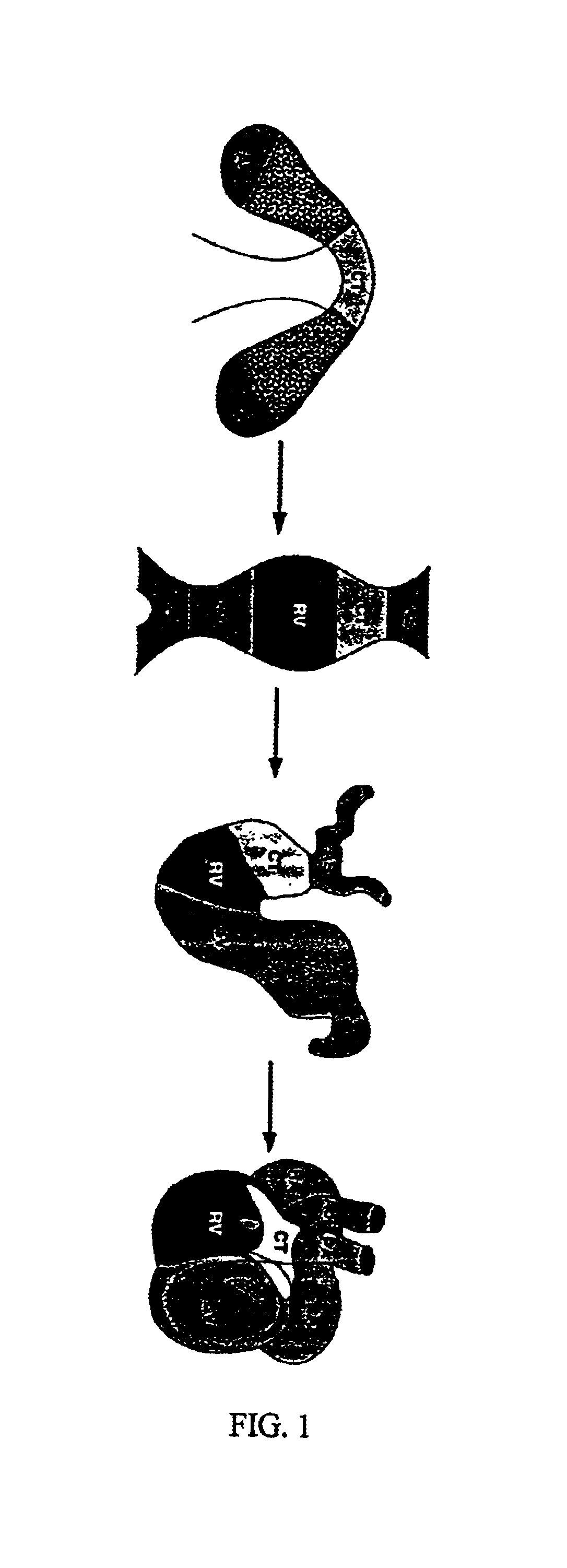
[0270]The heart is the first organ to form during mammalian development. Cardiac muscle cells originate from a region of the embryo known as the cardiac crescent and develop into a primitive heart tube along the midline of the embryo (FIG. 1). Because this is the only region of the embryo that can give rise to cardiac muscle cells, it would be expected to express a unique set of genes responsible for cardiogenesis. By identifying the genes that are uniquely expressed in this region, master control genes for heart formation can be identified. Subsequent embryonic events of looping, chamber maturation and alignment with the vascular system give rise to the mature four-chambered heart (Olson et al., 1996 and Fishman et al., 1997).

[0271]Expression of myocardin 1 was determined by whole-mount (FIG. 3A) or section (FIG. 3B and FIG. 3C) in situ hybridizations to mouse embryos at E7.75 (FIG. 3A), E8.0 (FIG. 3B), and E12.5 (FIG....

example 2

Expression Pattern of Myocardin 1 in Adult Mouse Tissues

[0275]The expression of myocardin 1 transcripts in adult mouse tissues was analyzed by Northern blot, utilizing techniques well known in the art. RNA was isolated from adult mouse heart, brain, spleen, lung, liver, skeletal muscle, kidney and testis according to standard RNA isolation procedures, e.g., phenol / chloroform / isoamyl alcohol (RNAzol, Life Technologies, Inc.) or guanidine thiocyanate.

[0276]Briefly, fractionated RNA was transferred from an agarose gel to a membrane by upward capillary action. The transferred RNA is cross-linked to the membrane. Next, a radiolabeled probe (DNA or RNA) was hybridized to the membrane in a formamide solution. After hybridization, autoradiography was performed to detect the transcripts.

[0277]The results in FIG. 4 show that the transcripts were detected only in the heart. A RNA molecular marker illustrates the size of the transcripts.

example 3

Nuclear Localization of Myocardin 1 Protein

[0278]Cos cells were transiently transfected with an expression vector encoding myocardin 1 with a Flag-epitope tag. Transient transfection assays were performed using standard methods, such as LifofectAMINE plus (Life Technologies, Inc.), calcium phosphate or electroporation. Briefly, the cells were plated 12 hr before transfection in tissue culture dishes. They were transfected with a total of about 0.5-1.0 μg of plasmid DNA. The subcellular location of myocardin 1 protein was determined by immunostaining with anti-Flag antibody.

[0279]All myocardin 1 protein is localized to the nucleus as illustrated in FIG. 5. The inset in the lower right comer shows an enlargement of a single cell, with strong myocardin 1 staining in the nucleus, but excluded from the nucleoli.


PUM


Abstract

The present invention relates to a novel cardiac-specific transcription factor, myocardin. This molecule modulates the development and differentiation of cardiomyocytes and is a potent inhibitor of cell growth. Methods to exploit these observations are provided and include respecifiying non-cardiac cells into cardiac cells, stimulating cardiac tissue regeneration, and methods for treating cardiomyopathies, myocardial infarction.

Description

[0001]This application claims benefit of priority to U.S. Provisional Application Ser. No. 60 / 257,761 filed Dec. 21, 2000.[0002]The government owns rights in the application pursuant to NIH Grant Nos. P01 HL49953 and HL63926.BACKGROUND OF THE INVENTION[0003]1. Field of the Invention[0004]The present invention relates generally to the fields of developmental biology and molecular biology. More particularly, it concerns proteins involved in the regulation of cardiomyocyte cell growth and development.[0005]2. Description of Related Art[0006]The leading cause of morbidity and mortality in industrialized countries is heart disease, particularly heart disease that is associated with myocardial infarction. Myocardial infarction results in the loss of cardiomyocytes. Cardiomyocytes are post-mitotic cells and generally do not regenerate after birth. Furthermore, it has been discovered that they respond to mitotic signals by cell hypertrophy (Kodama et al., 1997; Pan et al., 1997) rather than...

Claims


Application Information

Patent Timeline
IPC IPC(8): C07K14/435C07K14/47C12N5/06A61K38/00C12N5/077
CPCC07K14/4705C12N5/0657A01K2217/05A61K38/00C12N2501/60C12N2510/00
Inventor OLSON, ERIC N.WANG, DA-ZHI
Owner BOARD OF RGT THE UNIV OF TEXAS SYST
Why Eureka
Try Eureka