Nucleic acid vaccines for prevention of flavivirus infection

a vaccine and nucleic acid technology, applied in the field of vaccines and diagnostics, can solve the problems of serious health concerns, inability to easily interrupt or inability to prevent infection by wnv, etc., and achieve the effects of convenient preparation, stable storage, and easy administration

Inactive Publication Date: 2007-06-05
HEALTH & HUMAN SERVICES THE UNITED STATES OF AMERICA AS REPRESENTED BY THE SEC +1
View PDF22 Cites 33 Cited by
  • Summary
  • Abstract
  • Description
  • Claims
  • Application Information

AI Technical Summary

Benefits of technology

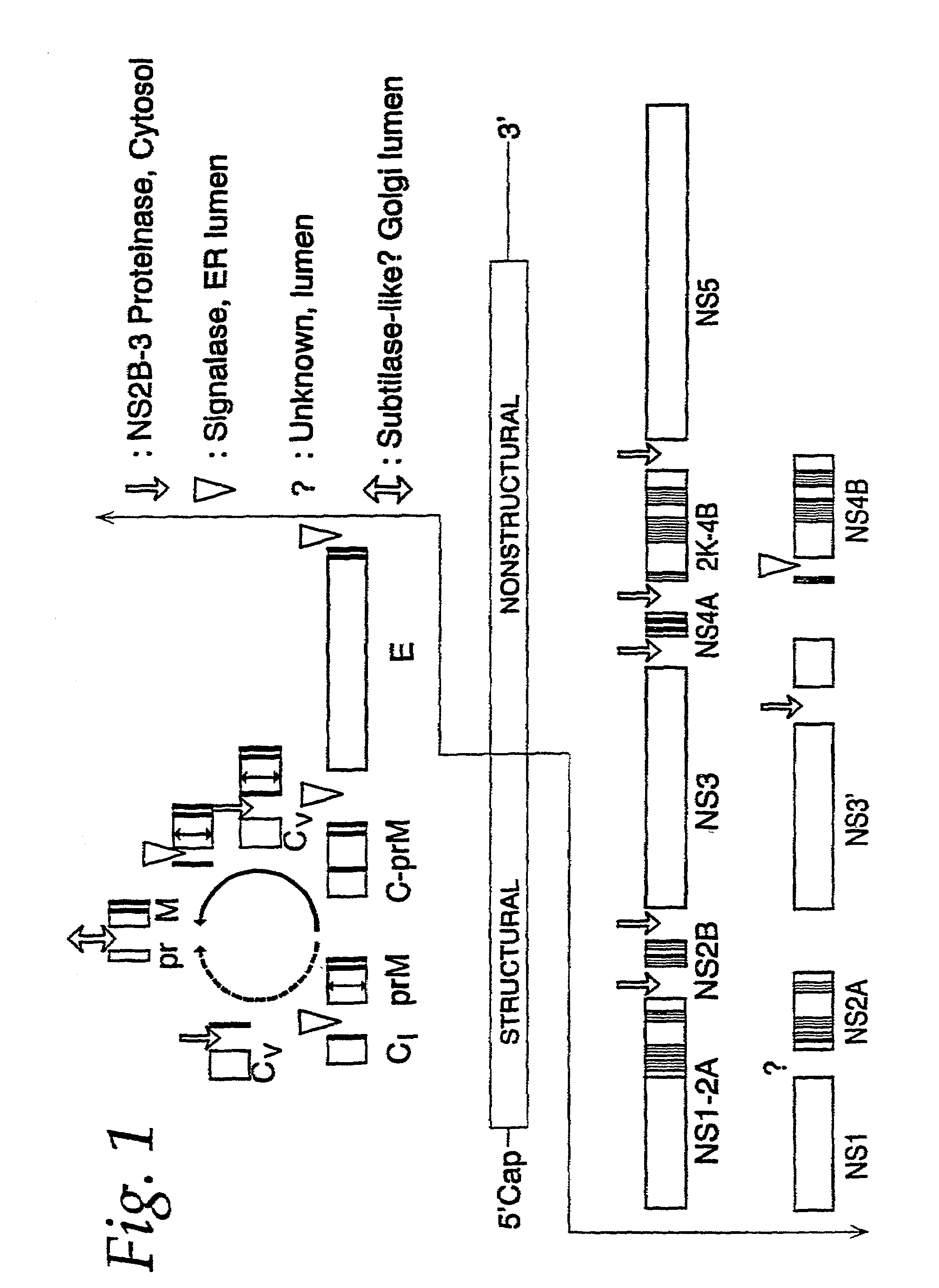
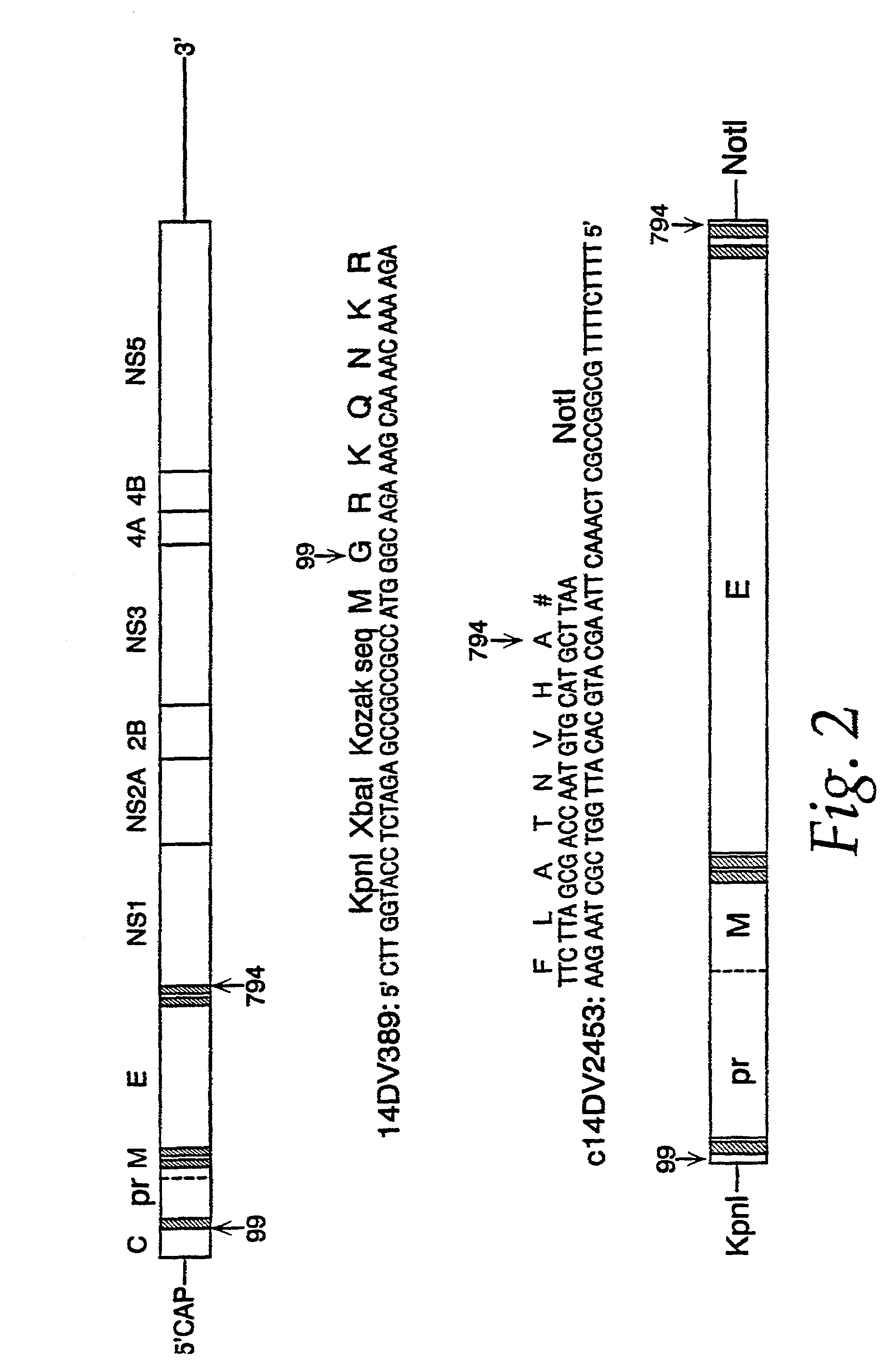
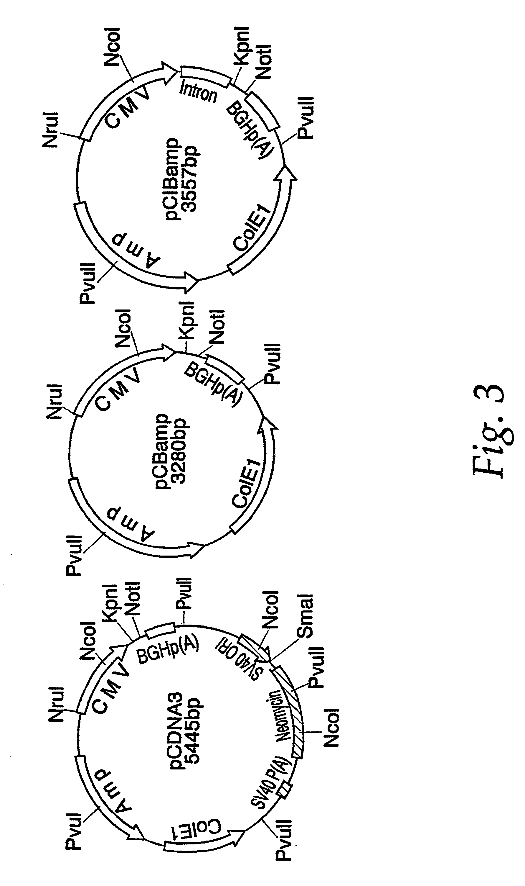
[0026]The present invention provides a nucleic acid molecule which contains a transcriptional unit (TU) for an immunogenic flavivirus antigen. The TU directs a host cell, after being incorporated within the cell, to synthesize the antigen. In an important aspect of the invention, the flavivirus can be yellow fever virus (YFV), dengue serotype 1 virus (DEN-1), dengue serotype 2 virus (DEN-2), dengue serotype 3 virus (DEN-3), dengue serotype 4 virus (DEN-4), St. Louis encephalitis virus (SLEV), Japanese encephalitis virus (JEV), West Nile virus (WNV), Powassan virus or any other flavivirus. In important embodiments of the present invention, the antigen can be the flavivirus prM / M protein, the E protein, or both. In particular, when the TU includes both the prM / M and E proteins, the host cell secretes subviral particles containing the prM / M and E antigens. In a further important aspect of the invention, the nucleic acid is a DNA molecule. In additional significant embodiments, the nucleic acid TU includes a control sequence disposed appropriately such that it operably controls the expression of the prM / M and E antigens and this control sequence can be the cytomegalovirus immediate early promoter. In an additional embodiment, the nucleotide sequence of the TU is engineered to optimize eukaryotic translation by minimizing large hairpin structures in the 5′-end untranslated region of an mRNA produced by the TU and / or the inclusion of a Kozak consensus sequence at the translational start site of an mRNA produced by the TU. In an additional embodiment, the transcriptional unit also includes a poly-A terminator.
[0030]These aspects and embodiments of the invention are the basis for its distinct attributes and advantages. Being a nucleic acid construct involving only portions of the flavivirus genome rather than the sequence encompassing the complete genome, the nucleic acid TU-containing vaccine is completely nonviable. It therefore poses no danger of infection by the flavivirus to those involved in its manufacture or to subjects receiving the vaccine. The nucleic acid vaccine is easy to prepare and easy to administer and is stable in storage prior to use. Unexpectedly it has been found that the nucleic acid vaccine of the invention is essentially 100% successful in conferring protective immunity in mammals after administering only a single dose. A further unexpected result is that the nucleic acid TU is able to engender immunity to a flavivirus in a female mammal which can be transmitted to its progeny through the milk. Without wishing to be limited by theory, the inventor believes that a possible mechanism for the success of the nucleic acid in conferring protective immunity is that a host cell harboring the nucleic acid, such as the cell of a subject to whom the vaccine is administered, produces subviral particles containing the flaviviral prM / M and E antigens. These particles mimic the immunogenic attributes of native flavivirus virions.

Problems solved by technology

They therefore occur over widespread areas and their transmission is not easily interrupted or prevented.
Thus, infection by WNV is a serious health concern in many regions of the world.
Concurrent with this outbreak, local health officials observed increased mortality among birds (especially crows) and horses.
Currently, no human or veterinary vaccine is available to prevent WNV infection and mosquito control is the only practical strategy to combat the spread of the disease.
ng survivors, there are serious neurological consequences, related to the symptoms of encephalitis, that persist after infection. In
le virus vaccines, although effective, do have certain problems and / or disadvantages. The
h culture methods are cumbersome and expensive. Fur
thermore, there is the attendant risk of incorporating antigens from the host cells, i.e., the brain or other host, into the final vaccine product, potentially leading to unintended and undesired allergic responses in the vaccine recipients. The
re is also the risk of inadvertent infection among workers involved in vaccine production. Fin
ally, there is the risk that the virus may not be fully or completely inactivated or attenuated and thus, the vaccine may actually cause disease.
Nevertheless, despite decades of effort, safe and effective vaccines to protect against dengue virus infection are not yet available.
Infection leads to fever, chills, severe headache and other pains, anorexia, nausea and vomiting, with the emergence of jaundice.
Also, monkeys failed to develop fully protective immunity to dengue virus challenge when immunized with recombinant baculovirus expressing the E protein (Lai et al.
Challenge experiments using a two-dose schedule, however, failed to protect mice against lethal dengue-2 virus challenge.
Inactivated vaccine is costly and inconvenient to prepare.
In addition, any such vaccine entails the risk of allergic reaction originating from proteins of the host cell used in preparing the virus.
Furthermore, such vaccines present considerable risk to the workers employed in their production.
Also, like the whole virus preparations, these vaccines carry the risk of adverse allergic reaction to antigens from the host or to the vector.
Vaccine development against dengue virus and WNV is less advanced and such virus-based or recombinant protein-based vaccines face problems similar to those alluded to above.
Particularly, the concurrent risks and costs involved in the production of antigens from viruses and the failure of most currently available recombinantly expressed antigens to elicit effective immune responses are paralleled in the field of immunodiagnostics by the same risks, high costs and a corresponding lack of sensitivity.
Thus, because of the high costs, risk of accidental infection with live virus and the lower than desired levels of sensitivity of the previously available tests, there exists a need for rapid, simple and highly sensitive diagnostic tests for detecting flavivirus infection and / or contamination.

Method used


Image

  • Nucleic acid vaccines for prevention of flavivirus infection
  • Nucleic acid vaccines for prevention of flavivirus infection
  • Nucleic acid vaccines for prevention of flavivirus infection

Examples


example 1

Preparation of Recombinant Plasmids Containing the Transcriptional Unit Encoding JEV prM and E Antigens

[0108]Genomic RNA was extracted from 150 μL of JEV strain SA 14 virus seed grown from mouse brain using a QIAamp™ Viral RNA Kit (Qiagen, Santa Clarita, Calif.). RNA, adsorbed on a silica membrane, was eluted in 80 μL of nuclease-free water, and used as a template for the amplification of JEV prM and E gene coding sequences. Primer sequences were obtained from the work of Nitayaphan et al. (Virology 177: 541–552 (1990)). A single cDNA fragment containing the genomic nucleotide region 389–2478 was amplified by the reverse transcriptase-polymerase chain reaction (RT-PCR). Restriction sites KpnI and XbaI, the consensus Kozak ribosomal binding sequence, and the translation initiation site were engineered at the 5′ terminus of the cDNA by amplimer 14DV389 (nucleotide sequence, SEQ ID NO:1; amino acid sequence, SEQ ID NO:2). An in-frame translation termination codon, followed by a NotI re...

example 2

Evaluation of JEV prM and E Proteins Expressed by Various Recombinant Plasmids Using an Indirect Immunofluorescent Antibody Assay

[0112]The expression of JEV specific gene products by the various recombinant expression plasmids was evaluated in transiently transfected cell lines of COS-1, COS-7 and SV-T2 (ATCC, Rockville Md.; 1650-CRL, 1651-CRL, and 163.1-CCL, respectively) by indirect immunofluorescent antibody assay (IFA). The SV-T2 cell line was excluded from further testing since a preliminary result showed only 1–2% of transformed SV-T2 cells were JEV antigen positive. For transformation, cells were grown to 75% confluence in 150 cm2 culture flasks, trypsinized, and resuspended at 4° C. in phosphate buffered saline (PBS) to a final cell count 5×10⁶ per mL. 10 μg of plasmid DNA was electroporated into 300 μL of cell suspension using a BioRad Gene Pulse™ (Bio-Rad) set at 150 V, 960 μF and 100 Ω resistance. Five minutes after electroporation, cells were diluted with 25 mL fresh med...

example 3

Selection of an In vitro Transformed, Stable Cell Line Constitutively Expressing JEV Specific Gene Products

[0115]COS-1 cells were transformed with 10 μg of pCDJE2-7 DNA by electroporation as described in the previous example. After a 24 hr incubation in non-selective culture medium, cells were treated with neomycin (0.5 mg / mL, Sigma Chemical Co.). Neomycin-resistant colonies, which became visible after 2–3 weeks, were cloned by limited dilution in neomycin-containing medium. Expression of vector-encoded JEV gene products was initially screened by IFA using JEV HIAF. One JEV-IFA positive clone (JE-4B) and one negative clone (JE-5A) were selected for further analysis and maintained in medium containing 200 μg / mL neomycin.

[0116]Authenticity of the JEV E protein expressed by the JE-4B clone was demonstrated by epitope mapping by IFA using a panel of JEV E-specific murine monoclonal antibodies (Mab) (Kimura-Kuroda et al., J. Virol. 45: 124–132 (1983); Kimura-Kuroda et al., J. Gen. Virol....


PUM

PropertyMeasurementUnit
MWaaaaaaaaaa
MWaaaaaaaaaa
resistanceaaaaaaaaaa

Abstract

The present invention encompasses isolated nucleic acids containing transcriptional units which encode a signal sequence of one flavivirus and an immunogenic flavivirus antigen of a second flavivirus. The invention further encompasses a nucleic acid and protein vaccine and the use of the vaccine to immunize a subject against flavivirus infection. The invention also provides antigens encoded by nucleic acids of the invention, antibodies elicited in response to the antigens and use of the antigens and / or antibodies in detecting flavivirus or diagnosing flavivirus infection.

Description

[0001]This application is a continuation-in-part of, and claims the benefit of, U.S. application Ser. No. 09 / 701,536, filed Jun. 18, 2001, which status is pending and which is a national stage application of international application serial. No. PCT / US99 / 12298, filed Jun. 3, 1999 from U.S. provisional application Ser. No. 60 / 087,908, filed Jun. 4, 1998, which applications are hereby incorporated herein in their entirety by reference.FIELD OF THE INVENTION[0002]This invention relates to novel vaccines, diagnostics and methods of using both in the treatment and prevention of the diseases caused by flaviviruses. In particular, the vaccines are recombinant nucleic acids which contain genes for structural proteins of flaviviruses, such as Japanese encephalitis virus (JEV), West Nile virus (WNV) or related flaviviruses. These vaccines serve as a transcriptional unit for the biosynthesis of the virus protein antigens when administered in vivo. The diagnostics are compositions containing an...

Claims


Application Information

Patent Timeline
Patent Type & Authority Patents(United States)
IPC IPC(8): C07H21/04A61K39/12C12N15/09A61K31/711A61K39/00A61K48/00A61P31/14C07K14/18C07K16/10C12N1/15C12N1/19C12N1/21C12N5/10C12N15/40C12Q1/70G01N33/53G01N33/569
CPCA61K39/12C07K14/005C07K16/1081C12Q1/701G01N33/56983A61K48/00A61K2039/53C07K2319/02C12N2770/24134C12N2770/24122G01N2333/18A61K2039/70C12N2770/24022A61P31/12A61P31/14A61P37/04Y02A50/30C12N15/11
Inventor CHANG, GWONG-JEN J.
Owner HEALTH & HUMAN SERVICES THE UNITED STATES OF AMERICA AS REPRESENTED BY THE SEC
Why Eureka
Try Eureka