Device and method for treating central nervous system pathology

a technology a device, applied in the field of devices and methods for treating tissues of the central nervous system using subatmospheric pressure, can solve the problems of cns parenchyma death, compromise of physiological recovery, complications of drugs, etc., and achieve the effects of reducing the edema of the central nervous system, preserving neurologic function, and increasing the probability of recovery and survival

Active Publication Date: 2012-09-18
WAKE FOREST UNIV HEALTH SCI INC
View PDF549 Cites 19 Cited by
  • Summary
  • Abstract
  • Description
  • Claims
  • Application Information

AI Technical Summary

Benefits of technology

[0014]In use, the present invention can provide a pressure gradient to remove edema from the central nervous system, thus preserving neurologic function and increasing the probability of recovery and survival in a more physiologically preserved state. Decrease in central nervous system edema in turn can lead to a decrease in intracranial pressure, minimizing the risk of central nervous system compromise and herniation. In addition to the removal of edema, the present invention can remove mediators, degradation products, and toxins that enhance the inflammatory and neuropathological response of tissues in the central nervous system to injury.
[0015]The present invention can protect the central nervous system from exogenous infection and contamination, and facilitates and maximizes healing of the intracranial and adjacent structures when tissues are contaminated by central nervous system abscesses, meningitis, ventriculitis, and brain tissue infection. The central nervous system tissue may also be protected from adjacent infection, such as infection which exists subclinically in the sinuses, oral cavity, and other potentially infected spaces that exist in the normal human state, either by increased blood flow and directly decreasing bacterial load. Moreover, the device and method of the present invention can prepare central nervous system tissue to achieve a stage of healing and diminution of bacterial counts such that acceptance of secondary treatments (e.g., flaps, bone grafts) can be successful.
[0016]The present invention can also facilitate closure of pathologic openings communicating between the central nervous system and the extradural space, e.g. between the extradural space and the subdural / epidural, and / or subarachnoid space. Likewise, the progression of pathologic processes, disruption of physiological central nervous system integrity, the interference with central nervous system blood flow and nutrition can be minimized.
[0017]The devices and methods of the present invention can be used to treat the following conditions: exposure of the central nervous system as a result of trauma, surgery, infection, or any other pathologic process; treatment of any of the spaces and tissues surrounding the central nervous system, including the subdural / epidural and intraventricular spaces; treatment of edema of the central nervous system parenchyma secondary to any cause, including hemorrhage, trauma, tumor, infection or any other pathologic state; treatment of elevated intracranial and intraspinal pressure due to the any of the aforementioned causes; and treatment of cerebrospinal fluid pathology in which the spinal fluid is pathologically in communication with any non-anatomical and non-physiologic spaces. In addition, the present invention can be used to promote formation of granulation tissue in areas where central nervous system disruption has occurred, and to control cerebrospinal fluid leaks. Further, the modified present material can be used for control or closure of defects existing between the central nervous system, the cutaneous space, intranasal space, and intrasinus space.

Problems solved by technology

Among the clinical problems that threaten survival of CNS tissues, the control of central nervous system edema, infection, and blood supply are central.
Because the brain is enclosed in a closed space (the dura and skull), edema results in compression and compromise of the blood flood and nutritional performance of the CNS, which greatly impairs physiological recovery of the central nervous system and often of itself results in progression of compromise and death of the CNS parenchyma.
However, disadvantages to these treatments include poor results, complications from the drugs, and inconsistent results.
The need for rapid and effective treatment is also vital due to the disastrous consequences and high likelihood of rapid propagation of infection and edema in the CNS.
At present there are few successful methods available to treat pathologies affecting the intracranial and intraspinal space, CNS parenchyma, and the surrounding structures.
Where elsewhere tissues can be treated with dressing changes, the CNS is not amenable to this type of treatment because of its inaccessibility, precarious structure, propensity for infection, and progression of injury.
The response of the CNS to decreased blood flow secondary to edema results in hypoxia and ischemia / reperfusion-mediated injury.
Within three minutes of complete interruption of blood flow to the brain, irreversible brain damage results, though the brain can however remain viable and recover from reduced blood flow for more prolonged periods.
Cerebrovascular disease may be a result of: inadequate blood flow to the brain cells from decreased perfusion pressure, rupture of a blood vessel resulting in direct injury to the local brain area and by compression of adjacent tissue.
In addition, lifelong debility, inability to work and function in society and the family, and the frequent need for nursing home treatment often result.
People affected by strokes usually have significant impairments for the rest of their lives.
In cases of intracranial hemorrhage, the hemorrhage usually begins as a small mass that grows in volume by pressure dissection and results in displacement and compression of adjacent brain tissue.
Edema in the adjacent compressed tissue around the hemorrhage may lead to a mass effect and a worsening of the clinical condition by damaging a larger area of brain tissue.
The occurrence of edema in the week following the intracerebral hemorrhage often worsens the prognosis, particularly in the elderly.
Treatment of these conditions has been disappointing.

Method used


Image

  • Device and method for treating central nervous system pathology
  • Device and method for treating central nervous system pathology
  • Device and method for treating central nervous system pathology

Examples


examples

Rat Brain Injuries and Sub-Atmospheric Pressure Exposure

experiment 1

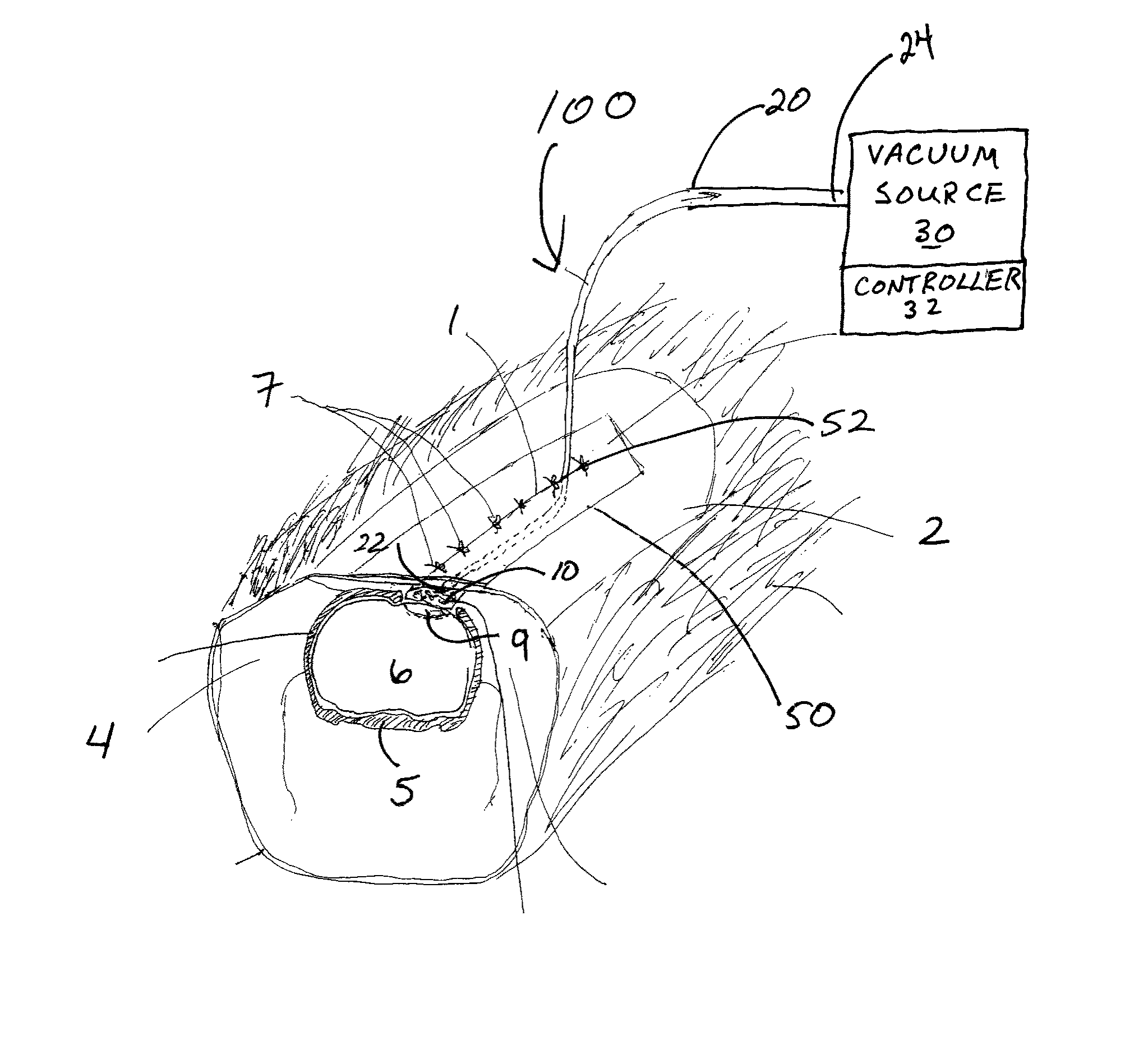
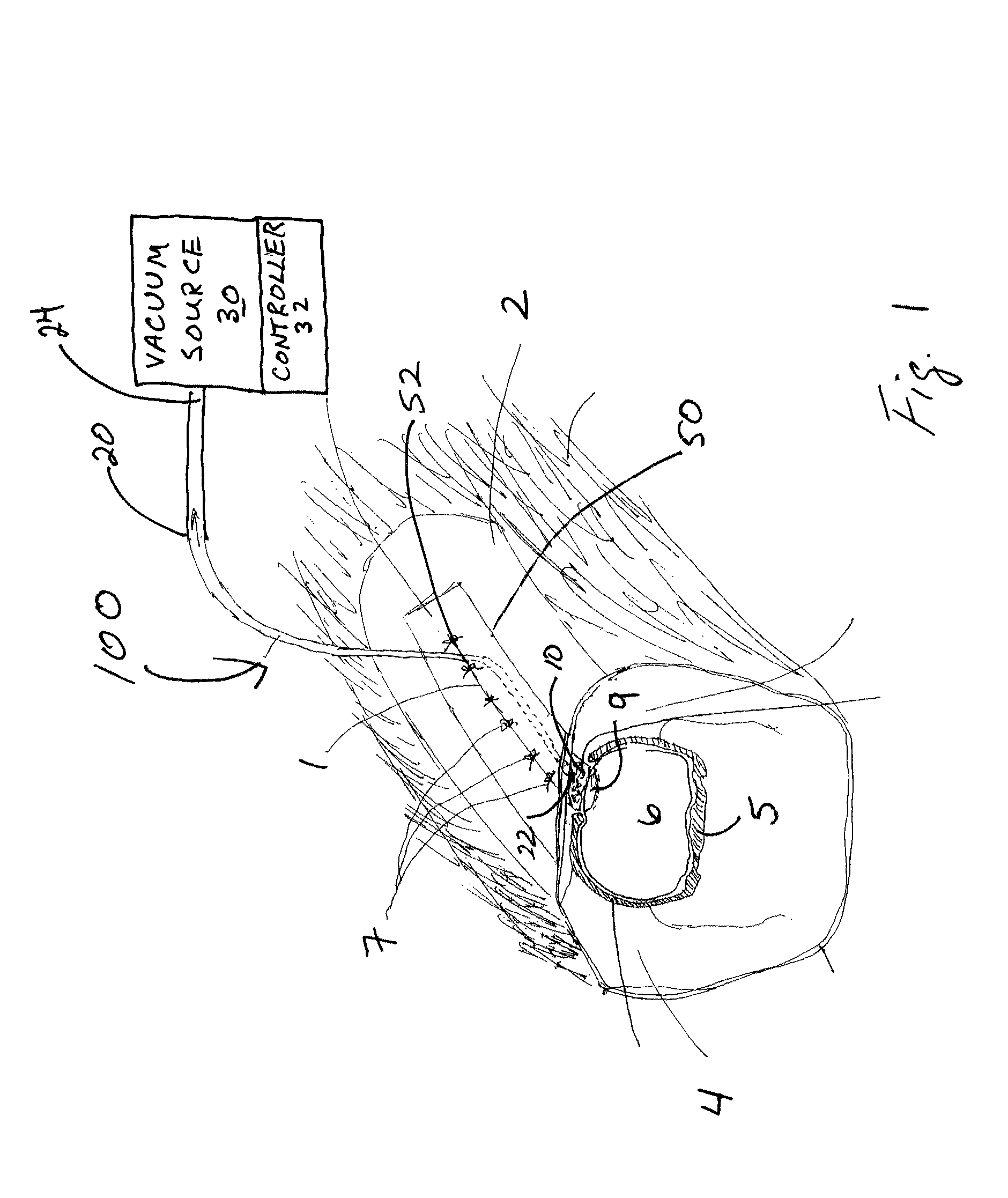
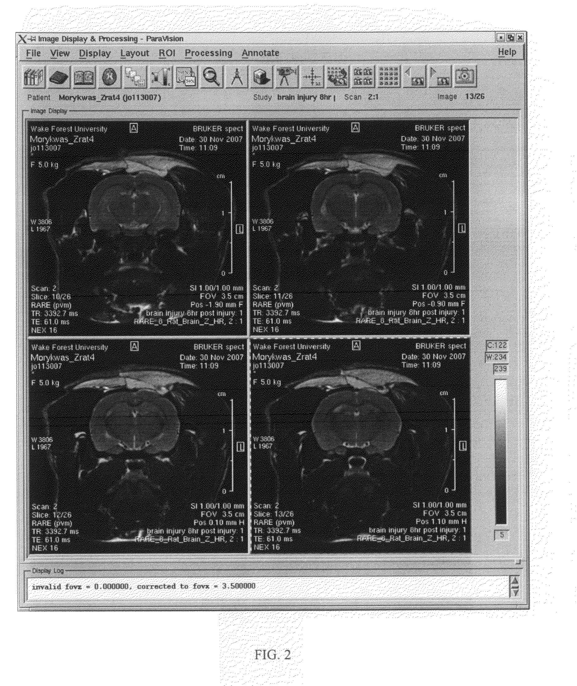
[0051]An experiment was conducted to develop a model of brain contusion and vacuum treatment of the contused brain. Twelve (12) 300 gram Sprague Dawley rats were procured and allowed to acclimated to the housing conditions. For two of the animals, a MRI scan (Bruker Biospin Horizontal Bore 7 Tesla small animal scanner, Ettlingen, Germany) of the brain was obtained before any other procedures were performed. The animals were sedated with isoflurane (2% inhalation) and the scan of the brain obtained. The animals were allowed to recover from anesthesia and returned to their cages. For creation of the injury, on the day of surgery the animals were sedated with isoflurane (2-2.5% inhalation). The top of the head was shaved and the hair removed with a depilatory agent. A midline incision 1 was made down to the bone 5, FIG. 1. The right side of the skull was removed exposing the right half of the brain; the dura was left intact. The animal was placed into the stereotaxic holder on the impa...

experiment 2

[0069]Cell death following traumatic brain injury is biphasic, with initial death due to the trauma itself, then an ongoing death as sequela to the release of excitatory amino acids, buildup of lactate, etc. The release of excitatory amino acids (glutamate, aspartate) cause a disturbance in ion homeostasis via agonist opened channel, thus increasing energy demand and increasing lactate production. Elevated levels of glutamate have been shown to be correlated with increased levels of lactate. This increase in lactate is reflective of increased energy demand during periods of impaired supply (ischemia), and is inversely related to patient outcome. Lactate production leads to apoptotic neuronal cell death.

[0070]In this preliminary study, anesthetized rats underwent an 8 mm diameter craniectomy between the bregma and lambda, 1 mm lateral to the midline. A controlled cortical impact injury with intact dura was created using the apparatus of Example 1. The impactor tip was 6 mm in diamete...


PUM

PropertyMeasurementUnit
pressureaaaaaaaaaa
pressureaaaaaaaaaa
sub-atmospheric pressureaaaaaaaaaa

Abstract

The present invention relates generally to a device and method for treating tissues of the central nervous system and more particularly, but not exclusively, to a device and method for treating the brain tissue.

Description

RELATED APPLICATIONS[0001]The present application claims the benefit of priority of U.S. Provisional Application 61 / 019,968, filed on Jan. 9, 2008 and U.S. Provisional Application No. 61 / 081,997, filed on Jul. 18, 2008, the entire contents of which are incorporated herein by reference.FIELD OF THE INVENTION[0002]The present invention relates generally to a device and method for treating tissues of the central nervous system using sub-atmospheric pressure and more particularly, but not exclusively, to a device and method for treating the brain tissue using sub-atmospheric pressure.BACKGROUND OF THE INVENTION[0003]The anatomy, physiology, and pathologic processes that involve the central nervous system (CNS) make CNS tissue unique. The preservation of both the three-dimensional structural anatomy and the microanatomical relationships of neurons (whose function depends specific on spacial relationships with other neurons and other supporting cells), as well as the maintenance of proper...

Claims


Application Information

Patent Timeline
Patent Type & Authority Patents(United States)
IPC IPC(8): A61B17/08
CPCA61L27/18A61L27/24A61L27/56A61M1/0088A61L27/20B29C47/0076A61H7/00B29C67/202B29C67/0059C08L67/04C08L5/08B29L2031/753A61M2210/0693B29K2995/006A61M2207/00B29K2089/00B29C64/112B29C48/142A61M1/962A61M1/916A61L27/58A61M1/00A61M37/00
Inventor ARGENTA, LOUIS C.CARROLL, DAVID L.LEVI, NICHOLE H.LIU, JIEMORYKWAS, MICHAEL J.TATTER, STEPHENWAGNER, WILLIAM D.
Owner WAKE FOREST UNIV HEALTH SCI INC
Why Eureka
Try Eureka