Fusion of peptidoglycan hydrolase enzymes to a protein transduction domain allows eradication of both extracellular and intracellular gram positive pathogens

a technology of peptidoglycan hydrolase and protein, which is applied in the direction of enzyme stabilisation, peptide/protein ingredients, antibacterial agents, etc., can solve the problems of high frequency of treatment failure, inability to effectively treat s. aureus, and deep-seated intramammary abscesses, so as to facilitate the translocation of full-length proteins and effective antimicrobial treatment

Inactive Publication Date: 2013-02-26
UNITED STATES OF AMERICA
View PDF0 Cites 15 Cited by
  • Summary
  • Abstract
  • Description
  • Claims
  • Application Information

AI Technical Summary

Benefits of technology

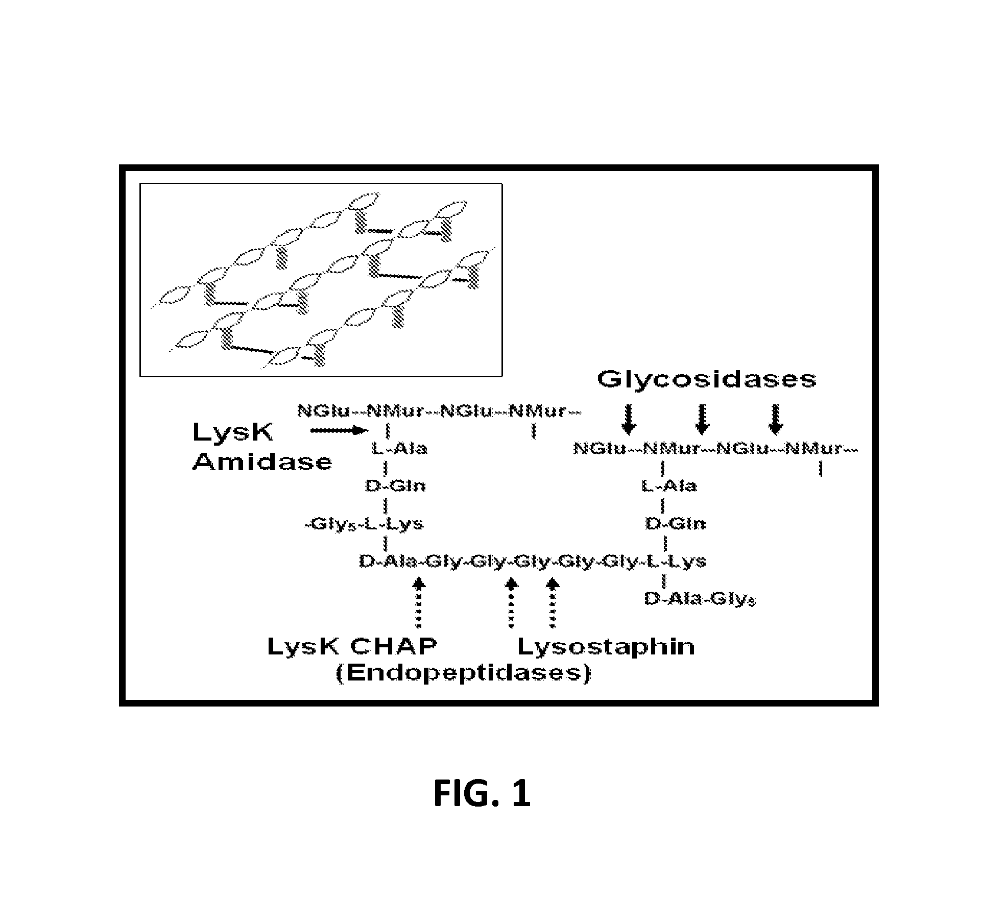
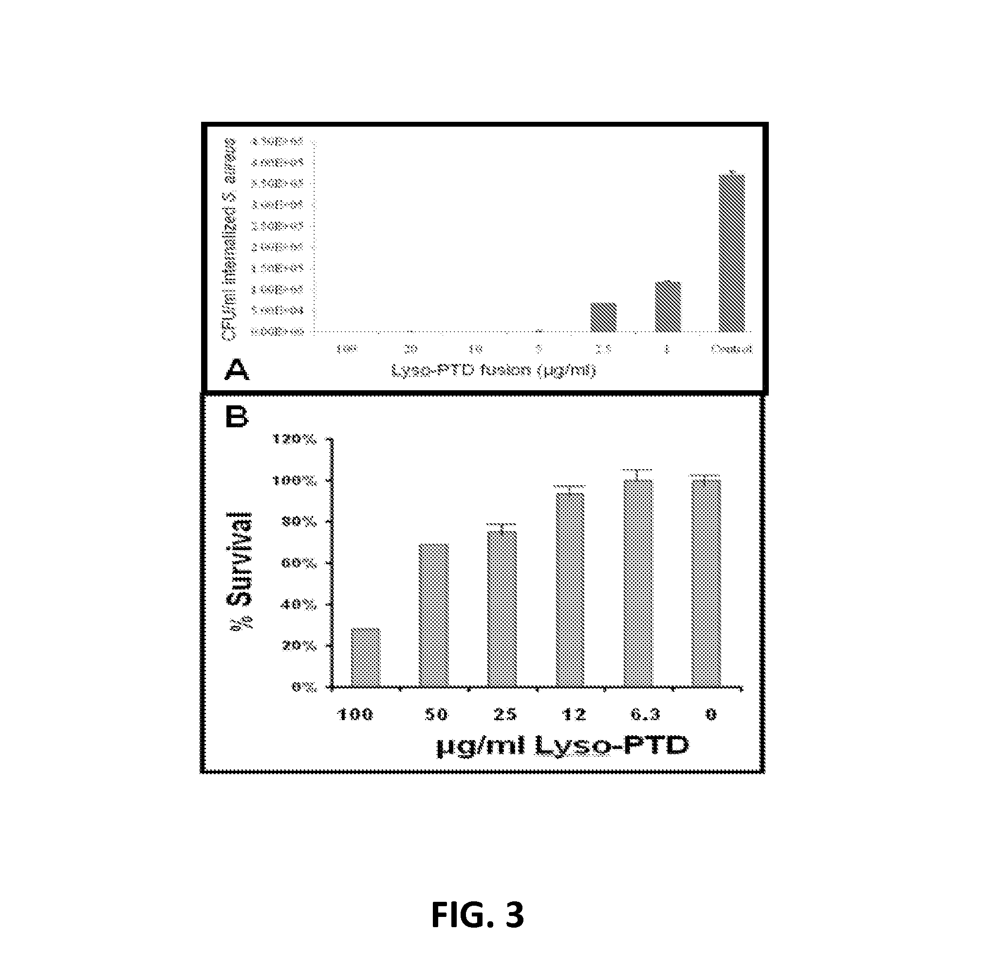
[0026]We have discovered that a fusion polypeptide comprising lysostaphin, a protein that specifically digests the peptidoglycan cell wall and lyses live, untreated S. aureus, and a protein transduction domain (PTD), which facilitates translocation of full length proteins across the eukaryotic cell plasma membrane into the eukaryotic cytoplasm from outside the cell, can be used as an effective antimicrobial treatment for both extracellular and intracellular infections caused by S. aureus, by multidrug-resistant staphylococci, and other gram positive pathogens.
[0029]An added object of the invention is to provide a lysostaphin-PTD fusion polypeptide according to the invention, which allows both extracellular and intracellular Staphylococcus-induced disease and infection to be treated, including those caused by MRSA.

Problems solved by technology

However, S. aureus infections are chronic and prone to a high frequency of treatment failures.
Treatment failures result from deep-seated intramammary abscesses, resistance to antibacterials, and intracellular survival of the pathogen.
In the absence of pathogen-specific antimicrobials, treatment of mastitis has historically been limited to the use antibiotics such as tetracycline, penicillin, and pirlimycin which are often less than 50% successful (Deluyker et al.
Vaccines have been ineffective.
The complexity and variety of virulence factors as well as the diversity of strains that cause intramammary infections hamper the development of effective vaccines.
Several lines of research using novel approaches to S. aureus vaccination have been investigated, but the results of in vivo efficacy trials in large mammals have not yielded promising results.
In addition, the ability of S. aureus capsular material to mask surface protein epitopes hampers antibody and complement binding making the use of cell wall antigen vaccines questionable (Middleton et al., supra).
Nonetheless, many treated quarters relapsed after treatment with either lysostaphin or antibiotic treatments ceased.
These antibodies were not inactivating, did not eliminate the antimicrobial properties of the enzyme, nor elicit any observable effects on the host animal (Daley and Oldham.

Method used


Image

  • Fusion of peptidoglycan hydrolase enzymes to a protein transduction domain allows eradication of both extracellular and intracellular gram positive pathogens
  • Fusion of peptidoglycan hydrolase enzymes to a protein transduction domain allows eradication of both extracellular and intracellular gram positive pathogens
  • Fusion of peptidoglycan hydrolase enzymes to a protein transduction domain allows eradication of both extracellular and intracellular gram positive pathogens

Examples


example 1

Plasmids, Constructs and Strains

[0078]All subcloning was performed in E. coli DH5α (Invitrogen, Carlsbad, Calif.) for plasmid DNA isolation and sequence verification of all constructs. pET21a constructs were induced in E. coli BL21(DE3) (EMD Biosciences, San Diego, Calif.). Staphylococcus aureus Newbolt 305 capsular polysaccharide serotype 5 (ATCC 29740), S. aureus UAMS-1 (ATCC 49230), S. aureus strain Newman, and the MRSA strains: NRS194, NRS 271, and NRS 384 were grown at 3TC in Brain Heart Infusion broth (BD, Sparks, Md.) or Tryptic Soy Broth (BD, Sparks, Md.).

example 2

PCR Cloning

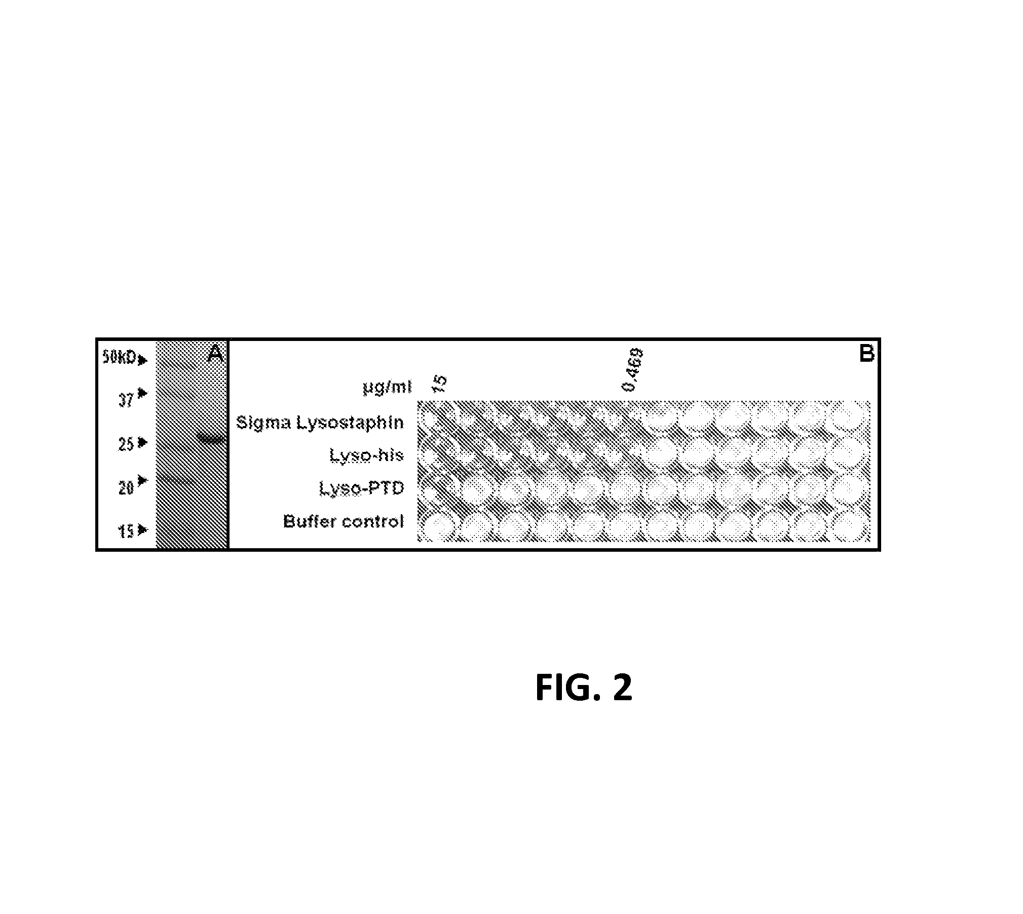
[0079]PCR cloning was used to create the Lyso-PTD construct. The reverse PCR primer Lyso-TAT-XhoR (5′-GTG GTG CTC GAG GCG GCG GCG CTG GCG GCG TTT TTT GCG CTT TAT AGT TCC-3′ (SEQ ID NO: 5) containing sequences encoding a 9 amino acid derivative (italics) of the 13 amino acid HIV-TAT PTD sequence (Table I), the last four codons (bold) of the C-terminus of the lysostaphin gene and an engineered XhoI site (underlined), together with a generic pET21a forward primer harboring an NdeI site at the translational start site, were used to amplify just the mature lysostaphin (256 aa) in a pET21a derived vector. The amplified Lyso-PTD DNA fragment was restriction enzyme digested (XhoI and NdeI) and ligated into similarly digested pET21a using conventional molecular techniques. BL21(DE3) E. coli (EMD Biosciences, San Diego, Calif.) were transformed with the characterized ligated coding sequences (SEQ ID NO:1) and the expressed protein products were purified (SEQ ID NO:2).

example 3

Extract Preparation and Protein Purification

[0080]E. coli BL21(DE3) cells harboring plasmid constructs were grown in 500 ml Superbroth (Becton Dickenson, Franklin Lakes, N.J.) supplemented with 100 ug / ml ampicillin at 37° C. with shaking. At mid log phase (OD600nm of 0.4-0.6), cultures were induced with 1 mM IPTG (isopropyl-beta-D-thiogalactopyranoside) followed by four hours shaking at 37° C. Cells were pelleted, washed with lysis buffer (50 mM NaH2PO4, 300 mM NaCl, 10 mM Imidazole, pH 8) and pellets frozen at −80° C. Extracts were prepared according to a modified procedure of Pritchard et al. (2004. Microbiology 150: 2079-2087). For nickel column-purified protein, cell pellets from 500 ml cultures were resuspended in 10 ml lysis buffer (50 mM NaH2PO4, 300 mM NaCl, 10 mM Imidazole, pH 8) and disrupted with 15×10 second pulses of sonication on ice with 10 second rest periods between pulses. Lysates were centrifuged at 6800 rpm in a Sorvall HS4 rotor (8500×g) and the supernatant deca...


PUM

PropertyMeasurementUnit
MWaaaaaaaaaa
pHaaaaaaaaaa
Tmaaaaaaaaaa

Abstract

Lysostaphin is a bacteriocin secreted by S. simulans to kill S. aureus, and has been shown to also be a potent antimicrobial for many antibiotic-resistant strains of S. aureus. By adding a ˜13 amino acid protein transduction domain (PTD) from the HIV-TAT protein to lysostaphin to form lysostaphin-PTD, both extracellular and intracellular forms of S. aureus and MRSA are killed in all (multiple) cell types examined.

Description

REFERENCE TO RELATED APPLICATION[0001]This application claims the benefit of U.S. Provisional Application No. 61 / 216,779, filed May 21, 2009, which is incorporated herein by reference in its entirety.BACKGROUND OF THE INVENTION[0002]1. Field of the Invention[0003]This invention relates to a pathogen-specific fusion protein comprising a peptidoglycan hydrolase, lysostaphin, and a protein transduction domain. The lysostaphin specifically degrades the peptidoglycan cell wall of S. aureus including methicillin-resistant Staphylococcus aureus (MRSA). In addition to lysing bacterial cells in broth, fusion of a protein transduction domain to a peptidoglycan hydrolase enzyme allows delivery of such protein antimicrobials to the intracellular locations of persistent pathogens. These staphylococcal antimicrobials have both extracellular and intracellular activity. They can be used to treat chronic staphylococcal mastitis, and any disease caused by intracellular S. aureus (e.g. MRSA osteomyeli...

Claims


Application Information

Patent Timeline
Patent Type & Authority Patents(United States)
IPC IPC(8): A61K38/54C07H21/04C12N5/10C12N15/63
CPCC07K14/31A61K38/00C07K2319/03A61P31/04
Inventor DONOVAN, DAVID
Owner UNITED STATES OF AMERICA
Why Eureka
Try Eureka