Thin-film material for optical fiber biosensor probe and preparation method thereof

A biosensor and thin-film material technology, applied in the field of optical fiber biosensor probe thin-film materials and their preparation, can solve the problems of reducing the sensitivity and accuracy of the sensor, disorderly arrangement, affecting the catalytic activity of enzymes, etc. Excellent biocompatibility, favorable effect for separation and recovery

Inactive Publication Date: 2009-10-28
WUHAN UNIV OF TECH
View PDF0 Cites 24 Cited by
  • Summary
  • Abstract
  • Description
  • Claims
  • Application Information

AI Technical Summary

Problems solved by technology
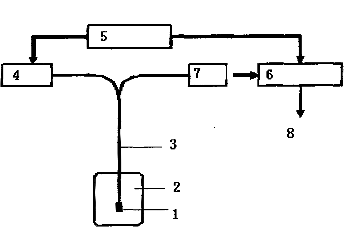
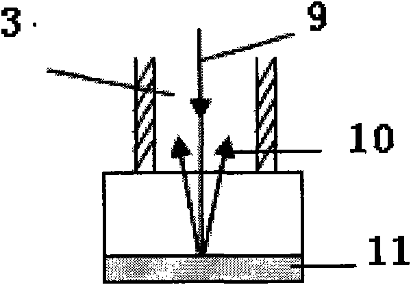
However, this biosensor has limitations: (1) only glucose oxidase is embedded and fixed by sol-gel technology, and the enzyme is easy to leak
Since the catalytic performance of the enzyme directly affects the performance of the sensor, this enzyme immobilization method will greatly reduce the sensitivity a...

Method used


Image

  • Thin-film material for optical fiber biosensor probe and preparation method thereof
  • Thin-film material for optical fiber biosensor probe and preparation method thereof

Examples


Embodiment 1

[0029] Mix tetraethyl orthosilicate, 0.05mol / L hydrochloric acid, and formamide at a volume ratio of 8:4:1, and then add Ru(bpy) with a concentration of 8mg / ml to 1.75ml of the mixture 3 Cl 2 Fluorescent indicator 140 μl, sealed and stirred at room temperature until uniformly mixed, left still to obtain a sol solution, aged in an incomplete seal at room temperature for 2 days to obtain a sol-gel solution. Take 5ml of Fe immobilized with glucose oxidase (GOD) 3 o 4 @SiO 2 Nanocomposite magnetic particles (the concentration of the nanocomposite magnetic particles is 10 mg / ml) are dissolved in pH7.0 phosphate buffer solution, and then fully mixed with the aged sol-gel solution at a volume ratio of 1:1, and 0.1 ml of mixed sol solution was added dropwise on the surface of the treated glass substrate (the glass substrate was washed with hot concentrated sulfuric acid, absolute ethanol, and distilled water in sequence and dried), and it could be cured into a condensate of about 5...

Embodiment 2

[0032] Under mechanical stirring, in a 250mL three-necked flask, add Fe at a concentration of 10mg / ml 3 o 4 @SiO 2 Nanocomposite magnetic particle solution 3ml, 80 μ l APS (aminopropyltriethoxysilane), at room temperature, continue to mechanically stir for 12h, the reaction solution is separated by a combination of permanent magnet and centrifugation, and the composite particles are separated with absolute ethanol and secondary Wash with distilled water, and then disperse with double distilled water after sonication to obtain aminated Fe 3 o 4 @SiO 2 Nanocomposite magnetic particles. Take 5ml concentration of 10mg / ml aminated Fe 3 o 4 @SiO 2 Nanocomposite magnetic particles were activated by adding 4ml of glutaraldehyde aqueous solution with a concentration of 3% by volume, mechanically stirring for 2 hours at room temperature, and then washed three times with a phosphate buffer solution with a concentration of 0.01mol / L and pH=7.0. Add the activated carrier to (GOD) p...

Embodiment 3

[0036] Prepared Fe 3 o 4 Disperse with tetramethylammonium hydroxide to obtain Fe 3 o 4 ferrofluid. Mix cyclohexane, n-hexanol and surfactant Triton X-100 evenly at a mass ratio of 9:1:2, add 1.5ml of secondary distilled water to 60ml of the mixture as the dispersed phase, and stir for 5min to obtain a transparent and stable Water-in-oil microemulsion. In the microemulsion, add 2ml of magnetic fluid (10mg / ml), stir for 1h, add 1ml of ammonia water, and then add tetraethyl orthosilicate and amination reagent (TEOS and EPTES) with a volume ratio of 3:1, the total volume is 500μl , after continuous stirring for 24 hours, the surface aminated SiO 2 Magnetic nanocomposite particles. Silicon-shell magnetic nanocomposite particles react with glutaraldehyde to generate a C=N bond. In this way, an aldehyde group is introduced on the surface of the nanocomposite particle, and the aldehyde group can react with the amino group in the glucose oxidase molecule to make the enzyme mole...


PUM

PropertyMeasurementUnit
Diameteraaaaaaaaaa

Abstract

The invention relates to a thin-film material for an optical fiber biosensor probe and a preparation method thereof. The material has catalytic characteristic and photosensitivity and is made from Fe3O4/SiO2 magnetic nanocomposite particles, bioactive enzyme, fluorescent material and sol-gel. The bioactive enzyme is fixed on the Fe3O4/SiO2 magnetic nanocomposite particles and the fixed bioactive enzyme and the fluorescent material are embedded in a sol-gel grid. The preparation method comprises the following steps: a Fe3O4/SiO2 magnetic nanocomposite particle carrier is synthesized; the carrier is activated by glutaraldehyde or by glutaraldehyde, bovine serum albumin and glutaraldehyde in sequence and then has a crosslinking reaction with the bioactive enzyme to fix the enzyme; the Fe3O4/SiO2 magnetic nanocomposite particles fixed with the bioactive enzyme are dripped on the surface of a treated glass substrate after being mixed with the sol-gel liquid blended with a Ru(bpy)3Cl2 fluorescent indicator; after cleaning, drying and aging, a sol-gel optical bio-composite sensing film embedding the bioactive enzyme and the fluorescent indicator is obtained. The optical fiber biosensor probe made from the thin-film material is especially applicable to the detection of blood sugar, blood cholesterol and the like.

Description

technical field [0001] The invention relates to an optical fiber biosensor probe film material and a preparation method thereof, in particular to a multifunctional composite optical biosensing film material with catalytic and photosensitive properties and a preparation method thereof. The optical fiber biosensor probe is especially suitable for the detection of blood sugar. Background technique [0002] Fiber optic sensors have many unique advantages, represent the development trend of a new generation of sensors, and have broad and important application prospects in many fields. Optical fiber biosensor is a large category of optical fiber sensors. Its probe can sense the biological substance to be measured, and generate corresponding optical signal, which is then converted into electrical signal output by photoelectric conversion device. Due to the advantages of small probe diameter, large information transmission capacity, low energy loss, strong anti-interference ability...

Claims


Application Information

Patent Timeline
IPC IPC(8): G01N21/64G01N33/50
Inventor 黄俊王海袁银权丁莉芸赵荣赵文琪王超
Owner WUHAN UNIV OF TECH
Why Eureka
Try Eureka