In-vitro screening and culturing method for tissue engineering skin seed epidermal keratinocyte high-simulation in-vivo cell extracellular matrix attachment

A technology for tissue engineering skin and extracellular matrix, which is applied in the field of tissue engineering, can solve the problems of low fibroblast adhesion, loss, and short plating cycle, and achieve the effects of high cell purity, simple preparation, and strong specificity

Inactive Publication Date: 2016-01-13
AFFILIATED HUSN HOSPITAL OF FUDAN UNIV
View PDF2 Cites 4 Cited by
  • Summary
  • Abstract
  • Description
  • Claims
  • Application Information

AI Technical Summary

Problems solved by technology

Therefore, if the trophoblast is not used, it is possible that the differentiation of skin cells during long-term culture may cause uncontrollable development of subsequent research;
[0017] Method 6 (homologous fibroblast attachment screening method): Scholar Jin Yan et al. used references to improve scholars HagarB and Dunnwald mouse fibroblasts, changed the donor fibroblasts pre-expanded in vitro as the attachment substrate as a trophoblast, and cultured homologous fibroblasts. Skin cells, but the patent does not specify the detailed steps of how to prepare the trophoblast plate, the plating cycle is short, the fibroblast adhesion force is low, and the repeated PBS cleaning and purification process of screening will lose part of the cultured fibroblasts that successfully adhere to the target, thus Reduce the yield of target skin cells. Skin cells are theoretically homogeneous. Another technical problem is that the two cells share the same culture environment. Considering the complexity of the culture medium, the two cells may grow competitively. Proliferation inhibition and other phenomena, the subsequent separation of target cells is technically difficult, and it is not superior to the existing six common methods. It is suggested that this method can be changed into a compound recipe and added to other screening materials with good covering effect and relatively strong adhesion behavior. Increase the yield of target skin cells, or inactivate fibroblast plated cells or add essential factors during culture

Method used


Image

  • In-vitro screening and culturing method for tissue engineering skin seed epidermal keratinocyte high-simulation in-vivo cell extracellular matrix attachment
  • In-vitro screening and culturing method for tissue engineering skin seed epidermal keratinocyte high-simulation in-vivo cell extracellular matrix attachment
  • In-vitro screening and culturing method for tissue engineering skin seed epidermal keratinocyte high-simulation in-vivo cell extracellular matrix attachment

Examples


Embodiment 1

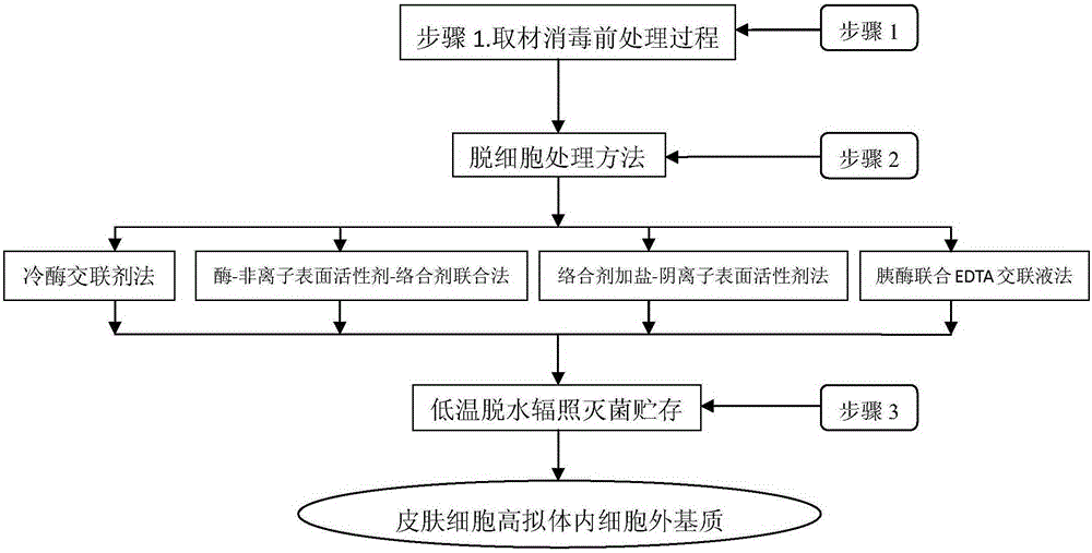
[0082] Embodiment 1 The specific implementation steps and methods of the preparation of the extracellular matrix in the hypermimetic body of the present invention:

[0083] Step 1: Skin disinfection process: take the donor skin including the following (embryonic skin, adult prepuce outer plate, scalp, armpit skin, and trunk skin can all be used) faulty skin with epidermis and dermis structure, cut into tissue pieces and soak in Dilute it in a solution containing 0.05-0.1% benzalkonium bromide with normal saline for 10-15 minutes, disinfect it, and wash the residual benzalkonium bromide repeatedly with sterile normal saline.

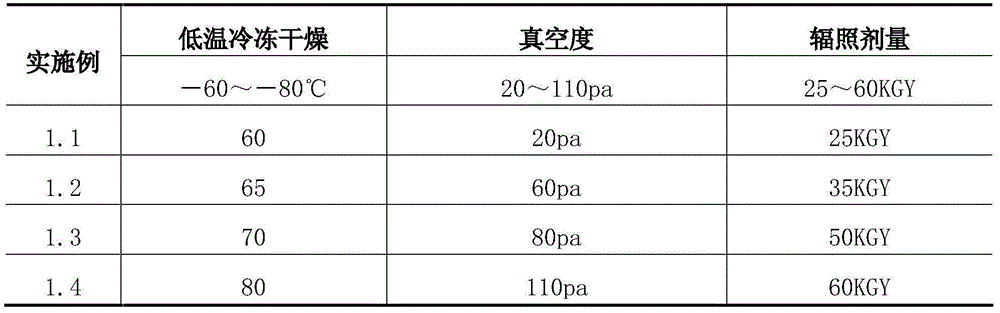
[0084] Step 2: Freeze-dry at low temperature -60-80°C, vacuum up to 20-110pa, remove more than 90-95% of the water in the tissue block, take it out, seal it and pack it in vacuum, and sterilize it by irradiation at a dose of 25-60KGY. The finished product can be stored at room temperature for up to 5 years. To start, you only need to soak at 37°C, recove...

Embodiment 2

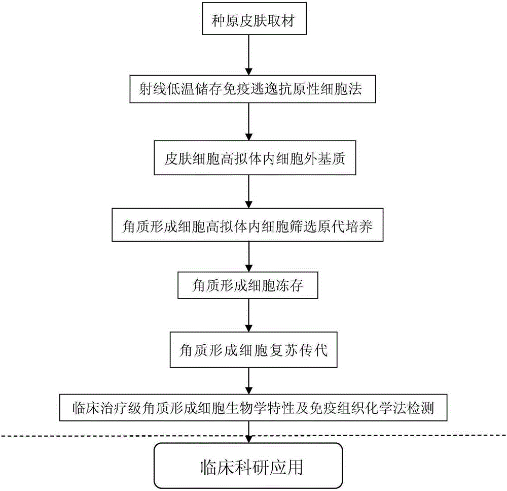
[0109] Embodiment 2 The method for culturing epidermal keratinocytes in a highly mimicked in vivo extracellular matrix of the present invention includes:

[0110] Screening of skin samples:

[0111] Skin cell screening sites include fetal skin, foreskin outer plate, scalp, and armpit skin.

[0112] Highly simulated extracellular matrix parts in the body: fetal skin, foreskin outer plate, scalp, armpit skin, and skin from other parts can be used.

[0113] When receiving it, fully soak it in PBS containing antibiotics (penicillin 80-100u / ml and 80-100μg / ml streptomycin) for 3-5 minutes, and trim the specimen to remove the subcutaneous tissue as much as possible.

[0114] Skin cell screening culture steps:

[0115] Because epidermal keratinocytes have strong adhesion to basement membrane, differential speed of adhesion to extracellular matrix, and habit of growth and expansion of adhesion, this characteristic is used for culture and screening.

[0116] Epidermal keratinocyte s...


PUM

PropertyMeasurementUnit
diameteraaaaaaaaaa

Abstract

The invention discloses the culturing, screening and amplification technology for keratinocytes. The bottlenecks are always hot spots and difficulty in biological cell field research. At present, separation and purification are conducted through basilar membrane significant adhesion characteristics, and the keratinocytes are obtained through a three-dimensional culturing and amplification means. The technology is suitable for depending on amplified anchorage-dependent cells and providing a three-dimensional high-simulation in-vivo cell extracellular matrix system for screening and separation of target cell levels. The in-vivo attachment environment is highly simulated, and a novel biological cell culturing technology for culturing adult (embryo skin) skin cells is provided. Adhering, screened and cultured target keratinocytes in the environment of high-simulation substrates have high passage capacity and multiplication capacity and can form a cortex structure under the in-vitro environment. Immunogenicity cells are screened out to safely obtain immune escape, transplanting time is prolonged, tissue engineering clinical application is improved, wound healing is promoted, skin diseases are treated, and a clinical treatment-grade cell solution with absolute advantages is provided.

Description

1. Technical field: [0001] The invention belongs to the field of tissue engineering skin seed keratinocyte (KC) technology and tissue engineering. It relates to an in vitro construction of a highly simulated in vivo attachment environment for separation and screening, and a technology for culturing and expanding skin keratinocytes (KC) cells. The KC cells obtained in the present invention are used as active seed cells in tissue engineering skin construction technology. 2. Background technology: [0002] The skin is the largest organ of the human body. It plays a very important physiological role in resisting biological invasion, ultraviolet radiation, preventing water loss, regulating body temperature and maintaining individual appearance. It is an important part of the human immune system, and skin cells are the composition The basic unit of this organ, the basic research field of skin cells is an important scientific research way to reveal skin health, development, preven...

Claims


Application Information

Patent Timeline
Patent Type & Authority Applications(China)
IPC IPC(8): C12N5/071C12N5/073
Inventor 朱宁文王凌仪李伟亓发芝贾正刘天一任捷苏丽娜刘建兰
Owner AFFILIATED HUSN HOSPITAL OF FUDAN UNIV
Why Eureka
Try Eureka