Cross-linking microspheres, and preparation method and application of injecting type chondrocyte composite body of cross-linking microspheres

A technology of cross-linked microspheres and chondrocytes, which is applied in prostheses, pharmaceutical formulations, and medical sciences. Accurate and controllable range, high repair efficiency

Inactive Publication Date: 2020-01-24
SOUTHERN MEDICAL UNIVERSITY
View PDF3 Cites 6 Cited by
  • Summary
  • Abstract
  • Description
  • Claims
  • Application Information

AI Technical Summary

Problems solved by technology

This process not only increases the risk of infection of the prosthetic stent, but also increases the secondary trauma of the patient undergoing transplantation.

Method used


Image

  • Cross-linking microspheres, and preparation method and application of injecting type chondrocyte composite body of cross-linking microspheres
  • Cross-linking microspheres, and preparation method and application of injecting type chondrocyte composite body of cross-linking microspheres
  • Cross-linking microspheres, and preparation method and application of injecting type chondrocyte composite body of cross-linking microspheres

Examples


Embodiment 1

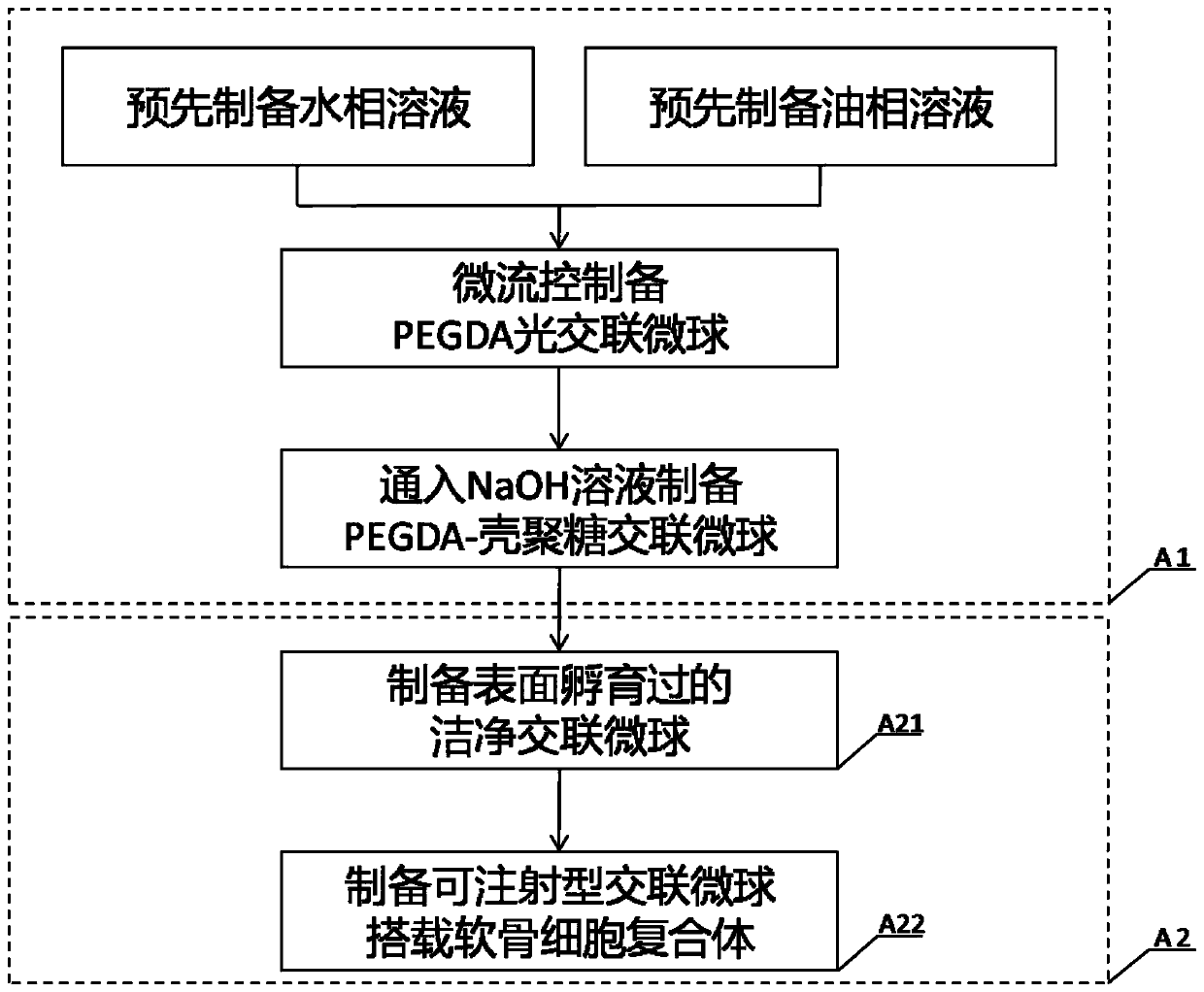
[0044] A method for preparing cross-linked microspheres based on microfluidics, the steps are as follows:

[0045]The injection pump device is used to push the syringes of the water-phase solution and the oil-phase solution, and the water-oil two-phase solution forms water-in-oil emulsion droplets in the outlet channel of the microfluidic chip, and is irradiated under a 90-120W, 350-400nm ultraviolet curing lamp. 1 to 2 minutes to prepare PEGDA photocrosslinked microspheres; the PEGDA photocrosslinked microspheres are passed into 0.3 to 0.6 mol / L sodium hydroxide solution for physical crosslinking of chitosan to obtain a double network PEGDA-chitosan cross-linked microspheres in hydrogel system.

[0046] It should be noted that PEGDA (polyethylene glycol diacrylate) has the characteristics of low viscosity, low skin irritation, good adhesion and water solubility, and the hydrogel made of it is an ideal biological tissue scaffold material. Chitosan is a natural alkaline polysa...

Embodiment 2

[0066] A method for preparing cross-linked microspheres based on microfluidics, the operation steps of which are the same as in Example 1, except that FITC fluorescein is added to the pre-prepared aqueous solution, and its mass in the aqueous solution The fraction is 0.015 to 0.030%.

[0067] The cross-linked microspheres containing FITC fluorescein prepared according to this example were centrifuged with deionized water at a speed of 800-1000 r / min for 3-5 minutes, and then the supernatant was removed. The obtained cross-linked microspheres could be directly placed in Observed under a fluorescence microscope, such as figure 2 shown.

[0068] The cross-linked microspheres after the above centrifugal cleaning were dehydrated with 30%, 50%, 70%, 90% ethanol gradient, soaked in isoamyl acetate, and subjected to supercritical CO 2 Fluid extraction freeze-drying process, the dried cross-linked microspheres can be observed and characterized under scanning electron microscope, suc...

Embodiment 3

[0071] A preparation method of an injectable chondrocyte complex is carried out through the following steps:

[0072] A1, adopt the method for embodiment 1 to prepare cross-linked microspheres;

[0073] A2, cross-linked microspheres carrying chondrocytes:

[0074] A21, washing and detoxifying the cross-linked microspheres prepared in step A1, putting them into the complete medium for soaking for 3 to 4 hours, and then removing the complete medium to obtain clean cross-linked microspheres incubated on the surface;

[0075] A22, mix the surface-incubated clean cross-linked microspheres with chondrocytes at a ratio of 1:50 to 1:200, supplement complete medium, and then place the whole in an incubator for 2 to 3 hours to obtain an injectable form Cross-linked microspheres loaded with chondrocyte complexes.

[0076] Such as Figure 5 As shown, the cross-linked microspheres with a diameter of 240 μm were co-cultured with chondrocytes for 1 day before live / dead staining, and the c...


PUM

PropertyMeasurementUnit
diameteraaaaaaaaaa
diameteraaaaaaaaaa
diameteraaaaaaaaaa

Abstract

The invention provides a preparation method of cross-linking microspheres based on microfluidics. The preparation method comprises the following steps of pushing an injector of a water phase solutionand an oil phase solution which are prepared in advance through a pushing and injecting pump device, enabling a water-oil two-phase solution to form water-in-oil type emulsion liquid drops at an exitchannel of a micro-fluidic chip, and performing illumination at an ultra-violet curing lamp to prepare PEGDA photo-crosslinking microspheres; and enabling the PEGDA photo-crosslinking microspheres tobe charged into a sodium hydroxide solution so as to obtain PEGDA-chitosan cross-linking microspheres, wherein the diameter range of the cross-linking microspheres is 240-440[mu]m, and is controlled through the flow velocity and the flow velocity ratio of the water phase solution and the oil phase solution. The preparation method is flexible and easy to operate. A preparation method of an injecting type chondrocyte composite body comprises the following steps of A1, preparing the cross-linking microspheres; and A2, enabling the cross-linking microspheres to carry chondrocyte. Biological cellsare carried on the cross-linking microspheres having a dual-network hydrogel system, and through proliferation and disintegration of the biological cells, the injecting type cell composite body can beprepared, and is a novel tissue repair material which is widely applied.

Description

technical field [0001] The invention relates to the field of tissue engineering, in particular to a preparation method and application of a cross-linked microsphere and an injectable chondrocyte complex. Background technique [0002] The articular cartilage of the human body ensures the stability of the joints and the movement ability of the human body. Diseases such as trauma or arthritis destroy the normal tissue structure of cartilage, resulting in cartilage defects of various sizes. The cartilage covering the joint surface is a special tissue that ensures the flexibility and accuracy of movement. Its self-repair ability is poor. Once the cartilage is damaged or diseased, the condition will gradually aggravate and directly cause joint pain and limited movement function. Articular cartilage damage caused by various trauma and pathological factors is extremely common in orthopedic clinics. Due to the poor self-repair ability of articular cartilage, it can accelerate its de...

Claims


Application Information

Patent Timeline
Patent Type & Authority Applications(China)
IPC IPC(8): A61L27/18A61L27/20A61L27/38A61L27/50A61L27/52
CPCA61L27/18A61L27/20A61L27/3817A61L27/50A61L27/52A61L2400/06A61L2430/06C08L5/08C08L71/02
Inventor 黄文华吴耀彬曾春林林
Owner SOUTHERN MEDICAL UNIVERSITY
Why Eureka
Try Eureka