Tissue engineering scaffold assembled by cell-loaded microgel as well as preparation method and application thereof

A tissue engineering scaffold and microgel technology, applied in the field of biomedical engineering, can solve the problems of difficult and effective transmission, single cell, insufficient flux, etc., and achieve the improvement of material transmission efficiency, high activity and function, and high material transmission efficiency. Effect

Active Publication Date: 2022-01-07
DALIAN UNIV OF TECH
View PDF10 Cites 2 Cited by
  • Summary
  • Abstract
  • Description
  • Claims
  • Application Information

AI Technical Summary

Problems solved by technology

However, there are many bottlenecks in the tissue-engineered tissue obtained by this design method: (1) Although the bulk hydrogel is rich in water and porous, the dense pores at the nanometer scale make the signal exchange between cells difficult. Active proteins or macromolecules, nutrients, and cell excretions are difficult to effectively transport, and the material diffusion efficiency is low, which eventually leads to the apoptosis of cells inside the block scaffold, which makes it difficult to reproduce and organize normally; (2) due to the The diffusion efficiency of internal drug molecules is low and difficult to control, and it is difficult to realize the controlled release of drug factors; (3) it is difficult to repair irregularly shaped tissue defects, and it is impossible to achieve injectability and plasticity; (4) in the construction of complex tissue structures During the process, it is difficult to accurately immobilize different types of cells on three-dimensional scaffolds (Jiang W, Li M, Chen Z, et al.Cell-laden microfluidic microgels for tissue regeneration[J].Lab on a Chip, 2016, 16( 23): 4482-4506.)
However, there are problems in the existing technology: the size is difficult to reach the single-cell level, the throughput is not high enough, the biocompatibility of the method and the module assembly are still difficult problems (Dubay R, Urban J N, Darling E M.Single-Cell Microgels for Diagnostics and Therapeutics[J]. Advanced Functional Materials, 2021: 2009946.)
However, there are still many challenges in the use of microgels encapsulating biologically active substances as bioinks for 3D bioprinting: (1) There are still gaps in the printability of microgels loaded with single cells as bioinks and the verification of their biological functionality as tissue-like tissues; (2) There are still challenges in cell viability and functionality in gel microspheres

Method used


Image

  • Tissue engineering scaffold assembled by cell-loaded microgel as well as preparation method and application thereof
  • Tissue engineering scaffold assembled by cell-loaded microgel as well as preparation method and application thereof
  • Tissue engineering scaffold assembled by cell-loaded microgel as well as preparation method and application thereof

Examples


Embodiment 1

[0075] 1. Cell culture:

[0076] Taking the culture of human bone marrow-derived stem cells (hMSCs) as an example, the proliferation medium is composed of α-MEM (α-minimum Eagle's medium), 10% fetal bovine serum (FBS, Gibco), and the culture conditions are 37°C and 95% relative humidity with 5% CO 2 . Cell culture medium was changed every three days. Before use, the cells were washed with phosphate-buffered saline (PBS), placed in trypsin / EDTA solution for 5 minutes, and suspended in the medium for use.

[0077] 2. Preparation of cell-loaded microgel particles:

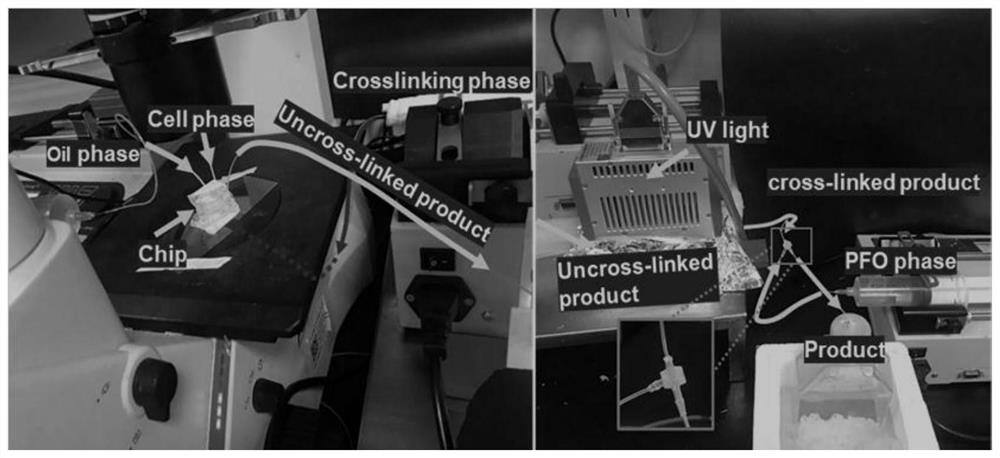
[0078] 0.1wt% LAP (lithium phenyl-2,4,6-trimethylbenzoylphosphonate), 1wt% AlgMA (sodium methacryloyl alginate) and 2.5wt% GelMA (methacrylic acylation Gelatin) precursor mixture was dissolved in α-MEM serum-free medium. Dispersing the hMSCs cells in the polymer prepolymer solution is called the cell phase (the concentration of the cell-loaded microgel is 9 × 10 6 Each / mL), connected to the sampling port of subs...

Embodiment 2

[0081] The cell-loaded microgel prepared in Example 1 was collected by centrifugation (1000 rpm), and then the cell-loaded microgel was densely packed by means of suction filtration. At this time, the dry weight of the microgel was 4 wt%. Then, rheological tests were carried out on the close-packed microgels, and the results showed that the close-packed microgels had shear-thinning properties and self-healing properties (such as Image 6 A and 6B), the rheological properties of the microgel meet the properties of extrudable printing as a bioink. Image 6 C demonstrates the extrudable advantage of microgels as inks. With the help of 3D printing equipment, microgels can be used as bioprinting inks to obtain different tissue structures, and biomimetic tissue printing can be performed, which will be beneficial to tissue construction (such as Figure 7 shown). The above results indicate that the microgels have printable properties.

Embodiment 3

[0083] After the close-packed microgels were printed in Example 2, in order to maintain the stability of the density structure, the microgels were extruded through a syringe and tested for stability. When larger, bending occurs (as in Figure 8 Shown in A), after secondary crosslinking (after calcium ion crosslinking), the close-packed microgel has structural stability (such as Figure 8 shown in B). A constant frequency (1 Hz) strain sweep was further tested to represent the shear yield characteristics of the shear modulus in response to shear strain ( Figure 9 A). It can be found that the microgels exhibit crossover at different strain amplitudes before and after crosslinking. The modulus (G′=G″) at the intersection point, the crosslinked microgel is significantly higher than that of the uncrosslinked microgel. The above results show that in the low strain range, the crosslinked microgel has Higher shear modulus. The shear modulus of the uncrosslinked microgel is an ord...


PUM

PropertyMeasurementUnit
sizeaaaaaaaaaa
diameteraaaaaaaaaa
thicknessaaaaaaaaaa

Abstract

The invention discloses a tissue engineering scaffold assembled by cell-loaded microgel as well as a preparation method and application thereof, and belongs to the technical field of biomedical engineering. Firstly, gel microspheres carrying bioactive substances prepared from a biological material with good biocompatibility by adopting a micro-flow liquid drop control technology are used to serve as basic units, and the cell-loaded gel microspheres are assembled into the tissue engineering scaffold by adopting a biological 3D printing mode. The micro-flow liquid drop control technology adopted by the invention can effectively shorten the preparation time of the cell-loaded gel microspheres, improve the preparation efficiency and ensure the cell activity; and the construction of a three-dimensional complex structure can be realized by utilizing a biological printing mode, and the application prospect is good.

Description

technical field [0001] The invention belongs to the technical field of biomedical engineering, and in particular relates to a tissue engineering scaffold assembled with cell-loaded microgel, a preparation method thereof, and an application in the field of bone repair. Background technique [0002] For the repair of human tissues / organs with loss of function or defects, the traditional clinical solution still relies on tissue and organ transplantation. However, the shortage of donated human tissue and the potential risk of infectious diseases have caused social ethical and moral issues, making tissue / organ transplantation face many challenges. The provision of engineered tissues by tissue engineering technology brings hope for organ repair and reconstruction, and is expected to alleviate the practical problem of organ donor shortage. At present, tissue engineering technology has realized the repair of tissues including skin and cartilage and entered clinical application. Ho...

Claims


Application Information

Patent Timeline
Patent Type & Authority Applications(China)
IPC IPC(8): A61L27/20A61L27/56A61L27/52A61L27/54A61L27/38A61L27/22A61L27/02B33Y70/10B33Y80/00B33Y10/00
CPCA61L27/20A61L27/56A61L27/52A61L27/54A61L27/3834A61L27/3847A61L27/222A61L27/025B33Y70/10B33Y80/00B33Y10/00A61L2430/02A61L2300/412A61L2300/602A61L2300/102C08L5/04C08L5/08
Inventor 王华楠安传锋张玉洁张昊岳
Owner DALIAN UNIV OF TECH
Why Eureka
Try Eureka