
Magnetic separating direct chemical illuminating reagent and testing method using the same reagent

A chemiluminescent reagent and magnetic separation technology, applied in chemical instruments and methods, luminescent materials, organic chemistry, etc., can solve the problems of unstable reagents, hindered by trace active substances, insufficient sensitivity, etc., so as to avoid the easy decline of enzyme activity and simplify the Immunological design to overcome the effect of easy hydrolysis

Active Publication Date: 2007-01-03
SHENZHEN NEW INDS BIOMEDICAL ENG
View PDF1 Cites 58 Cited by
  • Summary
  • Abstract
  • Description
  • Claims
  • Application Information

AI Technical Summary

Problems solved by technology

[0003] radioimmunoassay using the radioisotope I 125 As a tracer, labeled on the antigen or the corresponding antibody, on the gamma ray detector, through the I 125 The measurement of radiation intensity is used to determine the content of the substance to be tested. Although this method solves the quantitative analysis purpose of trace active substances well, its defects are obvious: radioisotope I 125 As a tracer technology, during the manufacture, storage and application of reagents, it pollutes the environment and causes serious harm to the human body; at the same time, due to the 125 The half-life limit determines that the validity period of radioimmunoassay reagents is only one month; the validity period of radioimmunoassay reagents is also only one month; the non-specificity of the separation process leads to poor accuracy of the results, and it cannot be used as emergency specimens
[0004] The enzyme immunoassay method uses HRP horseradish peroxidase as a luminescent marker, plastic microplate as a carrier and separation technology, OPD o-phenylenediamine as a chromogenic substrate, 1MH 2 SO 4 As the termination solution of the enzyme color reaction, the concentration of the substance to be tested is analyzed by measuring the absorbance value of the enzyme reaction product wavelength at 492nm. This method is mainly used for qualitative screening of a large number of human serum samples. The defect is that it is obvious The color intensity of the chromatic product changes with time and cannot be fixed. The chromogenic substrate OPD, etc. are strong carcinogens. At the same time, the sensitivity to a considerable part of trace hormones is not enough, the reaction time is too long, and it is difficult to standardize during the operation.
[0005] The enzyme-catalyzed chemiluminescent immunoassay method uses alkaline phosphatase AKP or horseradish enzyme HRP as a marker, and uses AMPPD (adamantane) or luminol, H 2 o 2 As a luminescent substrate; its advantage is that the analyte is converted into a measurable light signal, which significantly improves the sensitivity of the analysis. Its disadvantage is that many factors that affect the enzyme activity, such as ambient temperature, incubation temperature and time, storage conditions, etc. It will directly affect the measurement results, and the stability of the reagents is not good; at the same time, the luminescence reaction of this method starts slowly, and it needs to be incubated at 37°C for a certain period of time to reach the luminescence platform, which will more strongly affect the results of luminescence determination
[0006] In addition, the direct chemiluminescence method labeled with acridinium ester and the electrochemiluminescence method labeled with ruthenium terpyridine bring defects such as unstable reagents, low accuracy of analysis results and insufficient test speed during the analysis process, which hinders the accurate analysis of trace amounts. The amount of active substance presents a hindrance

Method used


Image

  • Magnetic separating direct chemical illuminating reagent and testing method using the same reagent
  • Magnetic separating direct chemical illuminating reagent and testing method using the same reagent
  • Magnetic separating direct chemical illuminating reagent and testing method using the same reagent

Examples


Embodiment 1

[0032] 1. Preparation of monoclonal antibody labeled with isoluminol derivatives:

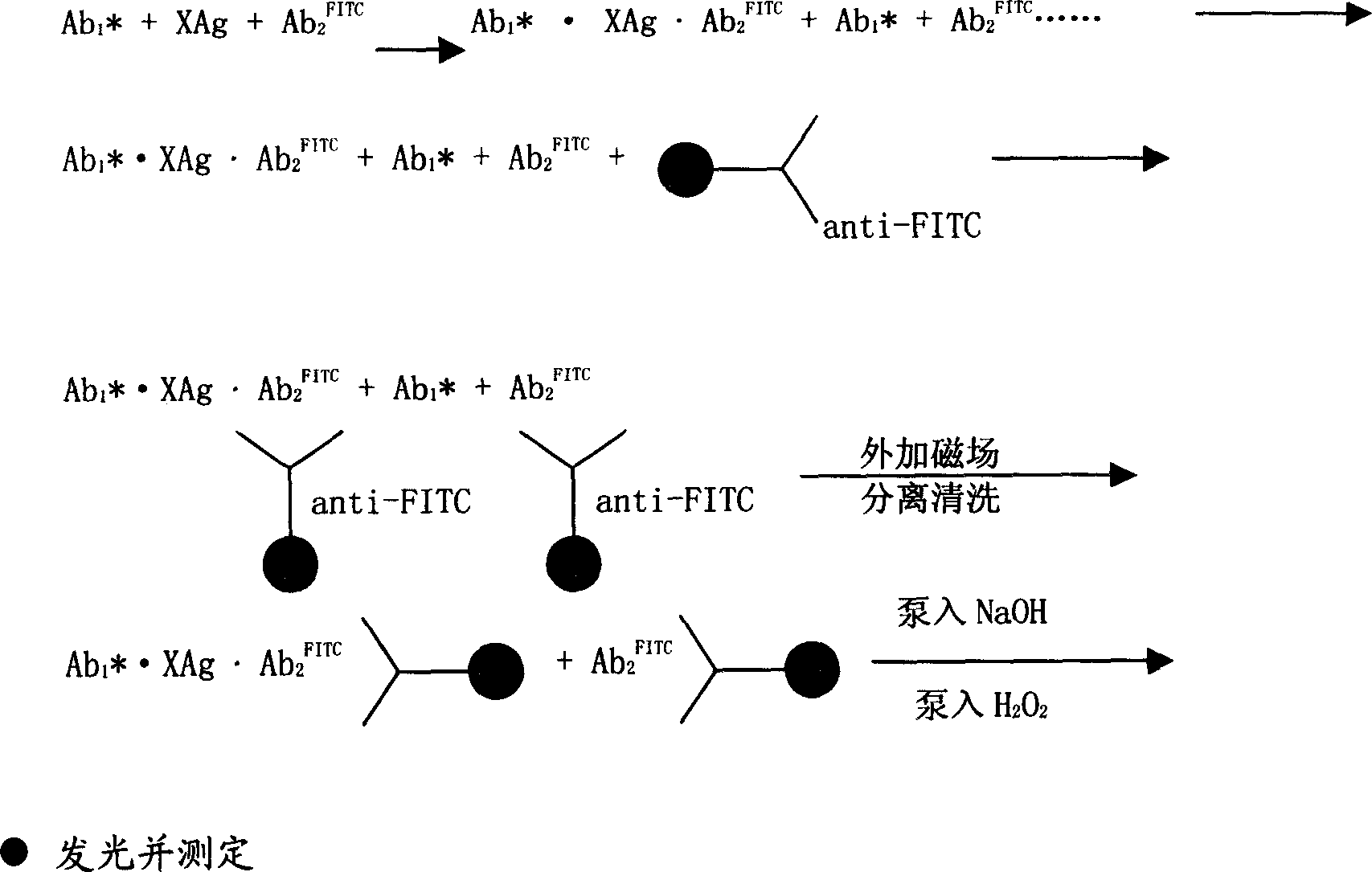
[0033]Take three test tubes of 3.2mmol ABEI, dissolve them in 0.2ml of secondary water, 3.5mmol of CSCl 2 Dissolve in 0.3ml DMF, mix the two evenly, react at room temperature for 2 hours, take 10mg anti-CEA-α, anti-AFP-α and 10mg anti-PSA-α monoclonal antibody respectively, adjust the volume with pH9.5 carbonate buffer to 1ml, add the above-mentioned activated ABEI solution, mix well, react at room temperature for 20h, and purify by G-25 gel column;

[0034] 2. FITC-labeled CEA, AFP, PSA monoclonal antibody

[0035] Take 10mg of anti-CEA-β, AFP-β and PSA-β monoclonal antibodies, slowly adjust the volume to 1ml with pH9.5 carbon, add FITC100ug, react at room temperature for 20 hours, and purify through G-25 gel column;

[0036] 3. Preparation of goat anti-FITC polyclonal antibody coated on the surface of nano-magnetic microbeads:

[0037] The preparation of nano-magnetic micro-beads is prepar...

Embodiment 2

[0046] The preparation of monoclonal antibody of isoluminol derivatives and the preparation method of goat anti-FITC polyclonal antibody coated on the surface of nano-magnetic microbeads are the same as in Example 1. The luminescent marker used in this example is AHEI, and the surface of the magnetic beads is The group containing -COOH, its content is 0.3eq / g;

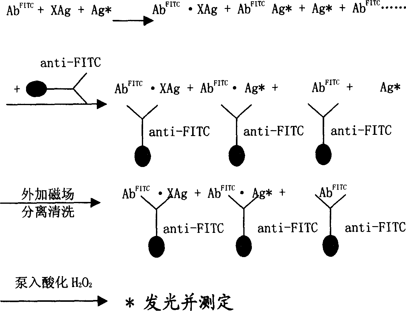
[0047] Take TSH, T3, T4, FT3 and FT4 standard products, 20 μl each of serum samples, add 40 μl each of AHEI and FITC monoclonal antibodies of the luminescent marker monoclonal antibody, mix well and incubate at 37°C, in a water bath 15 minutes; after the immune reaction occurs, add 40 μl of immune nano-magnetic beads separation reagent coated with goat anti-FITC polyclonal antibody on the surface, mix well, and bathe in water for 5 minutes. Under the action of an external magnetic field, separate on the upper separator for 4 minutes, pour Supernatant; add 400 μl of washing solution, mix well, separate for 4 minutes, po...


PUM


Abstract

A magnetic separation direct chemiluminescence reagent and the testing method, which uses the isoluminol derivatives as the immunity nano-magnetism tiny bead enwrapped by anti-goat FITC multiclonal antibody as the separating reagent, uses the NaOH (sodium-hydroxide) and H2O2 (oxydol) as the magnetization direct chemiluminescence reagent and the testing method of the illuminant marker; the invention is: the reagent is stabilization, non-polluted, non-decomposed and can not affect by the environment.

Description

technical field [0001] The invention relates to a magnetic separation direct chemiluminescence reagent and a test method using the reagent, in particular to a reagent using isoluminol derivatives as markers and nano magnetic microbeads as separation materials. Background technique [0002] The rapid development of modern clinical medical technology requires the precise quantitative determination of trace active substances in more and more people's blood or urine to provide accurate basis for clinicians to diagnose diseases, formulate effective treatment plans, and evaluate treatment effects. , the methods to achieve accurate quantitative analysis of trace active substances in human serum or urine mainly include radioimmunoassay, enzyme immunoassay, enzymatic chemiluminescence or acridinium ester-labeled direct chemiluminescence method and triple pyridine ruthenium-labeled electrochemical Luminous method. [0003] radioimmunoassay using the radioisotope I 125 As a tracer, l...

Claims


Application Information

Patent Timeline
IPC IPC(8): G01N33/52G01N21/64G01N33/577C07D237/32
CPCG01N33/532C07D237/36C09K11/07G01N33/58G01N33/54326
Inventor 饶微宋洪涛李婷华邵佳阎颖王丽蓉黄国光刘海燕
Owner SHENZHEN NEW INDS BIOMEDICAL ENG
Learn More