Prosthetic implantable antibacterial surgical mesh

a surgical mesh and implantable technology, applied in the field of implantable surgical mesh, can solve the problems of high incidence of mesh infection, difficult treatment, and large postoperative complications of mesh infection, and achieve the effects of reducing the risk of infection, reducing the number of surgical meshes, and improving the quality of surgical meshes

Active Publication Date: 2020-06-23
KING ABDULAZIZ UNIV
View PDF8 Cites 4 Cited by
  • Summary
  • Abstract
  • Description
  • Claims
  • Application Information

AI Technical Summary

Benefits of technology

[0017]In a more preferred embodiment, the polysaccharide is chitosan.

Problems solved by technology

However, postoperative mesh infection is a major complication that is difficult to treat.
The incidence of mesh infection is higher when a mesh is used in a contaminated surgical field, such as in cases of strangulated hernias.
However, the instability, the whipping of the fiber, and bead formation remain important problems in the electrospinning process.
Such conventional drug-impregnated materials are not, however, effective for use in surgical treatment of hernias.
Conventional materials are unable to provide drug release profiles that provide a steady and predictable threshold dosage of active material.
In addition conventional materials do not exhibit good compatibility with live tissue to promote quick and lasting wound healing, especially for hernia surgery which occurs at a location on a human subject that is exposed to significant pressure and pulling strain.

Method used


Image

  • Prosthetic implantable antibacterial surgical mesh
  • Prosthetic implantable antibacterial surgical mesh
  • Prosthetic implantable antibacterial surgical mesh

Examples


example 1

Materials:

[0107]Unless otherwise specified, all chemicals including 85% degree N-deacetylated chitosan, PVP (polyvinypyrrolidone), PVA (polyvinyl alcohol), glacial acetic acid, potassium dihydrogen phosphate, potassium monohydrogen phosphate, orthophosphoric acid, ciprofloxacin (CP) and other reagents were fine grades purchased from Sigma Aldrich, USA. Ciprofloxacin-HCl was supplied from Fluka Biochemical, Germany.

[0108]Staphylococcus aureus (ATCC 12228), Escherichia coli (ATCC 25922), and Pseudomonas aeruginosa (ATCC 27853) were used for evaluation of the antibacterial activity of polymer coated electrospun nanofiber mat samples.

Methods:

Polymer Solutions:

[0109]Table 1 summarizes the compositions of the polymer solutions containing ciprofloxacin-HCl to prepare the corresponding electrospinning polymer solution. A 2 wt. % solution of chitosan in 2% (v / v) aqueous acetic acid was mixed with aqueous solution of 10 wt. % of PVP and / or PVA comprising 10 wt. % of ciprofloxacin / HCl.

[0110]

TA...

example 2

Preparation of Nanofibers by Electrospinning

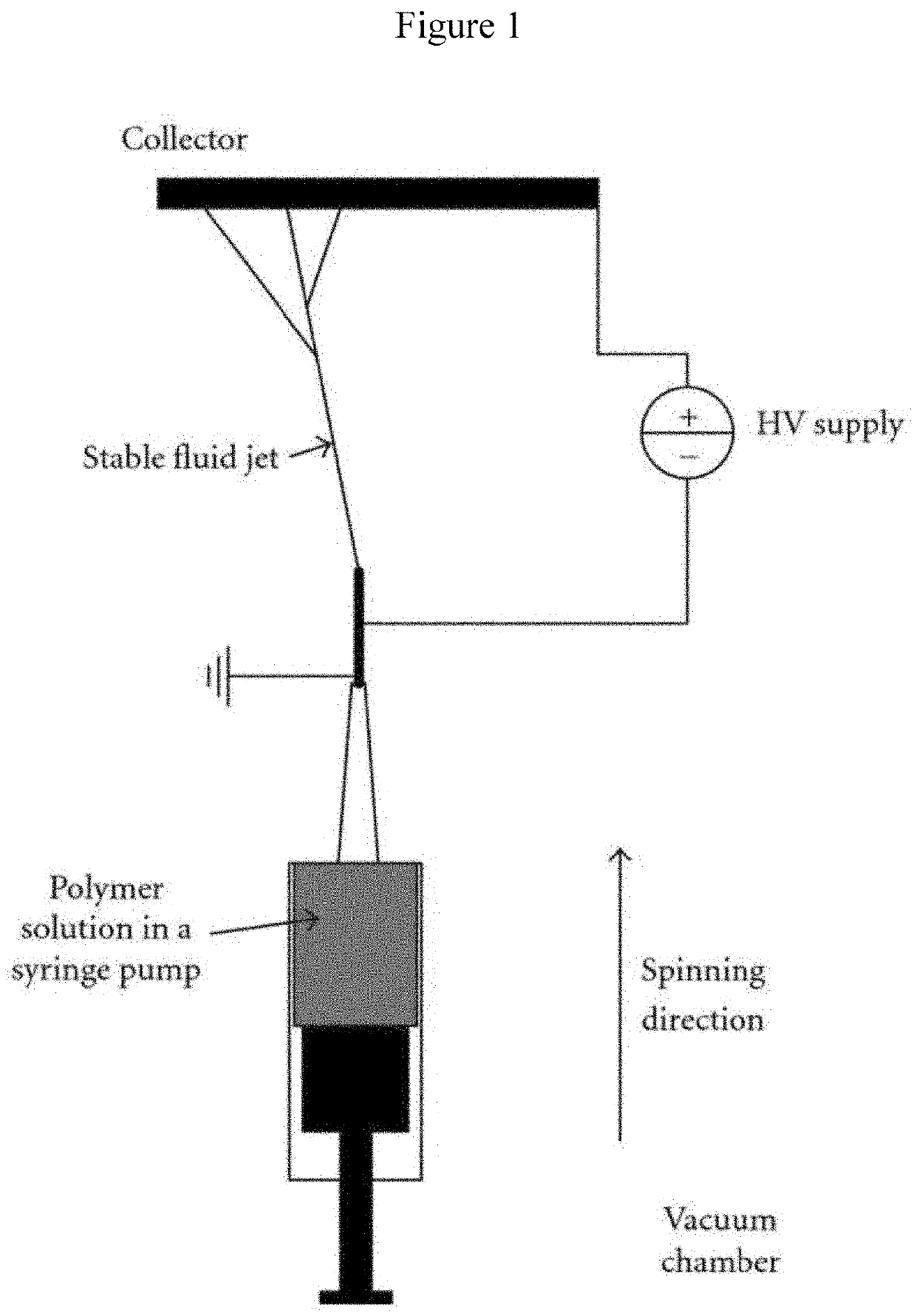
[0114]A custom design of an upward electrospinning apparatus diagramed in FIG. 1 was manufactured by Shenzhen Tong Li Tech, China. A set of experiments have been carried out in order to determine suitable system parameters such as the high voltage, distance between the two electrodes, and needle diameter as well as the solution parameters including percentages of the different component in the solution, viscosity, and surface tension to identify suitable solution for spinning.

[0115]Medicated and non-medicated CH solutions were prepared as described above. But, the concentration of CP was reduced to 0.25% (wt. / v) in 2% acetic acid solution (final concentration of CP in the nanofiber is approximately 3.35 wt. % in CH. The upward spinning method was used for production of the nanofibers from CH medicated or non-medicated solutions. The solutions were loaded into a glass syringe with a 28 gauge needle. The needle had outer diameter of 0.64 mm ...

example 3

Drug Release

Determination of Ciprofloxacine-HCl Calibration Curve

[0118]Column, Agilent Zorbax extend-C18 column (150 mm length×4.6 mm, i.d., 5 μm), mobile phase, 0.025 M σ-phosphoric acid adjusted to pH 3 with triethylamine:acetonitrile (75:25), UV detector set at A, =278 nm, flow rate of 1 mL / min, and injection volume of 20 μL. Approximately 20 mg of ciprofloxacin-HCl standard was weighed, transferred into a 100 mL volumetric flask, dissolved in methanol and volume was completed with phosphate buffer pH 7.4. The stock standard solution (0.2 mg / mL) were diluted to give a concentration range of 1.6 to 40 μg / mL using phosphate buffer pH 7.4 as diluting solvent.

In Vitro Drug Release Study

[0119]In vitro drug release studies from 2×2 cm2 pieces of nanofiber mesh were carried out over 12 days at specified times. The mesh of each formula of Table 1 was placed in bottles with 20 mL PBS, pH 7.4. The bottles were placed in oscillating water bath at 37° C.±2. Aliquots of 1 mL were withdrawn at...


PUM

PropertyMeasurementUnit
diameteraaaaaaaaaa
sizeaaaaaaaaaa
diameteraaaaaaaaaa

Abstract

The disclosed invention is directed to an implantable surgical prosthetic mesh having a nanofiber comprising one or more antibiotic and a polysaccharide, and a non-polysaccharide polymer deposited on the mesh. The mesh of the invention is shown to be effective in eliminating or minimizing the bacterial population in the mesh and surrounding tissue for at least 14 days from surgical implantation of the mesh.

Description

BACKGROUND OF THE INVENTIONField of the Disclosure[0001]The present invention relates to an implantable surgical mesh having a nanofiber comprising one or more antibiotic deposited on the surface of the mesh.Description of Related Art[0002]The “background” description provided herein is for the purpose of generally presenting the context of the disclosure. Work of the presently named inventors, to the extent it is described in this background section, as well as aspects of the description which may not otherwise qualify as prior art at the time of filing, are neither expressly or impliedly admitted as prior art against the present disclosure.[0003]A hernia is an abnormal protrusion of intra-abdominal tissue or organ through a defect in the abdominal wall. Currently, the only effective treatment of a hernia is surgical repair of the defect. The use of mesh in surgical repair of hernias has dramatically decreased the incidence of hernia recurrence. However, postoperative mesh infectio...

Claims


Application Information

Patent Timeline
Patent Type & Authority Patents(United States)
IPC IPC(8): A61L31/04A61L31/10A61K31/47A61K31/497D04H1/728A61L31/16
CPCA61L31/10A61K31/47A61K31/497A61L31/16D04H1/728A61L2400/12A61L2430/34A61L2300/406
Inventor ALTAF, ABDULMALIK MOHAMMED SALEHABDULHADY, SEHAM ALSAYEDABDEL-HADY, FAISSALYASSIEN, MAHMOUD ABDUL MEGEAD
Owner KING ABDULAZIZ UNIV
Why Eureka
Try Eureka