
Gene regulation therapy involving ferritin

a technology of ferritin and gene regulation, applied in the direction of animals/human proteins, biocide, animals/human peptides, etc., can solve the problems of morphological distortion of red cells, inability of the molecule to absorb or release oxygen, and insufficient expression of one or more globin chains, so as to reduce the labile iron pool, reduce cancer cell line proliferation, and suppress the transcription of this gene

Inactive Publication Date: 2004-09-23
OKLAHOMA MEDICAL RES FOUND
View PDF0 Cites 7 Cited by
  • Summary
  • Abstract
  • Description
  • Claims
  • Application Information

AI Technical Summary

Benefits of technology

The patent is about a new method for treating genetic diseases caused by mismanagement of iron in the body. The method involves delivering a gene for ferritin-H, a protein that regulates iron levels, into cells. This can be done by either transferring a gene for ferritin-H or a peptide for ferritin-H into cells or by stimulating the endogenous ferritin-H gene. The ferritin-H gene can also be delivered using a vector or antisense oligonucleotides. Increasing or decreasing the expression of ferritin-H can lead to a change in the phenotype of the cell, either by repressing or activating the expression of other genes. The patent also describes how to use ferritin-H to treat sickle cell disease and neurodegenerative diseases such as Alzheimer's disease and Parkinson's disease. Overall, the patent provides a new way to regulate gene expression and treat genetic diseases caused by mismanaged iron.

Problems solved by technology

Some of these genetic mutations pose no adverse or only minor consequences to the person; however, most mutations prevent the formation of an intact or normal hemoglobin molecule through a functional or structural inability to effectively bind iron, an inability of the chains or chain pairs to effectively or properly interact, an inability of the molecule to absorb or release oxygen, a failure to express sufficient quantities of one or more globin chains or a combination of these malfunctions.
Upon deoxygenation, HbS molecules undergo aggregation and polymerization ultimately leading to a morphological distortion of the red cells which acquire a sickle or holly-leaf shape.
This process of polymerization-depolymerization is very damaging to red cell membranes and eventually leads to irreversibly sickled cells (ISC) which retain their abnormal shape even when fully oxygenated.
Crises are episodic and reversible, but may be fatal.
Such treatments may potentially decrease the frequency of symptomatic episodes caused by vaso-occlusive crises if enough of the chemical can be administered to bind all hemoglobin in the body.
The hemolytic consequences of deficient globin chain synthesis result from decreased synthesis of one chain and also an excess of the complementary chain.
Free chains tend to aggregate into insoluble inclusions within erythrocytes causing premature destruction of maturing erythrocytes and their precursors, ineffective erythropoiesis, and the hemolysis of mature red blood cells.
First, there is an inadequate formation of HbA and, therefore, an impaired ability to transport oxygen.
These inclusions damage cellular membranes resulting in a loss of potassium.
The cumulative effect of these inclusions on the red blood cells is an ineffective erythropoiesis.
Those that do escape immediate destruction are at increased risk of elimination by the spleen where macrophages remove abnormal cells.
Further, hemolysis triggers an increased expression of erythropoietin which expands populations of erythroid precursors within bone marrow and leads to skeletal abnormalities.
Another severe complication of .beta.-thalassemia is that patients tend to have an increased ability to absorb dietary iron.
As most treatments for thalassemia involve multiple transfusions of red blood cells, patients often have a severe state of iron overload damaging all of the organs and particularly the liver.
However, there is great concern since this antineoplastic ribonucleotide reductase inhibitor is carcinogenic.
Its carcinogenic properties make its widespread and long term use as a pharmaceutical a questionable practice.
Many of these agents including AZA, AraC and hydroxyurea are myelotoxic, carcinogenic or teratogenic making long-term use impractical.
Although promising in pilot clinical studies, treated patients were unable to maintain adequate levels of fetal globin in their system.
It was later determined that many of these forms of butyric acid had extremely short-half lives.
However, these studies also demonstrated that positive effects could only be maintained for very short periods of time.
It would be expected that this would lead to higher levels of the more harmful ferrous ion and have adverse affects on the cells.
Ferritin-L-subfamily peptides, on the other hand, are likely to cause even more harm in that they readily give up iron which would exacerbate the problem by increasing free iron and radical generation.
For in vivo gene transfer, the choices are currently limited because of the difficulty of efficiently targeting specific cells with sufficient gene copies.
Although some of the DNA elements in the ferritin-H gene promoter and nuclear proteins that bind to them (e.g., P / CAF-CBP, Bbf, and NF-E2) are known, the mechanism of activation of ferritin-H transcription is not understood sufficiently to be applied clinically.

Method used


Image

  • Gene regulation therapy involving ferritin
  • Gene regulation therapy involving ferritin
  • Gene regulation therapy involving ferritin

Examples


example 1

Materials and Methods

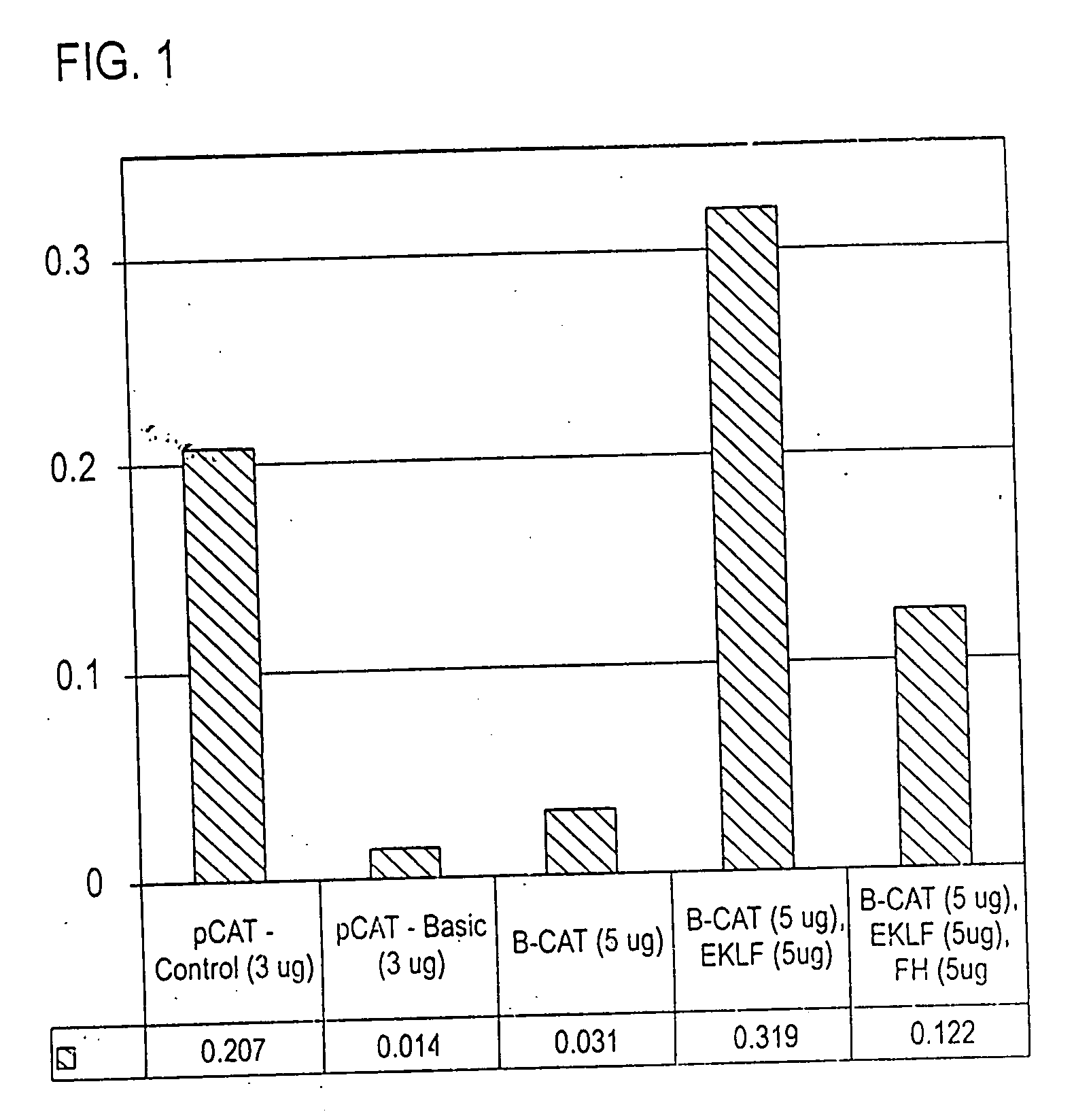
[0093] Cell lines. K562 (human erythroleukemia) cells were grown in suspension in RPMI 1640 medium with 10% or 15% fetal bovine serum (FBS) and antibiotics as described (Berg, P. E., Williams, D. M., Qian, R. L., Cohen, R. B., Cao, S. X., Mittelman, M. & Schechter, A. N. (1989) Nucleic Acids Res 17, 8833-52) and harvested at a density of 10.sup.6 cells / ml for making nuclear extracts. CV-1 (African green monkey kidney epithelial) cells (adherent cells used for transfections / transient gene expression assays) were grown in DMEM with L-glutamine, 10% FBS and antibiotics (Miller, I. J. & Bieker, J. J. (1993) Mol Cell Biol 13, 2776-86).

[0094] Clones, transfections, and gene expression assays. The upstream region (-610 / +20) of the human .beta.-globin gene, previously cloned in pSV2CAT (Berg, P. E., Williams, D. M., Qian, R. L., Cohen, R. B., Cao, S. X., Mittelman, M. & Schechter, A. N. (1989) Nucleic Acids Res 17, 8833-52), was subcloned through pGEM and pSELECT (now c...

example 2

Materials and Methods

[0113] Materials: Calf intestine alkaline phosphatase, T4-polynucleotide kinase, and Sau 96 I were obtained from Promega / Fisher. 32P-.gamma.-ATP was from Dupont / NEN. Polyclonal (rabbit) antiserum to human spleen ferritin was obtained from Sigma Chemical Company. All other reagents were molecular biology grade.

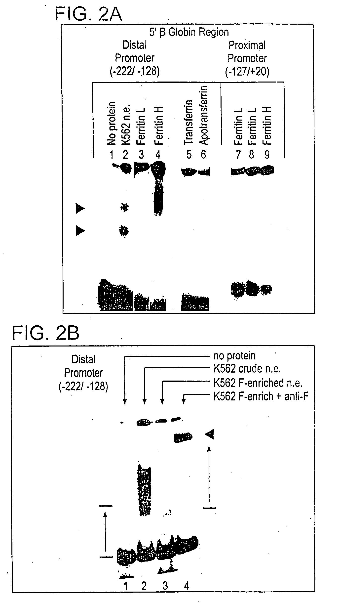
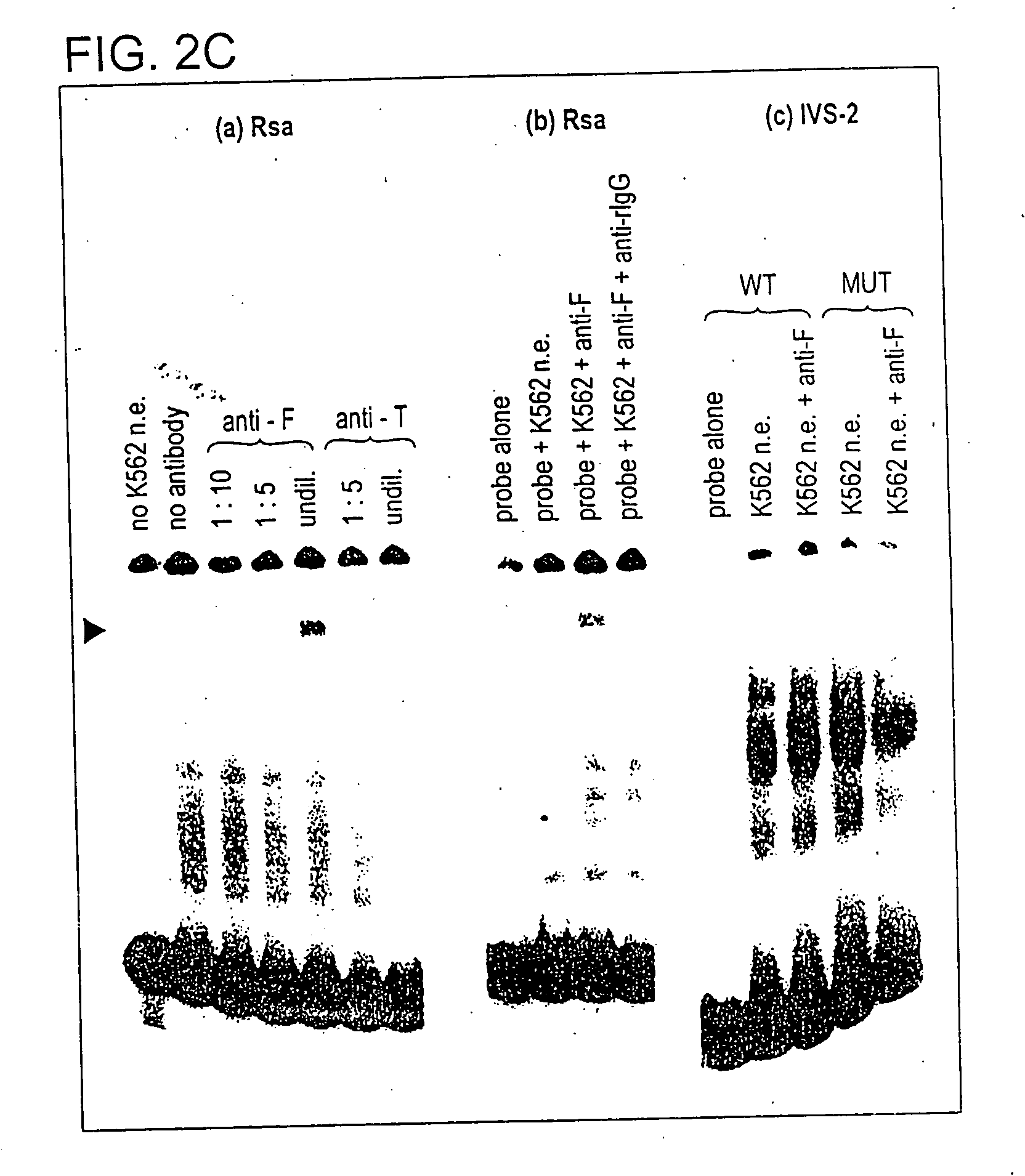
[0114] Restriction fragments and oligonucleotides: The 5' region of the human beta globin gene (from -610 to +20), previously cloned in pSVOCAT, was cut from the purified plasmid by digestions with Hind III and Bam H I. The 630 bp fragment was phenol / chloroform treated,: dephosphorylated, and end-labelled with 32-P. Synthetic oligonucleotides corresponding to the core / BP-1 binding site of NCR1 (-584 / -527), the more distal of the two 5'-.beta.-globin silencers, and -164 / -128 region of the promoter were purified and annealed, and the double-stranded oligos were end-labeled as above and / or used as unlabeled competitors in gel mobility shift assays.

[0115] Prepa...


PUM

PropertyMeasurementUnit
volumeaaaaaaaaaa
volumeaaaaaaaaaa
volumeaaaaaaaaaa

Abstract

A method is described for regulating gene expression related to iron metabolism to ameliorate diseases that include sickle cell disease, cancers, neurodegenerative diseases, Friedreich's ataxia and other neuromuscular disorders, and atherosclerosis. This approach is illustrated by recent findings that show that ferritin-H, an iron-binding protein that is present in cell nuclei, can repress the human beta-globin gene, the gene that is mutated in sickle cell disease. Increased expression of ferritin-H or a related ferritin-family peptide, given to effected cells either as the peptide itself (or a part thereof), as an expression clone of the ferritin-H-subfamily gene, or via a gene regulator that increases expression of the ferritin-H-subfamily gene itself, prevents or ameliorates expression of the disease state in disorders where increased availability of iron is implicated in the etiology of the disease, including those named above.

Description

[0001] This application is a continuation-in-part of U.S. provisional patent application No. 60 / 245,003, filed Nov. 1, 2000, which is hereby incorporated by reference.BACKGROUND OF INVENTIONField of the Invention[0002] The present invention relates to gene regulation therapy involving ferritin. More specifically, the invention relates to the use of Ferritin-H and derivative proteins thereof for regulation of genes related to iron metabolism and regulation.Background for Sickle Cell Disease[0003] Hematopoiesis, or the formation of blood cells, begins in the developing human embryo as clusters of stem cells called blood islands. These cells appear in the yolk sac at about the third week of development and, at about the third month, migrate to the developing liver which becomes the principal site of blood cell formation. Although the spleen, lymph nodes and bone marrow all make small contributions to blood cell development, not until the fourth month does the bone marrow become the pri...

Claims


Application Information

Patent Timeline
Patent Type & Authority Applications(United States)
IPC IPC(8): A61K31/7088A61K9/127A61K38/00A61K38/16A61K38/17A61K45/00A61K48/00A61P7/06A61P25/00A61P25/16A61P25/28A61P43/00C07K14/47
CPCA61K38/1709C07K14/47A61K48/00A61P25/00A61P25/16A61P25/28A61P43/00A61P7/06
Inventor BROYLES, ROBERT H.FLOYD
Owner OKLAHOMA MEDICAL RES FOUND
Learn More