Use of high wavenumber raman spectroscopy for measuring tissue

a raman spectroscopy and tissue technology, applied in the field of instruments, can solve the problems of limiting the applicability of raman spectroscopy, strong signal background, and inability to enable detailed in vivo analysis of the molecular composition of plaques

Active Publication Date: 2006-06-29
RIVER DIAGNOSTICS
View PDF7 Cites 50 Cited by
  • Summary
  • Abstract
  • Description
  • Claims
  • Application Information

AI Technical Summary

Benefits of technology

[0016] The invention provides an instrument and the use of an instrument comprising a laser, a signal detection unit for measuring the Raman signal, and a fiber optic probe, wherein the fiber optic probe comprises one or more optical fibers for directing laser light onto the tissue and for collecting light that is scattered by the tissue and guiding the collected light away from the tissue towards the signal detection unit, the fiber comprising a core, a cladding and optionally a coating, and the fiber or fibers for collecting light having substantially no Raman signal in one or more parts of the 2500-3700 cm−1 spectral region, and wherein the detection unit records the Raman signal scattered by the tissue in said spectral region.

Problems solved by technology

However, they do not enable detailed in vivo analysis of the molecular composition of the plaque.
However fluorescence spectra arc easily disturbed by light absorbing molecules in the tissue and in blood, limiting its applicability.
Unfortunately, in the 400-2000 cm−1 spectral region, the materials of the optical fiber itself generate Raman signal, resulting in a strong signal background.
Moreover, bending of the fiber leads to variations in the amount of signal obtained from the core, cladding and coating materials, further complicating signal detection and signal analysis.
This deteriorates the signal-to-noise with which the tissue Raman signal can be detected, and also otherwise complicates signal analysis, and therefore negatively affects clinical utility.
Fiber optic probes for Raman spectroscopy are therefore complex.
It is difficult to miniaturise fiber optic probes for Raman spectroscopy and to keep them flexible, which is necessary for instance for intravascular use and for use in the auxiliary channel of an endoscope.
The complexity is also an obstacle to the production of such probes at a price that they can be used as disposables in hospitals.
Moreover, signal intensity of tissue in the 400-2000 cm−1 is low, necessitating relatively long signal integration times, which may be impractical for clinical use.
All above mentioned problems and disadvantages hinder the actual implementation of Raman spectroscopy for clinical diagnostic purposes in general, and for intravascular use in particular.
This is caused by absorption, by light scattering (Rayleigh scattering, scattering / reflection at larger inhomogeneities or at sites at which the fiber material is damaged), and by micro and macro-bending losses.
This will lead to the same disadvantages as discussed above, e.g. with respect to noise.
Such a method is usually not applicable to tissue, especially not in the case of in vivo measurements.
Next to atherosclerosis, cancer is also an import health issue.
The same problems as encountered above apply for determining tumor cells by Raman spectroscopy via fiber optics.
Such use also leads to a signal to noise ratio which is not satisfying.

Method used


Image

  • Use of high wavenumber raman spectroscopy for measuring tissue
  • Use of high wavenumber raman spectroscopy for measuring tissue
  • Use of high wavenumber raman spectroscopy for measuring tissue

Examples


example 1

Raman Mapping of Atherosclerotic Artery

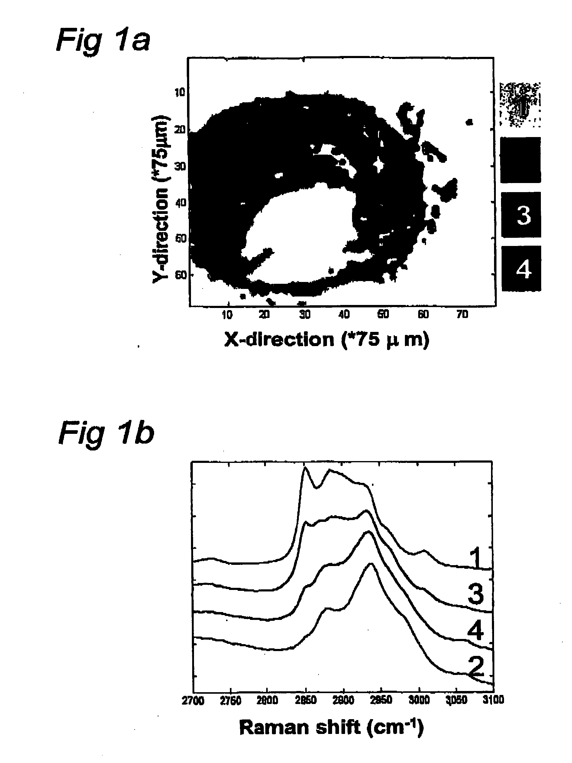
[0109] This experiment describes the possibilities of Raman spectroscopy in the spectral region of the invention for studying artherosclerotic plaque.

[0110] The human coronary artery sample used to create the Raman map shown in FIG. 1 was obtained at the time of autopsy (less than 24 hour post mortem). It was snap frozen in liquid nitrogen and stored at −80° C. until use. For the Raman measurements a 20 μm thick cryosection was placed on a calcium fluoride (CaF2) window (Crystan UK) and passively warmed to reach room temperature. After Raman measurements it was stained with a standard hematoxylin and eosin staining procedure.

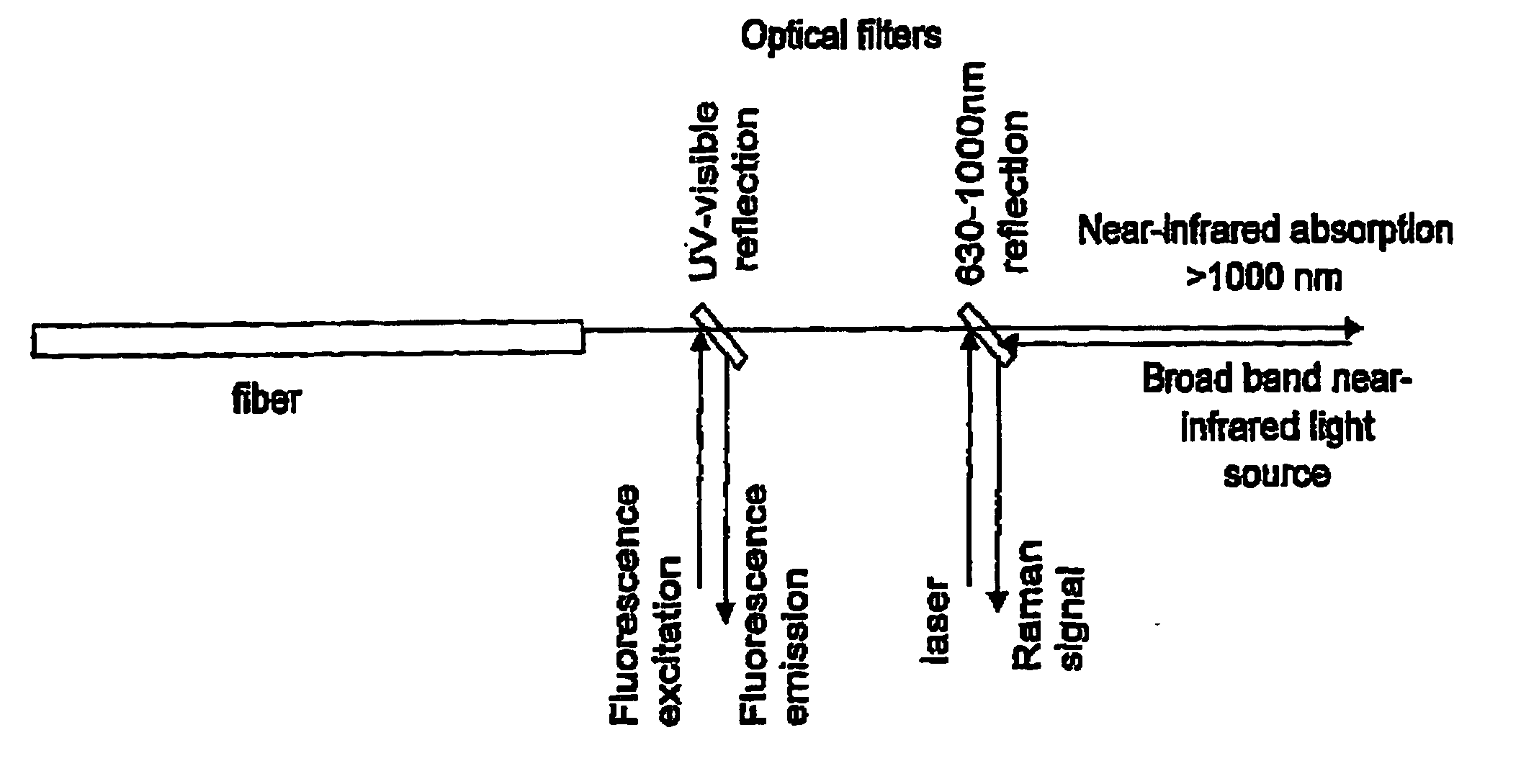
[0111] To collect Raman specta, 719 nm laser light from an argon-ion pumped Titanium: Sapphire laser system (Spectra Physics, Mountain View, Calif.) was used. The Raman microspectrometer system that was used has been described in detail in Van de Poll S W E, Bak Schut T C, Van der Laarse A, Puppels G J “In Situ Investiga...

example 2

Raman Mapping of Cancerous Tissue

[0124] This experiment describes the possibilities of Raman spectroscopy in the spectral region of the invention for studying cancerous tissue.

[0125] The high wavenumber region can also be used advantageously in various clinical oncology applications. For instance, FIG. 4A shows a Raman map obtained of a thin tissue section of human dura infiltrated by meningioma in a way similar to the map of an atherosclerotic lesion in FIG. 1A. Currently no good intra-operative assessment of excision margins is possible. However, it is known that meningioma tissue that is left behind may lead to recurrence of the tumor. FIG. 4B shows a picture of an adjacent tissue section after staining with hematoxylin and eosin (H&E stained). Surprisingly, the histopathological evaluation of this section and its comparison with the Raman map show that the light gray areas in the Raman map correspond to dura, while the dark areas correspond to meningioma (MG).

[0126] This expe...

example 3

Raman Mapping of Cancerous Tissue

[0127] This experiments describes the possibilities of Raman spectroscopy in the spectral region of the invention for studying cancerous tissue.

[0128]FIG. 5A shows a Raman map of a thin section of human glioblastoma with both vital tumor areas and areas with necrotic tissue. Surprisingly, comparison of the Raman map with the H&E stained adjacent section evaluated by a neuropathologist, shows that the light gray area corresponds to vital tumor tissue while the dark gray area in the Raman map corresponds to necrosis.

[0129] This experiment shows that Raman measurements in the spectral region of the invention give valuable information on cancerous tissue of the brain, enabling Raman spectroscopy for discriminating between vital tumor tissue and necrosis.


PUM

PropertyMeasurementUnit
wavenumberaaaaaaaaaa

Abstract

The invention is related to the instrument for measuring a Raman signal of tissue, the instrument comprising a laser, a signal detection unit for measuring the Raman signal, and a fiber optic probe, wherein the fiber optic probe comprises one or more optical fibers for directing laser light onto the tissue and for collecting light that is scattered by the tissue and guiding the collected light away from the tissue towards the signal detection unit, wherein the fiber or fibers for collecting light have substantially no Raman signal in one or more parts of the 2500-3700 cm−1 spectral region, and wherein the detection unit records the Raman signal scattered by the tissue in said spectral region. The invention enables ex vivo, in vitro and in vivo analysis and diagnosis of atherosclerotic plaque and detection of tumor tissue with great advantages over current state-of-the-art technology.

Description

FIELD OF INVENTION [0001] This Invention relates to an instrument and the use thereof for measuring a Raman signal of a tissue, comprising a laser, a signal detection unit, and a fiber optic probe. BACKGROUND OF INVENTION [0002] Atherosclerosis is an important cause of death in many parts of the world. Therefore, many techniques have been developed to obtain information about the plaque that develops in blood vessels. Image techniques such as angiography, magnetic resonance imaging, intravascular ultrasound, and optical coherence tomography provide information regarding the location of a plaque or blood vessel obstruction and about the morphology or internal structure of the plaque. However, they do not enable detailed in vivo analysis of the molecular composition of the plaque. Knowledge of the molecular composition of a plaque is important e.g. for determining the risk of acute cardiac events. So-called stable plaque and vulnerable plaque are distinguished, where it is thought tha...

Claims


Application Information

Patent Timeline
Patent Type & Authority Applications(United States)
IPC IPC(8): G01J3/44G01N21/65A61B5/00
CPCA61B5/0071A61B5/0075A61B5/0086A61B5/441A61B5/4848G01J3/44G01N21/65G01N2021/656
Inventor PUPPELS, GERWIN JANWOLTHUS, ROLFKOLJENOVIC, SENADA
Owner RIVER DIAGNOSTICS
Why Eureka
Try Eureka